Inflammation of the mammary glands, or mastitis, is predominantly a female disease that occurs between the ages of 16 and 35. Most often, breastfeeding is diagnosed in nursing mothers. A specific form develops in women and men, regardless of lactation.
In addition to the deterioration of well-being and “milk loss,” the risk of abscess and tissue necrosis increases, which sometimes cannot be stopped even with surgery. This is why self-medication is not recommended for mastitis. Timely contact with a doctor will allow you to stop the infectious infection before the onset of a critical condition.
Inflammation of the mammary gland: general questions
Mastitis is a nonspecific inflammation of breast tissue, mainly associated with childbirth and lactation, or with mastopathy (non-lactation mastitis). Mastitis is rarely chronic; it is usually an acute disease.
Causes of inflammation of the mammary gland:
- milk stagnation
- incomplete emptying of the gland during feeding
- cracked nipples
- improper attachment to the breast
- poor hygiene
- secretion stagnation
- calcifications in the ducts
Symptoms of breastfeeding
How to recognize the first signs of purulent inflammation? What symptoms should not be ignored?
| Stage | Symptoms |
| Serous | Most women consult a doctor at the stage of infiltration formation, when urgent measures have to be taken to “disinfect” the milk ducts. This is because they mistake the initial form of inflammation of the mammary glands for lactostasis (milk stagnation). The main sign of a purulent process is a deterioration in the general condition - increased body temperature (up to 37 degrees) or sharp jumps up to 39 degrees, weakness for 2 days. If there are no such symptoms, the breasts have become dense and painful, but when feeding, milk flows easily; to eliminate lactostasis, it is enough to express more often. |
| Infiltrative | Due to the formation of a compaction without clear boundaries, the mammary gland enlarges, but the skin remains unchanged. The temperature usually rises to 38 degrees. Lasts for 4-5 days. |
| The appearance of signs of intoxication: temperature 39-40 degrees, headache, insomnia. The breasts become dense, the skin turns red, veins appear, and inflammation of the lymph nodes of the mammary gland is noticeable. | |
| Abscess/gangrene | The abscess process is characterized by a sensation of overflowing fluid in the area of infiltration. With gangrenous purulent inflammation, blisters appear on the skin and tissue necrosis is noticeable. Blood pressure drops, pulse increases. |
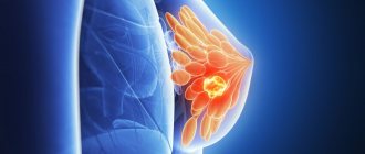
Symptoms of mastitis
Signs of inflammation of the mammary gland are classic: pain, redness, swelling, fever, dysfunction.
- engorgement, swelling
- skin redness
- bursting pain
- infiltrate
- pus in milk
- skin changes such as gangrene, infiltration
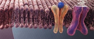
Inflammation is usually associated with a violation of the patency of the ducts and the addition of a secondary infection, which travels from the nipple into the gland tissue through the milk ducts and lymphatic vessels. With a normal level of immunity, the process is limited by a capsule, an abscess is formed, with immunodeficiency, pus spreads through the tissues - the phlegmonous form.
Classification of the disease
There are 2 types of mastitis:
- Lactational.
- Non-lactational (fibrocystic).
It occurs in the postpartum period as a result of infection entering the mammary gland - through microtraumas on the nipples and skin. In 90% of cases it occurs in an acute form. The main risk group is first-time mothers.
It develops due to hormonal disorders, low immunity, some chronic diseases, for example, radiculitis of the thoracic region, and previous viral infections. It is characterized by a less acute course, but more often becomes chronic.
Only in 10% of women the inflammatory process is activated in both mammary glands. It mainly affects one breast, more often the right one.
Stages of the disease
Serous (initial stage) - redness of the skin is noticeable, while the focus of inflammation does not have clear boundaries, the temperature rises.
Infiltrative - an accumulation of cells mixed with blood and lymph form a clearly palpable compaction. It is diagnosed in more than 50% of cases, because, having endured or not noticing the initial stage of mastitis, a woman decides to see a doctor. The appearance of infiltration is a borderline condition, when it is still possible to be cured with medication.
Purulent - the formation of one or more cavities filled with pus. In 80% of cases it is diagnosed at 2-3 weeks of lactation. If the abscess is not opened and drained in time, the infant will acquire a complex destructive form:
- phlegmonous, when inflammation penetrates the subcutaneous fat and covers more than 3 quadrants of the chest. In this case, the general condition sharply worsens, convulsions and loss of consciousness are possible;
- gangrenous, in which pus permeates not only the breast tissue, causing their necrosis, but also enters the blood and lymphatic vessels. As a result, the risk of blood clots and the development of toxic shock increases - factors leading to death.
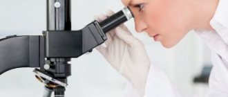
Diagnosis of inflammation of the mammary gland (mastitis)
Typically, mastitis is quite easily diagnosed by the patient herself, but it is still worth contacting a specialist. At the I. Medvedev Medical Center, appointments and emergency care are provided 363 days a year, and treatment focuses on minimally invasive and conservative techniques.
If a patient has pain in the mammary glands, treatment begins with diagnosis. It includes:
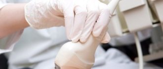
- inspection and palpation
- Ultrasound of the mammary glands
- if necessary, puncture with histology of the punctate contents
Treatment
At first, when mastitis is not yet purulent, but serous, you can get by with cold compresses, frequent pumping (or feeding), and antibiotics if the inflammation lasts longer than a day. In this case, penicillins, including inhibitor-protected ones, are preferred. When a woman experiences inflammation of the mammary gland, treatment differs depending on the stage of the process.
If an abscess has already formed, there is no point in prolonging it. Puncture of the abscess under ultrasound control can resolve the process conservatively; if not, then it is necessary to perform resection of the segment of the gland in which the abscess lies, with drainage of the cavity.
As for breastfeeding, it is contraindicated if there is pus in the milk; if not, then no restrictions are imposed.
Publications in the media
For the treatment of this disease, you can contact the Department of Resuscitation and Intensive Care of Generalized Surgical Infections with purulent surgery wards of the Clinic of Faculty Surgery named after. N.N. Burdenko
Mastitis (breast) - inflammation of the mammary gland. Periductal mastitis (plasmacytic mastitis, subareolar abscess) is inflammation of additional glands in the areola area. Neonatal mastitis is mastitis that occurs in the first days of life as a result of infection of hyperplastic glandular elements.
Classification • By course •• Acute: serous, purulent, phlegmonous, gangrenous, abscess •• Chronic: purulent, non-purulent • By localization: subareolar, intramammary, retromammary, diffuse (panmastitis). Etiology • Lactation (occurs in the postpartum period; see Breastfeeding) • Bacterial (streptococci, staphylococci, pneumococci, gonococci, often combined with other coccal flora, Escherichia coli, Proteus) • Carcinomatous.
Risk factors • Lactation period: impaired outflow of milk through the milk ducts, cracks in the nipples and areola, improper care of the nipples, poor personal hygiene • Purulent diseases of the skin of the breast • Diabetes • Rheumatoid arthritis • Silicone/paraffin breast implants • Taking HA • Removal breast tumors followed by radiotherapy • Long-term smoking history.
Clinical picture • Acute serous mastitis (can progress with the development of purulent mastitis) •• Sudden onset •• Fever (up to 39–40 ° C) •• Severe pain in the mammary gland •• The gland is enlarged in size, tense, the skin over the lesion is hyperemic, on palpation - a painful infiltrate with unclear boundaries •• Regional lymphadenitis. • Acute purulent abscess mastitis •• Fever, chills •• Pain in the gland •• Breast: redness of the skin over the lesion, sharp pain on palpation, softening of the infiltrate in the center with the presence of fluctuations •• Regional lymphadenitis. • Acute purulent phlegmonous mastitis •• Severe general condition, fever •• The mammary gland is sharply enlarged, painful, pasty, the infiltrate without sharp boundaries occupies almost the entire gland, the skin over the infiltrate is hyperemic, has a bluish tint •• Lymphangiitis, regional lymphadenitis.
Laboratory tests • Leukocytosis, increased ESR • Bacteriological examination is necessary to determine the sensitivity of microorganisms to antibiotics, bacterioscopy if fungal infections, tuberculosis are suspected. Special studies • Ultrasound • Mammography (if it is impossible to completely exclude breast cancer in patients with non-lactation mastitis) • Breast biopsy. Differential diagnosis • Infection of breast cysts (including parasitic ones) • Mastitis-like cancer • Tuberculosis • Actinomycosis • Suppurating atheroma • Syphilis.
TREATMENT Conservative therapy • Isolation of mother and child from other mothers and newborns • Bandage or bra that supports the mammary gland • Dry heat on the affected mammary gland • Expressing milk from the affected gland to reduce its engorgement • Stopping breastfeeding if purulent mastitis develops • If pumping is impossible and there is a need to suppress lactation, use drugs that suppress the formation of prolactin - cabergoline 0.25 mg 2 times / day for two days, bromocriptine 0.005 g 2 times / day for 4-8 days • Antimicrobial therapy with continuation breastfeeding - semisynthetic penicillins, cephalosporins: cephalexin 500 mg 2 times / day, cefaclor 250 mg 3 times / day, amoxicillin + clavulanic acid 250 mg 3 times / day; if anaerobic microflora is suspected, clindamycin 300 mg 3 times a day (in case of refusal of feeding, any antibiotics can be used) • NSAIDs • In case of stopping feeding, a solution of dimethyl sulfoxide in a dilution of 1:5, topically.
Surgical treatment • Fine-needle aspiration of the contents • If punctures are ineffective - opening and draining the abscess with careful separation of all bridges • Surgical incisions •• For subareolar abscess - along the edge of the parapapillary field •• Intramammary abscess - radial •• Retromammary - along the submammary fold • For small sizes a focus of fungal or tuberculous etiology, chronic abscess - it is possible to excise it with adjacent altered tissues • If the process progresses with the development of panmastitis - removal of the gland (simple mastectomy).
Complications • Fistula formation • Subpectoral phlegmon • Sepsis. The course and prognosis are favorable • Full recovery occurs within 8–10 days with adequate drainage. Prevention • Careful care of the mammary glands • Maintaining feeding hygiene • Using emollient creams • Expressing milk.
ICD-10 • O91.2 Non-purulent mastitis associated with childbirth • P39.0 Neonatal infectious mastitis • N61 Inflammatory diseases of the mammary gland • P83.4 Swelling of the mammary glands in the newborn
Breast inflammation and hyperplasia
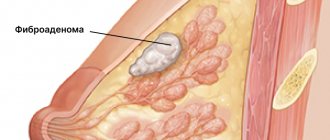
Dyshormonal processes, with a predominance of estrogen, also cause pain and discomfort in the gland, but, unlike mastitis, this is a non-inflammatory process. Treatment for breast hyperplasia is fundamentally different from mastitis; here hormonal therapy comes to the fore. Hyperplasia is the proliferation of gland tissue with the formation of cysts, cell swelling, and discharge from the nipple. It refers to mastopathy, fibroadenoma and adenomas.
Increased synthesis of prolactin plays a major role in this disease, so drugs that suppress its production, including herbal medicine, are widely used. Abraham tree - twig - stimulates the synthesis of dopamine, which reduces the production of prolactin; this component is contained in the herbal medicine Mastodinon.
Focal single formations can be removed surgically, by sectoral or atypical resection along with a block of adjacent tissue. Economical resection is carried out at the I. Medvedev Medical Center with a good cosmetic effect, so the patient does not have to worry about the aesthetics of appearance and the outcome of the disease.