Breast diseases in women are very common. Today, every third representative of the fair sex is faced with some kind of breast pathology. Diseases develop due to many factors: lifestyle, genetic predisposition, endocrine disorders, gynecological diseases, etc., so it is difficult to avoid them.
Painful sensations, lumps, vague discharge from the nipples and discomfort in the mammary glands are alarming symptoms that indicate the need to immediately consult a specialist - a mammologist. It is timely diagnosis and treatment that is the key to a woman’s recovery. According to statistics, in 80% of cases the identified diseases are benign and can be successfully treated.
Normal changes in the mammary glands
The content of the article
It is important to be able to differentiate the symptoms of pathological conditions from the symptoms of natural physiological processes.
The internal structure of the mammary glands is the same in all women, which cannot be said about external indicators. Each woman's breast has an individual shape, size, and asymmetry indicators. The condition of the mammary glands is affected by the menstrual cycle, pregnancy and menopause.
The following changes are considered normal:
- slight increase or decrease in mammary glands before menstruation;
- hardening or softening of breast tissue during ovulation or menstruation;
- pigmentation of nipple halos during pregnancy.
All other symptoms indicate an incipient disease, so they cannot be ignored. To understand how dangerous pathologies are, even if they do not cause severe pain or significant visual changes, let's look at the most common breast diseases in women.
Breast ultrasound or mammography? What to go through first?
Breast ultrasound and mammography (breast x-ray) are two complementary and popular research methods, each of which has its own advantages and disadvantages. An ultrasound examination is always prescribed first and is preferable because it is non-invasive, completely painless and, unlike x-rays, in which the patient receives a small but dose of radioactivity, an absolutely safe technique.
An ultrasound machine can easily identify cysts, which is impossible to do with an x-ray. But there are some subtleties here too. Using an ultrasound, it is ideal to examine small breasts with elastic, dense skin and a small amount of fatty tissue, while in women over 45 years of age, in whom the glandular tissue is replaced by fatty tissue, mammography will show the condition of the mammary glands no worse.
Palpation of the breast (feeling with hands) does not reveal microscopic neoplasms, but the disease begins with a tumor of 1-2 mm. But with the help of ultrasound, 42% of such pathologies are detected.
For these and other reasons, the gynecologist will always prefer to do an ultrasound first, immediately receiving the results, and only then refer the patient for additional examination. If, of course, it is needed.
Mastopathy
Mastopathy occupies a leading position among all pathologies of the mammary glands. This disease has several types and is characterized by fibrocystic changes in the tissues of the mammary glands in the form of individual nodes or dense fine-grained formations of various sizes, often painful. Mastopathy is a benign disease, but extremely dangerous, since oncology often develops against its background. Among young women, the incidence rate reaches 30-45% and increases significantly after 40-45 years.
There are two main forms of mastopathy:
- diffuse - the appearance of fine-grained neoplasms scattered throughout the entire structure of the gland;
- nodular - the formation of large formed nodes.
In the early stages of development, the disease may be asymptomatic. The first manifestation of the pathology is tenderness of the mammary glands and their increase in volume, which occurs before menstruation. A woman may also experience the following characteristic symptoms:
- Diffuse compactions and tumor nodes identified by palpation (the entire gland may feel compacted). They can be painful, and the pain becomes more noticeable in the second phase of the cycle and immediately before menstruation. With a long course of mastopathy, the pain syndrome persists throughout the entire cycle. Moreover, pain can radiate to the armpits, shoulders or back;
- Periodic appearance of aching pain in the chest area;
- The appearance of cracks in the nipples, they become less convex;
- Discharge from the nipples of varying degrees of abundance and of varying nature. They can be transparent, white, green or brown, and bloody. The latter are especially dangerous because they are an alarming sign of the development of cancer.
There are many factors contributing to the development of mastopathy:
- early first menstruation;
- early menopause;
- menstrual irregularities;
- hormonal imbalance disorder;
- hormone therapy;
- no history of pregnancy or childbirth;
- prolonged absence of childbirth;
- more than three terminations of pregnancy;
- irregular sex life (or prolonged abstinence);
- gynecological diseases with increased estrogen levels;
- short lactation period (less than 6 months);
- disorders of the endocrine system (diabetes mellitus, dysfunction of the adrenal glands, liver, pancreas, etc.);
- genetic predisposition;
- gastrointestinal diseases;
- psychological factor – frequent stress, neuroses, depression;
- injuries in the chest area (impacts, compression);
- excess body weight;
- unhealthy lifestyle (bad habits, lack of sleep, poor diet);
- unfavorable environmental conditions.
Mastopathy must be treated immediately, since advanced stages of mastopathy are very dangerous.
Top 11 most frequently asked questions from women about breast health.
ANSWERS OF OUR MAMMOLOGIST, EKATERINA NIKOLAEVNA MAKAROVA TO THE TOP 11 MOST FREQUENT QUESTIONS OF WOMEN ABOUT BREAST HEALTH.
QUESTION #1 – WHY CAN ONE BREAST BE BIGGER THAN THE OTHER?
Asymmetry of the mammary glands can be detected in healthy women.
A slight difference in the volumes of the right and left breasts is observed quite often and is considered a normal variant. This asymmetry occurs in many women and should not be a cause for concern.
Fear and alarm should be caused by a sudden increase in the volume of one of the breasts, which occurred in a short time. When such an increase occurs, a woman needs to undergo an examination such as an ultrasound of the mammary glands (on the 5-7th day from the start of menstruation) and an examination by a mammologist. This way you can exclude a severe pathological process (including oncopathology), which can provoke an increase in the volume of one of the mammary glands.
QUESTION No. 2 – WHY CAN THE MAMMY GLANDS BE THICK BEFORE MENSTRUGGLES?
Before the onset of menstruation, women may experience pain (in the nipples or breasts as a whole) and engorgement of the mammary glands for several days or 1-2 weeks. In most cases, the appearance of such symptoms is associated with mastopathy, which is provoked by a hormonal imbalance that occurs due to various reasons.
To identify this common pathology of the mammary glands, a woman needs to undergo a number of standard examinations:
- examination by a mammologist;
- examination by a gynecologist;
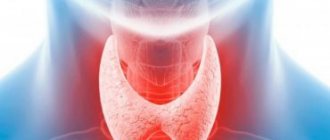
- Ultrasound of the mammary glands, uterus, ovaries and thyroid gland;
- blood tests for hormones (FSH, progesterone, estradiol, TSH, T3 and T4) and analysis for tumor markers;
- cytological analysis of nipple discharge (if any);
- puncture biopsy (if necessary).
One month before the examination, it is advisable to stop taking oral contraceptives - this issue should be discussed with your doctor.
After analyzing all the data received, the mammologist will be able to make a diagnosis. As a rule, such symptoms are characteristic of one of the forms of mastopathy (fibrous, nodular, mixed, cystic, etc.). After this, the woman can be prescribed the necessary treatment.
QUESTION #3 – WHY DOES HAIR GROW AROUND NIPPLES?
Throughout their lives, women may experience periods of hormonal surges, which may manifest as hair growth around the nipples. This symptom is considered a variant of the norm, because normally there are hair follicles in the areola, which in some women can be activated.
However, we should not forget that the symptoms of hirsutism should be a cause for concern; this is a sudden excess growth of male-pattern hair (on the chest, back, chin or upper lip). This disease indicates a significant imbalance in hormones and requires clinical observation and treatment.
QUESTION No. 4 – WHY DO SERUS OR BLOODY DISCHARGE APPEAR FROM THE NIPPLES?
The presence of transparent yellowish or brown discharge indicates a pathological process occurring in the mammary gland. Typically, such a symptom indicates the development of one of the forms of mastopathy, but sometimes discharge from the breast can be a sign of more dangerous diseases. If such a symptom appears, a woman should definitely contact a mammologist to clarify the diagnosis.
QUESTION #5 – WHAT ARE THE MOST COMMON SYMPTOMS OF BREAST DISEASES?
The most common signs of pathological processes in the mammary glands are the following symptoms:
- feelings of tension;
- painful sensations;
- skin changes in the breast, nipple or areola area;
- nipple discharge;
- determination by palpation of areas of compaction of gland tissue.
QUESTION No. 6 – HOW AND WHEN TO CONDUCT A BREAST SELF-EXAMINATION?
Breast self-examination can help detect cancer in the early stages.
Every woman should know that once a month (about the same days, 6-7 days after the end of her period) she should conduct a breast self-examination: this is the best way to early detect any changes in the condition of the mammary gland tissue .
The procedure should be carried out as follows:
- Inspect the bra in those places where it comes into contact with juices and make sure there are no stains.
- Examine the nipples and areola, making sure there are no changes: redness, retractions, peeling, rashes or ulcerations.
- Stand in front of the mirror, raise your hands behind your head and pay attention to the shape of your breasts and the presence of any irregularities, protrusions or depressed areas in certain parts of the chest.
- Examine the skin of the breast for changes in its color or changes in the appearance of “lemon peel”.
- Perform alternate palpation of the mammary glands. To do this, you need to lie down on the bed and place a bolster or small pillow under the shoulder blade (on the side of the gland being examined) so that the chest is slightly elevated and the mammary gland is most spread out. The right mammary gland can be felt with the fingertips of three or four fingers of the left hand. The fingers should be placed flat and moved in a circle (from the nipple to the outer edge of the gland), moving them centimeters and feeling the entire area of the gland. In the same way (only with the right hand) the left breast is felt.
- If you are unable to palpate your breasts this way, you can do it in the bathroom - wet fingers with soapy suds will glide over the skin better, and it will be easier for you to feel the lumps in the gland.
Remember that identifying any lumps, lumps or painful areas should always be a reason to immediately contact a mammologist. And don’t put off visiting your doctor until later, because malignant tumors of the mammary glands can be rapidly progressing and extremely aggressive, and you may miss your chance for recovery.
QUESTION No. 7 – HOW OFTEN DO YOU NEED TO HAVE A PREVENTIVE EXAMINATION WITH A MAMMOLOGIST?
Remember that even regular breast self-examination does not free a woman from the need to undergo a preventive examination once a year with a mammologist or breast oncologist.
Only a specialist who has experience in diagnosing diseases of the mammary glands and works in a medical institution with sufficient technical equipment for professional examination of the mammary glands will be able to professionally assess the condition of the breast and give you the necessary recommendations.
It is better to schedule a visit to the doctor in the first days after menstruation: approximately from 1 to 10 days.
QUESTION No. 8 – WHAT DIAGNOSTIC METHODS ARE USED TO STUDY THE BREAST GLANDS?
The main methods for diagnosing breast pathologies include the following types of studies:
- examination and palpation of the mammary glands;
- Ultrasound of the mammary glands;
- x-ray mammography;
- blood test for hormones (prolactin, FSH, progesterone, estradiol, TSH, T3 and T4);
- blood test for breast tumor markers CA 15-3, CA 27-29 and CEA;
- microwave radiothermometry (RTM research);
- electrical impedance tomography (EMM);
- CT;
- MRI;
- biopsy of tumor tissue followed by cytological or histological examination;
- axillography;
- ductography;
- scintigraphy.
The scope of breast examinations is determined by a mammologist individually for each patient.
For the purpose of preventive examination, women are recommended to conduct the following regular studies:
- Ultrasound of the mammary glands (for women under 40 years old - once a year, and for people with an increased risk of the disease, mammography is recommended);
- mammography (for women over 40 years old - 1-2 times a year, over 50 years old - annually).
Such preventive instrumental methods for examining the mammary glands are the gold standard for screening breast diseases and are recognized throughout the world. They make it possible to identify various diseases at the earliest stages and significantly reduce the risk of uncontrolled progression of tumor diseases of the mammary glands.
QUESTION No. 9 – WHICH EXAMINATION IS MORE RELIABLE – BREAST ULTRASOUND OR MAMMOGRAPHY?
Ultrasound of the mammary glands is a non-invasive, safe (in terms of dose load), painless and informative procedure, which is usually prescribed to young women and pregnant or nursing mothers. However, most doctors, when treatment is necessary, prefer to rely on mammography data, because they consider this diagnostic method to be more informative and accurate.
Some mistrust of the data obtained from ultrasound is due to the fact that the female breast is one of the most difficult objects for examination, since the structure and density of gland tissue constantly changes throughout a woman’s life and depends on age, body weight, physiological periods life, phase of the monthly cycle and the presence of pathological foci.
QUESTION #10 – WHAT IS THE PROBABILITY OF GETTING BREAST CANCER?
The risk of malignant neoplasms in the mammary glands is individual and depends on various factors. Taking into account the fact that the number of patients with breast cancer is steadily increasing from year to year, it should be remembered that, starting from the age of 40, every woman should regularly visit a mammologist and undergo preventive mammography.
The main risk factors for the development of breast cancer are:
- age – the risk increases after 40-45 years and reaches its peak at 65 years;
- heredity - especially if there are cases of illness in the mother or siblings;
- presence of pathologies of the mammary glands in the anamnesis;
- earlier onset of menstruation – before the age of 11-12 years;
- late onset of the first pregnancy (or complete absence of childbirth) - after 30-35 years;
- the onset of menopause is too early - before 45 years of age;
- too late menopause - after 55 years;
- refusal to breastfeed;
- prolonged contact with carcinogenic substances or ionizing radiation;
- Frequent chest X-ray examinations;
- obesity.
Research by scientists to study other factors that contribute to the degeneration of breast tissue cells into cancer cells continues to this day. These include: taking hormonal oral contraceptives and hormone replacement therapy for perimenopausal disorders, smoking, drinking alcohol and eating high-fat foods.
QUESTION No. 11 – HOW TO PREVENT THE DEVELOPMENT OF BREAST CANCER?
Despite the fact that scientists have not yet been able to create an effective vaccine for breast cancer, a woman can take care of its prevention by following simple rules:
- regularly conduct breast self-examination and contact a mammologist if any changes in its tissue are detected;
- regularly conduct preventive ultrasounds of the mammary glands and mammography;
- wear a properly selected bra - it must fit perfectly, properly hold the breasts in a normal anatomical position (it is not recommended to choose underwire or push-up models);
- to refuse from bad habits;
- eat right - introduce more vegetables and cereals into your diet, eat fresh fruits and vegetables that have antioxidant properties, do not abuse fatty and fried foods;
- maintain normal weight and get rid of extra pounds;
- do not give up breastfeeding;
- strengthen the chest muscles with gymnastics and lead an active lifestyle;
- to be a self-sufficient person - according to the observation of many scientists, doctors and psychologists, breast cancer often develops in those women who neglect themselves, deny themselves care and attention to themselves and are often offended by the people around them who treat them badly.
Dear women and girls, please take care of your health, love, take care of yourself! We are waiting for you at our Medical Center!
Lactostasis
Lactostasis is the name given to milk retention, which occurs when there is significant formation of milk or when its outflow is disrupted. The disease predominantly develops in primiparous women in the first 10 days after birth.
Lactostasis does not pose a threat to the life and health of a woman. However, ignoring the disease or its illiterate elimination can lead to dangerous consequences - the development of mastitis (inflammation of the mammary gland). Hypothermia (feeding in a draft) or overheating (compresses, hot baths) can also lead to complications.
Lactostasis manifests itself with a number of symptoms:
- breast lump;
- pain on palpation;
- feeling of heaviness and fullness of the chest;
- expansion of the saphenous veins on the chest in the area of stasis;
- feeling of heat and local hyperemia;
- increase in body temperature to 38°C.
Symptoms may either improve after emptying the mammary glands or persist. During the feeding process, a woman may experience severe pain. The stagnation zone can shift and increase.
The main reasons for the development of lactostasis:
- insufficient emptying of the breast;
- compression of the mammary glands by tight clothing or fingers during feeding;
- infrequent feedings (excess milk forms in the mammary glands);
- incorrect position of the woman or child during breastfeeding;
- aggressive pumping (provokes the production of additional
- milk);
- premature introduction of artificial formulas into the child’s diet (the baby may refuse breastfeeding, which will lead to stagnation of unused milk);
- refusal to breastfeed;
- stress and physical exhaustion;
- sagging of the mammary glands (impedes the flow of milk from the lower part of the large breast).
Lactostasis is easily eliminated in the first stages, so it is important to contact a gynecologist immediately.
Mastitis
Mastitis is a fairly common pathological condition that has varying degrees of severity and is characterized by the development of an inflammatory process in the tissues of the mammary glands. As a rule, the disease occurs during lactation. However, there are cases of mastitis that are not associated with feeding and childbirth - in women over 50 years of age and in newborns. Therefore, there are two main types of mastitis: non-lactation and lactation.
Mastitis is dangerous due to possible multiple complications: abscess, necrosis, phlegmon, etc. That is why, when you notice the first symptoms of the disease, you should immediately contact a specialist. With timely diagnosis, conservative treatment can be used; in advanced cases, surgery cannot be avoided.
In the early stages of development, the pathology manifests itself:
- redness, warming and swelling of the mammary glands;
- tension and feeling of chest fullness;
- painful sensations in the chest when pressed;
- pain and burning during breastfeeding;
- a sharp increase in body temperature;
- chills, headache, muscle and joint pain, general malaise;
- retraction of the nipple.
In the absence of appropriate treatment, symptoms worsen, new signs of the disease are observed:
- enlarged lymph nodes located in the axillary region on the side of the affected mammary gland;
- rapid pulse;
- further increase in body temperature, sweating;
- purulent or bloody discharge from the nipple.
The main cause of mastitis is an infection that enters the mammary glands through microtraumas on the chest. The most common culprit (in 70% of cases) is the pathogenic bacterium Staphylococcus aureus, which is usually transmitted from the nasopharynx of newborns during breastfeeding. Also, the causative agents of the disease can be streptococci, E. coli, tuberculosis bacteria, and fecal enterococcus.
Infection can occur through underwear and bedding, personal hygiene items, or people carrying bacteria. Also, in a nursing woman, mastitis can be caused by lactostasis.
Factors that increase the risk of developing the disease are:
- thyroid dysfunction;
- weaning the baby;
- improper feeding;
- physical exhaustion;
- weakened immunity;
- side effects of medications;
- stress;
- use of silicone breast implants;
- hypothermia (hypothermia);
- injuries to the breast and nipples, including piercings;
- infections of the sebaceous and sweat glands;
- syphilis;
- actinomycosis;
- mastopathy.
What a breast ultrasound sees and doesn’t see. Which ultrasound sensors and machines are better?
Regular clinics and hospitals are equipped with ultrasound machines of standard quality, equipped with several universal sensors, including for breast examination. They have high resolution clarity, display echogenicity and contours, but only detect fairly large neoplasms measuring 5 mm or more.
The tumor reaches this size 8 years after its appearance. This means that an important moment when it was still possible to cure the pathology quickly and without significant interventions may be missed. But other pathologies in the chest - cysts, fibroids, blocked ducts, etc. Ultrasound sees 5 points.
For this reason, ultrasound diagnostics of the mammary glands should be complemented by other methods if oncology is suspected (sunken nipple, breast asymmetry, lump upon palpation). In these cases, mammography or, in special cases, MRI is additionally prescribed.
Standard ultrasound clearly displays structural changes in breast tissue: the presence of nodules, cysts, deterioration of blood circulation, and the occurrence of infiltration. The doctor’s experience helps to suspect oncology and refer the patient for a more detailed examination.
Today, there is already an ultrasound method that detects tumors at an early stage. These are ultrasound machines of the ABVS (Automated Breast Volume Scanner) system, developed by Siemens specifically for mammological studies.
A special sensor installed on a woman’s chest allows you to obtain a 3D image on the monitor screen. Screening is carried out automatically and does not depend on the experience and qualifications of the doctor. The information received is processed by a special program for 15 minutes, while the sensor lies on the woman’s chest and takes pictures in different sections from the chest wall to the tip of the nipple. The ABVS system allows you to examine women who have a hereditary predisposition to breast cancer and who have several blood relatives who suffered from or died from this disease.
Lipoma
Breast lipoma is a benign disease characterized by the growth of fatty tissue in the breast area. The appearance of such tumors is typical for women after 40 years of age. Lipoma itself does not pose a threat to the health and life of a woman. It creates only a cosmetic defect: due to its ability to reach a large size, the lipoma deforms the mammary gland.
The only treatment option is surgery. Malignancy is extremely rare.
Breast lipoma can be either single or multiple (lipomatosis). It can include not only fat cells, but also other tissues. The following types of disease are distinguished:
- lipofibroma – predominance of adipose tissue;
- fibrolipoma – the presence of fibrous (connective) tissue;
- angiolipoma - riddled with small blood vessels;
- myxolipoma – the presence of mucus cells;
- myolipoma - muscle fibers are detected.
Typically, lipoma does not manifest itself with characteristic symptoms. It can be detected independently by palpation (palpation). It is inactive, painless and has a dough-like consistency. Often the lipoma reaches up to 2 cm in diameter, and can also be large in size - up to 10 cm. In some forms of the disease, a woman may feel pain and discomfort in the chest.
The following causes of the disease are identified:
- blockage of the outlet duct of the sebaceous glands;
- metabolic disorders;
- excess body weight;
- hereditary predisposition.
The following factors are predisposing:
- sedentary lifestyle (lymph movement is impaired);
- unbalanced diet, which is dominated by fats and carbohydrates;
- changes in hormonal levels;
- mechanical injuries of the mammary gland;
- long-term use of hormonal contraceptives;
- the appearance of stretch marks on the chest due to pregnancy and lactation;
- bad habits (smoking, drinking alcohol, drugs);
- X-ray and ultraviolet irradiation;
- incorrectly selected underwear;
- unfavorable environmental conditions.
Features of anatomy
In an adult woman, the mammary gland has a structured structure, consisting of several types of tissue, each of which performs its own function. Let's take a closer look at the anatomical features:
- The outside of the gland is covered with skin, on which the juice area is located. It is rich in pigment cells, which plays an important role in feeding the baby - the baby instinctively finds the nipple, since its surface is slightly different from smooth skin. The areola also contains a large number of nerve endings.
- The parenchyma of the breast has a segmental structure - dense strands extend from the skin, which form large cells (15-20), surrounded by fatty tissue. They contain lobules of the mammary gland, separated by connective tissue.
- Each lobule consists of glands, inside of which there are many alveoli that produce milk. Convoluted tubules extend from them, they gradually unite and form the nipple opening.
- The entire anatomical structure is supported by a special cord called the ligament that supports the mammary gland. Underneath it there is a layer of fat that prevents soft tissue injuries.
Normally, there should be no lumps in the breast; the entire structure is soft and can be painlessly palpated. The mobile ligament provides slight displacement without noticeable discomfort. Proportional sizes mainly depend on the volume of adipose tissue that fills the free space between the cells.
Breast cyst
A cyst is a type of benign tumor. It consists of single or multiple hollow neoplasms filled with fluid. These pathological cavities form in the ducts and have a round shape with clearly defined boundaries. The disease is dangerous due to the development of inflammation and suppuration of the cystic cavity.
Malignancy occurs quite rarely, but the disease increases the risk of developing cancer. In addition, large cysts deform the mammary glands.
The disease can be asymptomatic for a long time without bothering the woman. Then the following symptoms appear:
- small soft or hard round formations in the mammary gland;
- discomfort and pain when pressing on the chest;
- soreness, pulling sensations and burning sensations in the mammary gland, intensifying before and during menstruation;
- large cysts change the shape of the breast, causing a change in the color of the skin above them (redness, then cyanosis);
- when inflammation develops in the cyst - fever, redness of the mammary glands, enlargement of the axillary lymph nodes.
The causes of breast cysts are as follows:
- hormonal imbalance;
- mastitis;
- disorders of the thyroid gland;
- ovarian dysfunction;
- inflammatory diseases of the genitourinary system;
- psycho-emotional factor (excessive mental stress, frequent stress, nervous strain, anxiety, etc.).
What does pain indicate?
For most women, burning and mutual dull pain in the chest is a consequence of premenstrual syndrome. A burning sensation and pain in the chest is observed at the beginning of pregnancy.
Lumpiness and tenderness in the breasts occurs when milk accumulates in the mammary glands of a nursing mother. When cracks form in the nipples, there is a risk of infection and the development of mastitis.
Localized pain and burning in the chest can be the cause of the development of benign formations. This pain intensifies in a certain position of the body.
Burning and pain in one side of the chest may indicate the development of cancer. Tumor formations of the breast are most often located in the upper outer quadrant.
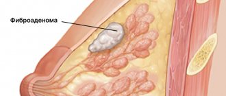
Fibroadenoma
Fibroadenoma (fibrosis) is a benign neoplasm that develops from connective tissue. The tumor occurs against the background of the production of hormones - estrogens. The disease is predominantly observed in women 20-30 and 40-50 years old, sometimes occurring at a young age (12-20 years). It does not pose a threat to the health and life of a woman. In most cases, fibroadenoma is not prone to progression and development of complications.
Sometimes it can reach large sizes and thereby deform the mammary glands. Only fibrosis of gigantic size is prone to malignancy. As a rule, the method of treating a tumor is surgical, but if it is small in size (up to 5-8 mm), therapeutic treatment is carried out aimed at its resorption.
Pathology is of the following types:
- Nodular - deformation of connective tissues near the glandular ducts or their ingrowth into the ducts;
- Leaf-shaped - characterized by an extreme speed of development and an increased threat of degeneration into sarcoma. This is a terrible disease that leads to breast amputation and even death.
Fibroadenoma is asymptomatic for a long time. The general symptoms are not pronounced. It is manifested by compactions in the body of the mammary glands, which have the following features:
- painlessness, density and sometimes mobility of the formation;
- clearly defined boundaries;
- variation in tumor size;
- lack of direct dependence of size changes on the phase of the menstrual cycle;
- intensive growth of education (sometimes).
In medicine, the exact causes of fibroadenoma are still unknown. There are a number of factors contributing to the development of pathology:
- genetic predisposition;
- puberty;
- diseases of the endocrine system and thyroid gland;
- pregnancy;
- frequent abortions;
- nervous and physical exhaustion;
- severe frequent stress;
- obesity;
- incorrect selection of oral contraceptives or their uncontrolled use;
- mammary gland injuries;
- overheating of the mammary glands;
- abuse of solarium and sunbathing.
Changes in the breasts during pregnancy
The mammary gland can function fully only during pregnancy and after childbirth. Natural physiological mechanisms are launched that ensure milk secretion.
The structural features are determined by three hormones:
- Progesterone - under its influence, the alveoli reach their final development and are ready for secretion. The milk ducts grow in the lobules, the glands prepare to produce milk.
- Prolactin - produced immediately after childbirth, triggers secretion in the alveoli and the release of colostrum through the ducts. It contains a large amount of vitamins, minerals and nutrients that a child needs.
- Oxytocin - supports the secretion and production of mother's milk for several months. Under its influence, the alveoli remain in a hypertrophied state until the end of feeding. When the hormone titer decreases, all structures of the mammary gland undergo reduction.
Against the background of all these changes, the mammary glands are exposed to a dozen hormones. The whole system works like a clock; even a small malfunction can lead to disruption of cell division in the breast and the appearance of problems.
Intraductal papilloma
Intraductal papilloma is a cyst-like formation with a disruption of the structure typical of secretory cells. Diagnosed in 10% of cases of breast diseases in women. It can develop at any age - from puberty to postmenopause. This is a benign tumor that can progress to malignant. Its size varies from 1 mm to 3 cm in diameter.
There are two main types of intraductal papilloma:
- central – location of cystadenomas in the areola area;
- peripheral - the occurrence of papillomas in any peripheral area of the mammary gland ducts, often multiple.
Often the disease is asymptomatic for a long period, and its diagnosis occurs by chance during an examination by a doctor. Symptoms of the disease include:
- discharge from the nipples of various types, increasing when pressing on the chest. May be transparent, bloody, whitish or greenish in color;
- pain in the area where the tumor develops, aggravated by pressure or when wearing tight clothing;
- palpation of a small elastic knot.
With the development of inflammation of the tumor and surrounding tissues, signs are observed:
- increased body temperature;
- severe tenderness of the mammary glands;
- swelling, breast enlargement;
- redness of the mammary glands;
- change in color and consistency of discharge.
The causes of intraductal papilloma are:
- hormonal imbalance;
- changes in hormonal homeostasis (ovarian dysfunction, oophoritis, adnexitis, abortion, obesity, stress, etc.);
- long-term uncontrolled use of hormonal contraceptives;
- smoking;
- influence of viruses;
- ionizing radiation;
- no history of pregnancy.
Embryological and comparative anatomical data.
The first rudiment of the milk secretion organs appears in the form of a paired epithelial strip on the ventral wall of the body. This rudiment is called the milky stripes. The milky stripes thicken and turn into milky plates (milky lines). The individual projections developing from them are called milk tubercles (primary nipples). These latter, becoming flatter and growing deeper like protrusions, give this name. milky points, or points; through the immersion of individual glandular areas, they lead to the formation of milky pockets.
The first germ of mamma
Guldberg (1899) found in dolphin embryos, 18 millimeters long, in the form of vaguely demarcated elevations of the epidermis, which in this place forms a crescent-shaped protrusion. The rudiment appears just at the time when the temporary hind limbs begin to show a tendency to disappear.
In cattle embryos, according to G. Burckhardt, “accessory” or “anal” nipples are found very often in both sexes (in approximately 37% of cases). They are always located between the normal nipples (on one or both sides), or behind the back pair of normal ones; but they are never ahead of the first pair. The mammary organs of modern cattle are subject to the process of caudocranial reduction.
Both in humans and in cattle, they distinguish between hypermastia, with all the signs of a normal, but only very small, mammary organ, and hyperthelia (false nipples).
False nipples and mikromammae
disappear, for the most part, already before birth.
In his article on the morphology and history of the development of normal and accessory mammary glands, G. Schickele
) (1889) comes to the following conclusions: in some mammals (mouse, rat, rabbit, cat) the nipples are arranged in 2 groups, which are characterized by cranial and caudal convergence, as well as the presence of a typical significant gap between the most caudal and the most cranial pair of glands.
Accessory areas of glands in these animals, of course, occur only to a limited extent. But broad-nosed monkeys have extra nipples more often than narrow-nosed monkeys. At Zebus
this condition seems to be observed very often.
In guinea pigs and mice, the milk line is the starting point for the formation of glands. Adult guinea pigs have never had more than 2 teats. But embryonic accessory rudiments of the mammary glands are found in varying numbers (from 2 to 10); but they are at a lower stage of development than the rudiments of the main glands ( Ztschr. fur Morph. u. Antroph. 1899
).
Schmidt, H, Uber normale Hyperthelie menschlicher Embryonen. Anat. Anz. XI, 1896; and Morphologische Arbeiten, herausg. v. G. Schwalbe. 7 Bd.
1896.
Humans do not have a milk line as noticeable from the outside as in mammalian embryos, or it is present only for a short distance. But microscopic examination reveals growths of the epithelium and the rudiments of the mammary glands. In one case there were 8 additional rudiments on each side, 4 above and 4 below the main rudiment. In other cases, a rudiment of 7 to 14 parts was found on one side. The cranial primordia were mostly oriented laterally, while the caudal primordia were oriented medially with respect to the normal primordium. In the lower abdomen, the rudiments were completely absent.
Callius ( Anat. Hefte, Bd. III
, 1897) found in a 15 mm human embryo a milky line 1.5 mm long.
Regarding the comparative anatomy of the mammary glands, the reader can find detailed data in the relevant textbooks. We will only point out here that Monotremata has a paired and “glandular area,” which generally corresponds to areola mammae
higher, is the only external apparatus; The glands here are built according to the tubular type. In echidna, such a device is hidden in a skin pocket. Alveolar mammary glands first appear in marsupials (Gegenbauer).
Bonnett, Die Mammaorgane. Ergebnisse der Anat. von Merkel und Bonnett. Bd. II and VII. — Breslau , E., Beitrage zur Entwicklungsgeschichte der Mammarorgane bei den Beuteltieren. Zeitschr. f. Morph. u. Antrop. IV
, 1902.
The pouch arises through the fusion of small pockets (marsupial pockets), each of which contains one lacteal rudiment (mammary pocket). Marsupialia bag
is homologous to the echidna bursa, in the same way the marsupial pocket of the first is homologous to the corresponding formation in the second;
the mamillary pouch of Marsupialia
corresponds to the glandular field of the echidna.
Marsupial pouches in marsupials can be demonstrated even in placental animals. They correspond to the pockets that surround the Murinae
nipples. In all mammals, the mammary glands arise in a homogeneous manner and should be classified as tubular skin glands. The possibility of diphyletic origin must be excluded.
Unger (1898) also subscribes to this conclusion. The mammary glands are derived from the glomerular glands. Normal, comparative and pathological anatomy, phylogeny and ontogeny unanimously point to precisely this origin of the mammary glands. Neither their structure nor their function gives us the right to compare them with the sebaceous glands.
H. Eggeling
) in his work on the skin glands of Monotremata (Verh. anat. Ges. 1900) criticizes previous attempts to subdivide the skin glands.
As a principle of division, one must use, firstly, the ratio of the epithelium to the lumen, and, then, the method of secretion formation. All glomerular glands, and together with them the mammary glands of higher mammals, should be considered as constantly canalized skin glands secreting in the intravital state; the sebaceous glands and peculiar glandular organs of reptiles should be considered temporarily canalized, secreting through necrobiosis by the skin glands; their secretion is released necrobiotically, as the secreting cells die. v.
Runge, G., Die zunehmende Unfahigkeit der Frauen, ihre Kinder zu stillen. Munchen., E. Reinhardt , 1900.
Oleogranuloma
Oleogranuloma is a benign tissue compaction altered by inflammation as a result of a foreign body entering the mammary gland. As a rule, it occurs as a result of surgery. Silicone implants, synthetic threads, etc. can act as a foreign body.
The symptoms of oleogranuloma and cancer are similar to each other, so it is often mistaken for oncology. The final verdict can be made by the doctor after a thorough examination.
There are the following types of breast oleogranuloma:
- injection (artificial) – formed as a result of the introduction of various fats and oils;
- post-traumatic – occurs after trauma to the chest (blow, fall, improper massage). A frequent companion is necrosis of fatty fibers;
- periinflammatory – location near the source of the inflammatory process, gradual movement to fatty tissue, which turns out to be destructive;
- spontaneous (cutaneous) – causes unknown.
Oleogranuloma can be identified by the following signs:
- the appearance of voluminous nodules under the skin of the breast;
- lumpy and dense consistency of the skin surface;
- redness in the area of formation;
- pain and discomfort in the area of compaction;
- retraction of the skin over the tumor;
- increasing education over time;
- formation of ulcers, discharge of pus through fistulas (in advanced forms of the disease).
Causes of oleogranuloma:
- breast injuries;
- surgical interventions (removal of the mammary gland or its lobe, breast augmentation by introducing implants);
- a sharp decrease in body weight;
- ingress of foreign bodies (silicone, gels, ointments, synthetic threads);
- infectious and inflammatory diseases;
- hormonal imbalance;
- radiation exposure.
Breast tuberculosis
Breast tuberculosis is a rare but extremely dangerous pathology. Often a consequence of pulmonary tuberculosis. The disease predominantly affects women of reproductive age, less often the elderly.
There are several clinical forms of breast tuberculosis:
- nodular – the most common. Characterized by the formation of one or several painful nodes, the appearance of local skin ulcerations, accompanied by an inflammatory process;
- abscess – the fusion of several nodes into one tumor, which gradually softens and is accompanied by a purulent process;
- ulcerative-fistula - the appearance of ulcers and fistulas with the release of purulent fluid;
- disseminated – the presence of multiple confluent foci with caseosis;
- sclerosing - observed mainly in older women. The predominance of fibrous changes, the absence (or weak expression) of the processes of caseous degeneration.
The disease is characterized by the following symptoms:
- painful lumps in the body of the mammary gland;
- swelling, redness at the site of formation;
- nipple retraction;
- weakness, chills, general malaise;
- increased body temperature;
- a sharp decrease in body weight;
- loss of appetite;
- increased sweating of the head and armpits;
- increased temperature in the affected area.
It is extremely rare that tuberculosis of the mammary glands develops in isolation. It is usually combined with genital tuberculosis, tuberculosis of the lungs, intestines, bones, and spine. Prerequisites for infection are:
- weak immunity;
- diabetes;
- breastfeeding period;
- chest injuries;
- HIV;
- long-term therapy with glucocorticoids and immunosuppressive drugs.
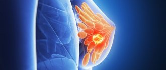
Mammary cancer
Breast cancer is the most common cancer in women, progressing more and more rapidly every year. This is a fast-growing malignant neoplasm with a high tendency to metastasize already in the early stages of the pathological process. Timely diagnosis significantly increases the likelihood of a full recovery.
There are 4 stages of breast cancer:
- Stage 1 – small (up to 2 cm) nodular neoplasms, no metastases, prognosis for recovery is favorable;
- Stage 2 – tumor from 2 to 5 cm, spread of metastases to regional axillary lymph nodes, prognosis is favorable if treatment is started early;
- Stage 3 – large tumor size (more than 5 cm), metastasis to internal organs and bones, poor prognosis;
- Stage 4 – total metastases in all organs and systems, the prognosis is extremely unfavorable (survival in 10% of cases).
Breast cancer manifests itself with a number of symptoms:
- painless dense formation with unclear boundaries in the mammary gland;
- a node or large formation in the armpit;
- deformation and change in breast size;
- bloody or purulent discharge from the nipples;
- the effect of “goose bumps” on the mammary gland, changes in its color, including nipples and areolas;
- redness;
- rash around or on the nipples;
- expression of blood vessels;
- the appearance of ulcers (as the disease progresses);
- changes in the appearance, size and sensitivity of the nipples, their retraction, itching;
- sharp asymmetrical enlargement of one breast;
- pain in bones and muscles;
- sudden weight loss;
- weakness, dizziness, general malaise.
Causes of breast cancer development:
- genetic predisposition;
- early onset of menstruation (before 12 years) and late completion (after 55 years);
- oncological diseases of the genital organs;
- bad habits (smoking, drinking alcohol, drugs);
- hormonal disbalance;
- chest injuries;
- mastitis;
- diabetes;
- hypertension;
- obesity;
- exposure to radiation;
- hormone replacement therapy;
- some professions (human contact with carcinogens and endocrine disruptors): gambling, plastics production, metalworking, agriculture.
Breast cancer is an insidious disease, so it can occur even without proper reasons.
What pathologies are detected on ultrasound of the mammary glands?
When examining the breast using an ultrasound machine, the doctor receives information about the structure of the organ (the ratio of glandular, fatty and fibrous tissues).
The mammary gland consists of three types of tissue: glandular, adipose and connective. With the help of connective tissue, the gland is divided into 15-20 lobes, the free space in which is filled with adipose tissue. The density of connective tissue is responsible for the shape of the breast; it attaches the gland to the chest. After childbirth, it stretches and loses tone.
Each lobe of glandular tissue is divided into smaller lobules, inside of which are the mammary glands. They turn into branching tubes with an expansion at the end in the form of alveoli - microscopic vesicles in which milk is formed during lactation. Tubes (lacteal ducts) extend from the alveoli, passing into the milky sinuses with a nipple at the end.
During an ultrasound examination, the doctor normally sees the following:
- the tissues must be well differentiated (they have different echogenicity, it should not be mixed);
- up to 40 years of age, glandular tissue predominates, after 40 - adipose tissue;
- the thickness of the glandular tissue in women under 40 years of age does not exceed 14 mm, but after 40 years of age it reaches 2 cm;
- milk ducts are homogeneous, without dilations;
- glandular and fatty lobules are not deformed;
- correspondence of morphotype to age.
The morphotype can be juvenile, early reproductive, prime, mature, premenopausal, postmenopausal, lactation. It is characterized by different ratios of glandular, connective and adipose tissue.
The specialist also looks at any deviations from the norm. These include identifying focal formations and their echogenicity
Areas with increased or decreased echogenicity indicate cysts, inflammation or tumors. If cysts, inflammations or tumors occur, the patient is additionally examined by ultrasound for nearby lymph nodes. If a tumor is suspected, its nature is determined using Doppler sonography. If the blood circulation is increased or uneven, the patient undergoes a fine-needle biopsy.
A focal formation is a single tumor with a clear contour. In 50-80% it is benign. Among the most common types of focal formations, the following are most often found:
- Mastopathy is a benign neoplasm that has many variations and needs treatment because it is prone to degeneration. On ultrasound, mastopathy is visible in hypoechoic areas with high density. With the diffuse type there will be many areas, with the fibrous type they will be single. The ducts of the mammary glands are dilated.
- Mastitis is an inflammation of the mammary gland caused by an infection or abscess. Ultrasound shows mixed anechoic areas and uneven contours of the glands.
- Fibroadenoma is a growth of fibrous tissue with clear edges caused by hormonal imbalance. On the monitor screen, fibroadenoma is displayed as a voluminous neoplasm of a round, oval or lobular shape. The echogenicity is heterogeneous and has posterior acoustic enhancement. The contours in some cases are smooth, but in most cases they have uneven edges and heterogeneous hypoechogenicity.
- An additional Doppler study can help differentiate fibroadenoma from cancer. With a benign formation, there is practically no blood flow, and single vessels have low blood flow rates. Oncology is well supplied with blood and has a network of vessels and capillaries.
- Lipoma is formed from adipose tissue, so it does not manifest itself for a long time, eventually degenerating into a sarcoma (malignant tumor). To the touch, a lipoma resembles a tight, inactive nodule. On ultrasound it is expressed as a hypoechoic formation in a thin capsule. It is not supplied with vessels, has clear contours and homogeneous echogenicity.
- A cyst is a capsule with fluid inside, located near the milk duct. The cyst capsule contains fluid, which appears as dark anechoic areas on the screen.
- Lymphoma is a malignant neoplasm caused by the spread of cancer cells from the lymph nodes.
- Sarcoma is a malignant tumor formed from connective tissue that can quickly spread metastases. This is a hypoechoic area with blurred contours. With sarcoma, nearby lymph nodes become enlarged and may also contain cancer cells.
- Dark areas indicate fluid stagnation and inflammation, and an unclear outline of the glands also indicates inflammation;
If a focal formation of any nature is detected, the patient will require a hormonal analysis, and in some cases, a fine-needle biopsy.
Diagnostics
The danger of breast diseases lies in their prolonged camouflage. Therefore, it is important not to neglect routine medical examinations and self-diagnosis.
Self-examination is recommended every month on days 5-7 of the menstrual cycle. It must be performed according to the following algorithm:
- checking your bra for nipple discharge;
- visual assessment of the size and shape of the breast in front of a mirror with arms down;
- examination of the skin and nipples for rashes, diaper rash, cracks, and swelling;
- placing your hand behind your head to observe whether the gland is moving evenly (lifting the chest with a delay or its deviation to the side, the presence of a depression or bulge after a change in position is a signal of an urgent need to visit a doctor);
- feeling the mammary gland with the fingertips of the opposite hand, moving from the base of the breast to the nipple;
- palpation of the axillary and supraclavicular cavities to check the lymph nodes;
- take a lying position and repeat the breast examination.
To diagnose breast diseases in women, the following methods are used in medicine:
- external examination and palpation;
- mammography - a survey x-ray examination with a low level of radiation;
- ultrasonography;
- computed tomography – used for breast cancer or if previous diagnostic methods are uninformative;
- biopsy - taking tissue samples for further cytological examination. Mainly used to clarify the nature of the tumor.
Which doctor should I contact for examination?
You should know that mastopathy and mastitis can cause the development of cancer. Regular examinations by a specialist will help avoid dangerous pathologies. If you have pain or lumps in the mammary glands, you should contact a mammologist. After an accurate diagnosis, he will prescribe appropriate treatment.
For preventive purposes, it is recommended to visit a mammologist annually. In case of complicated heredity, due to individual characteristics, in the presence of concomitant pathologies, additional visits to the doctor are required.
Prevention
The main preventive measure is regular and systematic breast examination by a mammologist or gynecologist:
- women 30-35 years old - ultrasound (annually), mammography (every 2-3 years);
- after 40 years – ultrasound of the mammary glands, mammography (annually);
- after 50 years - ultrasound, mammography (2 times a year).
The following measures will help prevent the disease:
- timely and complete treatment of inflammation of the genital organs;
- maintaining hormonal balance;
- timely implementation of childbearing function;
- absence of abortions;
- maintaining an active and healthy lifestyle;
- correct selection of underwear;
- avoiding stress, nervous tension and negative emotions;
- moderate physical activity;
- maintaining weight within recommended limits;
- balanced diet;
- complete rest;
- moderate passion for solarium and sunbathing;
- careful and attentive attitude towards your body.
Each woman is independently responsible for her health. It is always worth remembering that it is easier to prevent a disease than to start it and subsequently undergo hormonal therapy or, what is much worse, surgical intervention.
Diagnosis of the causes of lumps and nodes in the mammary gland
- Consultation with a mammologist. The mammologist will examine the patient, palpate, identify a lump or nodule, prescribe all the necessary tests and, if necessary, refer for consultation to other specialists in our center - oncologist, gynecologist, geneticist, endocrinologist, surgeon.
- Instrumental studies : Ultrasound of the mammary glands;
- ductography;
- digital and MR mammography.
- biopsy followed by histological examination;
If the examination revealed one or another pathology, the mammologist will draw up an individual treatment program.
Most breast lumps are not ultimately cancerous. However, to be sure of this, it is necessary to undergo a high-quality examination. By making an appointment now, you will have the opportunity to undergo diagnostics using the latest equipment at a time convenient for you.