The mammary gland is a complex organ of an alveolar-tubular structure, which consists of 15-20 lobes. Each of them contains small lobes from which the milk ducts emerge.
Each of the lobules has a cone-shaped shape, and their apices are located in the area of the nipple and the main excretory duct. Also in the mammary gland there may be “additional lobes”, which are atypically located in the sternum, axillary and clavicular areas.
The pigmented area around the nipple is called the areola. Here the skin is especially thin, and there is no layer of subcutaneous fat.
The mammary gland contains a large number of blood and lymphatic vessels, as well as nerve trunks.
Throughout life, the mammary gland changes under the influence of factors such as:
- age;
- hormonal background;
- phase of the menstrual cycle, etc.
At the age of 8–9 years, girls may experience unilateral enlargement of the mammary gland. This is a physiological condition that should not be a cause for alarm. By the age of 10–11 years, the size of both mammary glands becomes approximately the same.
By the age of 15, the formation of a woman’s breasts is almost completely completed. Further changes in it are associated with menstrual cycles, pregnancy, lactation, etc. That is why many doctors consider the mammary glands to be a “mirror” of a woman’s hormonal background.
What is breast fibroadenoma?
Breast fibroadenoma is a benign neoplasm of glandular origin with a predominance of connective tissue. Women of reproductive age are most often affected by the disease. The peak incidence occurs at the age of 20 years. As a rule, fibroadenoma is a benign single neoplasm. However, in some cases it can become multiple and affect both mammary glands.
The average tumor size is 2–3 cm, but sometimes giant fibroadenomas can form, reaching 6 cm or more.
The disease has an asymptomatic course: it is not characterized by pain and inflammation. Fibroadenomas have a minimal risk of degeneration into a malignant tumor. However, it is extremely important to receive timely assistance from a mammologist to prevent possible complications of the disease.
Over the past 40 years, the incidence of breast fibroadenoma has increased significantly.
Consequences of the operation and prognosis
After removal of fibroadenoma by surgical methods, a scar is formed, and with radical removal, a scar is formed. After breast resection, the patient needs prosthetics using a silicone implant.
Most patients do not have complications after surgery. Pain syndrome after the intervention is relieved by taking painkillers. A hematoma may appear. The sensitivity of the nipple decreases.
Surgery to remove fibroadenoma does not eliminate the causes of the disease, so relapses are possible in some patients. After surgery, a woman is recommended to regularly visit a mammologist and undergo an ultrasound of the mammary glands.
Causes and symptoms of breast fibroadenoma
Currently, there is no reliable information about the causes of the disease. Doctors identify only factors that contribute to the appearance of benign breast tumors.
The main role in the pathogenesis of the disease is given to hormonal levels. It is generally accepted that relative or absolute hyperestrogenism (excess estrogen) and progesterone deficiency are triggers in the development of pathology. Absolute hyperestrogenism is a condition in which the amount of estrogen in a woman’s body is increased. Relative hyperestrogenism is a condition in which estrogen levels are within the age norm, but its relationship with progesterone is disrupted.
Additional factors contributing to the development of the disease are:
- early onset of menstruation;
- no history of pregnancy or childbirth;
- the onset of the first pregnancy after 30 years;
- changes in the duration of lactation (less than 1 month and more than 1 year);
- multiple artificial terminations of pregnancy (abortions);
- overweight, obesity (body mass index more than 25 kg/m²);
- a history of gynecological diseases (endometriosis, endometrial hyperplasia, uterine fibroids, etc.);
- use of hormonal contraceptives before the age of 20;
- African-American race;
- chronic stress;
- chest injuries;
- operations in the chest area;
- late menopause;
- bad habits, etc.
Congenital conditions can also contribute to the development of the disease:
- Cowden's syndrome Includes multiple benign tumors of a nodular nature, the phenomenon of mastopathy arising from fibrous tissue, and early benign tumors of the uterus. Also with this syndrome, damage to the digestive tract is observed.
- Maffucci syndrome is accompanied by the appearance of multiple tumors in cartilage tissue, bone marrow, and other organs and systems.
- Beckwith-Wiedemann syndrome Already during the period of intrauterine development, disproportionate growth of organs and limbs and asymmetry of the body occurs. Also, even before birth, multiple tumors and hernias form.
Adolescence is one of the critical periods in the development of the female body. The formation of the menstrual cycle, which is invariably accompanied by hormonal changes, is itself a risk of developing benign breast tumors. Hyperestrogenism during this period is associated with imperfections in the activity of the hypothalamic-pituitary system. Hormonal imbalances can be suspected when dysfunctional uterine bleeding occurs.
Women with liver disease are also at risk. This is due to a violation of the metabolism of steroid hormones. Also, women with pathologies of the adrenal glands and thyroid gland should have increased alertness regarding tumors of the mammary glands, since these organs are indirectly involved in the synthesis of sex hormones.
A history of gynecological diseases significantly increases the risk of developing breast fibroadenoma. When the ovaries are damaged by inflammatory diseases or benign tumors, disturbances occur in one of the key links of neuro-endocrine regulation. A shortening of the luteal phase and menstrual irregularities over time lead to the formation of breast fibroadenoma.
Recently, women are often diagnosed with PCOS (polycystic ovary syndrome). In this condition, there is no ovulation, the corpus luteum does not show activity in the second phase of the menstrual cycle. All this leads to progesterone deficiency and the proliferation of glandular tissue in the mammary glands.
With hyperprolactinemia, ovulation is suppressed and at the same time the sensitivity of breast cells to estrogen increases. This also leads to the formation of benign tumors.
Breast fibroadenoma: main symptoms
Diagnosing the disease at an early stage is quite difficult. This is due to the fact that the pathology has an asymptomatic course and does not bother the woman in any way.
Most often, patients consult a doctor already at the moment when the fibroadenoma reaches 2-3 centimeters in size. A cause for concern is a large formation that can be palpated (felt) in the breast tissue.
The most common nodes are in the upper lateral part of the chest. In 20% of women, multiple fibroadenomas may be observed. In this condition, several nodes of equal size are palpated at some distance from each other.
The appearance of fibroadenomas is not accompanied by skin symptoms: there is no redness or deformation in the area of the mammary glands. Moreover, even the size of the breast may remain unchanged if the tumor is small. Fibroadenomas do not disappear if you try to palpate them while lying on your back.
Visually, fibroadenomas become noticeable in situations where they reach gigantic sizes (more than 6 cm). In this case, there is a unilateral enlargement of the mammary gland and noticeable asymmetry.
One of the ways to early diagnose fibroadenomas is self-examination. To do this, it is necessary to palpate the mammary glands after the end of menstruation. In this case, a woman can feel a dense formation in her breast, which easily moves under her fingers. When examining the mammary glands in a mirror, as a rule, no visual changes are observed. Please note that palpation should be carried out only during a certain period of the menstrual cycle! Otherwise, the research methodology will be of little information.
Due to changes in hormonal levels, fibroadenomas may cause mastopathy. This condition is accompanied by pain and tension in the affected breast. Also, mastodynia can be accompanied by a change in the sensitivity of the nipples: women complain of discomfort even with everyday contact with clothing. However, the benign breast tumor itself is absolutely painless when palpated.
In the luteal phase of the menstrual cycle (its second half), fibroadenoma can increase in size. Most often, this condition is observed in women suffering from premenstrual syndrome and is associated with fluid retention in the body, causing swelling in the mammary gland. Such cyclical changes are not a cause for alarm: they should not be mistaken for a phase of active tumor growth. However, any changes in the body are a reason to consult a specialist.
Discharge from the nipples with fibroadenoma is rare. If they appear, then only in the second phase of the menstrual cycle. In this case, the color of the discharge can vary from light yellow and whitish to brown. If blood appears in them, this is an unfavorable sign. Such discharge most often indicates the development of a malignant process in the breast and requires an immediate visit to the doctor.
Since fibroadenoma is not accompanied by signs of inflammation, it is not characterized by enlarged lymph nodes. Therefore, when palpating the sub- and supraclavicular, axillary groups, their increase or thickening is not observed. Changes in the lymph nodes require increased attention and further examination, as they may indicate an infectious or oncological process.
Please remember that the symptoms described are not a basis for making a diagnosis. Only a doctor, after conducting the necessary examinations, can make a diagnosis of breast fibroadenoma.
Fibroadenoma – treatment or control?
Any neoplasm requires special attention and treatment in specialized clinics. However, there are a number of cases when you can limit yourself to periodic observation by a doctor:
- non-palpable tumor;
- small size of the node, which is less than two centimeters;
- very slow growth of the node or complete absence of further growth;
- numerous small fibroadenomas, indicating a high probability of relapse.
In any case, the decision should be made only by the attending physician. In addition, if you discover a lump, you must immediately make an appointment. Specialists must make an accurate diagnosis and exclude the possibility of cancer.
Fibroadenoma and breast cancer: how to distinguish?
When a tumor is detected in the mammary gland, many women begin to worry: what if it’s cancer?
Although the exact causes of breast cancer are still unknown, there are a number of predisposing factors for this condition:
- Age Breast cancer in women most often occurs after 45 years of age and reaches its peak by 64 years of age. At an earlier age, breast cancer is less likely to develop.
- History of oncological diseases of the mammary glands or ovaries. Women who have already had cancer have a fairly high risk of relapse of the pathology. In addition, every year it increases by 0.5–1%. Therefore, such women belong to a special risk group and require careful follow-up.
- Hereditary predisposition If close relatives (mother, grandmother, sister) have had cancer of the mammary glands or ovaries, then you should pay careful attention to your health and be regularly monitored by specialists.
- Unstable hormonal levels Lack of pregnancies and childbirth, frequent abortions, early onset and late end of menstruation, prolonged absence of sexual intercourse - all these are risk factors for the development of breast cancer.
- Presence of concomitant diseases Many gynecological pathologies can create favorable conditions for the development of the oncological process.
It is not possible to differentiate fibroadenoma from breast cancer on your own. At a minimum, you need to see a doctor and conduct a series of instrumental examinations. One of the most reliable diagnostic methods is a columnar biopsy.
Remember that the sooner you contact a specialist, the more favorable the prognosis will be for any of these diseases.
How does fibroadenoma develop?
At its core, fibroadenoma is an increased proliferation of connective tissue and epithelial cells. This process is facilitated by the fact that breast tissue is “immature” before lactation.
The main reason for the development of breast fibroadenoma is an imbalance of estrogen in the body. They cause the following changes in breast tissue:
- increased proliferation of epithelial cells;
- active growth of mammary ducts;
- increased fibroblast activity;
- accelerating the growth of connective tissue.
Of course, fibroadenoma is not observed in all women with relative hyperestrogenism. Therefore, there is an opinion that tumor growth begins only when there is a sufficient number of estrogen receptors. In a healthy body, the activity of the receptor apparatus remains normal. And in women with pathologies of the neuro-endocrine apparatus, its sensitivity to estrogen increases.
Estrogens affect the growth of nodes not only directly, but also indirectly. Breast tissue is also affected by:
- epidermal growth factor;
- insulin-like growth factor types 1 and 2;
- transforming growth factor alpha;
- proto-oncogenes of several types.
Progesterone is the opposite of estrogen and acts as an antagonist. Acting gradually, it gradually converts active estrogens into inactive compounds. This hormone also helps to reduce the sensitivity of the receptor apparatus of the mammary glands and prevents tissue proliferation.
If the amount of progesterone is reduced in the luteal phase of the menstrual cycle, then it cannot restrain the proliferation processes in the mammary gland. This becomes the trigger for the increased proliferation of the fibroadenomatous node.
Thus, in women with hyperestrogenemia and progesterone deficiency, increased proliferation of connective tissue and epithelial cells is observed throughout the menstrual cycle. A dense node gradually forms in the area of the mammary gland where the receptors are most sensitive to changes in hormonal levels.
Non-palpable fibroadenoma can exist for a long time asymptomatically, without increasing in size. The onset of pregnancy or lactation can be a stimulus for its increased growth.
Despite the fact that the level of progesterone in the body of a pregnant woman is increased, the tumor grows due to the increasing level of estrogen and increased sensitivity of receptors to this group of hormones. During lactation, progesterone levels decrease significantly, but prolactin activity increases.
In most women, the maximum size of a benign breast tumor does not exceed 2–3 cm. After this, the development of fibroadenoma spontaneously stops. About 5–10% of benign tumors disappear on their own within a few years. This most often occurs in adolescence, as hormonal levels return to normal and the menstrual cycle is established. Statistically, breast fibroadenoma in teenage girls regresses on its own in 40% of cases.
In adulthood, the situation changes somewhat. Hyalinosis begins to develop in fibroadenoma tissues. In this case, calcifications (deposits of calcium salts) appear in the connective tissue cells. Therefore, such tumors detected during menopause indicate their long-term asymptomatic existence in the body.
Classification of breast fibroadenomas
According to histological data, the following types of breast fibroadenomas are distinguished:
- Pericanalicular tumor This neoplasm grows around the ducts of the mammary gland. Accounts for 51% of cases of all fibroadenomas. It is clearly demarcated from the surrounding tissues, and with age it can undergo atrophic changes. This type of tumor is most common in women over 45 years of age.
- Intracanalicular tumor The node grows inside the mammary duct. Accounts for about 47% of fibroadenomas. It has a lobular structure, the boundaries of the tumor can be erased.
- Mixed type of tumor The neoplasm combines the characteristics of previous tumors: it grows around and grows into the breast duct. Occurs in 2% of cases. It has blurred boundaries and a heterogeneous structure, as it combines the characteristics of two types of tumors.
- Juvenile fibroadenoma This type of tumor occurs in young girls who have not yet established their menstrual cycle.
- Leaf-shaped fibroadenoma One of the most dangerous types of tumor, as it has a tendency to malignancy (transformation into a malignant neoplasm). Combines three degrees of cellular changes. In it you can find both cells with a low degree of atypia and with a high one. It most often develops in women aged 40 to 60 years against the background of existing intraductal fibroadenoma. It progresses rapidly, growth can reach 10 cm in diameter. At first it can masquerade as other types of fibroadenoma, but over time it leads to severe deformation of the breast. The skin around it becomes pale and thinned. Even after high-quality and complete treatment, recurrence of leaf-shaped fibroadenoma is observed in 30–40% of cases.
The type of adenoma can be determined only after additional laboratory and instrumental studies. However, an experienced doctor can suggest one or another option based on medical history.
Complications of breast fibroadenoma
Breast fibroadenoma is a benign neoplasm, therefore it rarely leads to the development of complications and malignancy. The most dangerous types of tumors are intracanalicular and leaf-shaped, as they have a greater tendency to malignant transformation. Intracanalicular fibroadenoma malignizes 2.5–2.7% more often than pericanalicular fibroadenoma.
The risk of malignant tumor transformation increases in the presence of concomitant gynecological diseases. The risk of pathology progression over 5 years in patients with various types of diseases:
- endometriosis – 14.7% of women;
- uterine fibroids – 4% of women.
The most dangerous type of fibroadenomas is leaf-shaped. The risk of its transformation into sarcoma is 10%. And in 15% of cases it is capable of metastasis (the appearance of secondary tumors in the lymph nodes and distant organs). Malignant transformation of leaf-shaped fibroadenoma can reach 20–25% of cases.
Diagnosis of breast fibroadenoma
To diagnose fibroadenoma, it is important to see a doctor at a certain phase of the menstrual cycle. The best option would be to visit a mammologist on days 5–9 of the cycle (from 1 day of menstrual flow). This is necessary in order to exclude the influence of premenstrual factors on the condition of the mammary gland. If you visit a doctor in the second phase of the menstrual cycle, hardness and swelling of the glands may be mistaken for pathology.
If during a self-examination you find:
- any change in the mammary gland;
- change in the condition of the skin or nipple;
- swelling of the skin;
- lump in the chest;
- specific secretions.
This should be a reason to immediately contact a specialist.
To find out the factors that led to the development of fibroadenoma, the doctor conducts a detailed survey and finds out the life history.
The breast examination is carried out in two positions: standing and lying down. The main purpose of palpation is to detect a dense, delimited node that is easily displaced and does not cause pain. The predominant zone of localization of fibroadenomas is the upper lateral part of the mammary gland. The shape of the node can be round or resemble an egg, while its surface is uniform, but lumpy.
An instrumental method to confirm the diagnosis is ultrasound (ultrasound). Its use is informative in women under 45 years of age. Ultrasound diagnostics should also be performed on days 5–9 of the menstrual cycle.
The main signs of a benign tumor on ultrasound:
- regular oval or round shape;
- lobular structure of the neoplasm;
- homogeneous structure with low intensity, heterogeneous contents inside the node are acceptable;
- visualization of a clear acoustic rim or rear acoustic enhancement is possible;
- irregular, micro-lobed edges combined with posterior acoustic shadowing.
The ratio of length and width of a benign neoplasm is 1:1. When pressing on the node, an increase in homogeneity (homogeneity) is observed on the monitor. In this case, the internal structure does not change, the shape is flattened.
Ultrasound data can be supplemented with Doppler ultrasound. Intratubular fibroadenoma is often (in 33% of cases) accompanied by vascularization (formation of a vascular network). Pericanalicular neoplasms do not have internal vessels; only the circumflex branches are recorded on Dopplerography.
Mammography is the preferred method for diagnosing breast tumors in mature and elderly women. It is not recommended for young girls. In the image of elderly women, fibroadenoma looks like a homogeneous tumor of an oval or round shape with a lobular structure. In some situations, it must be differentiated from a breast cyst.
During menopause, calcifications may appear in the structure of the tumor: they arise from the periphery to the center. Quite rarely, the contour may be unclear and the structure heterogeneous.
If it is impossible to carry out other instrumental methods or if there are indications, MRI (magnetic resonance imaging) is performed. Despite the fact that the method is highly accurate, it does not always allow differentiating a benign tumor from a malignant one. On an MRI image, the tumor looks like an oval or round formation with smooth edges.
The most accurate method for differential diagnosis of benign and malignant neoplasms is cytological or histological examination. For this purpose, the following types of manipulations can be used:
- Puncture biopsy This study is necessarily carried out under ultrasound control. Cellular material is taken from the node with a thin biopsy needle. After this, a cytological examination is performed, which allows us to determine the characteristics of the tumor cells.
- Trephine biopsy is a more informative and reliable method for examining women with breast tumors. To do this, a column of cells is taken for analysis using a special needle. This allows a layer-by-layer assessment of the structure of the breast tumor.
Laboratory tests are carried out only in situations where surgery is to be performed. The study of hormonal levels is not a mandatory analysis when diagnosing fibroadenoma. However, it is desirable to conduct it to identify concomitant gynecological diseases.
Treatment of fibrous adenoma of the breast
The management of patients with breast fibroadenoma may differ significantly.
If the tumor was diagnosed in adolescence, then specific treatment is not required. To begin with, dynamic observation is necessary. If the tumor does not progress, its size does not increase, then in 40% of cases it regresses on its own after the establishment of a normal menstrual cycle and stabilization of hormonal levels. Some doctors allow similar tactics for managing patients under the age of 25 (only with a morphologically confirmed diagnosis). It is important to visit a doctor regularly in order to carry out dynamic monitoring (at least once every six months).
If fibroadenoma was diagnosed over the age of 40 years, then treatment is required. Waiting tactics in this situation is unacceptable.
Breast surgery is not the only way to remove fibroadenoma. Special ablative techniques have been developed that are not only less traumatic, but also highly effective.
Cryoablation
Cryoablation is a procedure that can be performed on an outpatient basis. It is performed under local anesthesia and does not require long-term hospital treatment. This method is suitable for women of all ages. It is especially often used in young girls who are planning a pregnancy. The indisputable advantage of cryoablation is the absence of a scar, so lactation after surgery is possible as usual.
During the operation, a probe is inserted into the tumor area, which is then cooled to a temperature of –180 C°. Low temperature causes the destruction of tumor cells, and their remains are resolved due to the body's immune function.
Another important advantage of the technique is its high aesthetics. The disadvantage of the procedure is that it is limited: it can only remove tumors up to 3–3.5 cm.
Laser destruction
During manipulation, heat is applied to the tumor after insertion of the probe. This technique has not yet been fully studied. Therefore, it is still too early to talk about its complete safety.
FUS ablation
During the procedure, the tumor cells are exposed to heat using focused ultrasound. FUS ablation is not only used on an outpatient basis, but also does not require local anesthesia or incisions on the skin. Ultrasound is capable of heating tumor cells through the skin: high temperature causes their destruction and necrosis. Within a few weeks, the node will dissolve under the influence of the body's defenses.
Surgery
There are a number of strict indications for surgical treatment of benign breast tumors. Among them:
- central location of the tumor (behind the areola);
- large neoplasm (more than 3.0–3.5 cm in diameter);
- leaf-shaped adenoma;
- questionable histological examination data;
- suspicion of a malignant neoplasm after histological examination.
Depending on the clinical situation, 2 main methods of surgical interventions are used:
- Sectoral tumor resection The sector, the area of the mammary gland in which the node is located, is removed.
- Enucleation of fibroadenoma Only the node and the surrounding capsule are removed from the mammary gland.
A mandatory step after surgical removal of any tumor is its histological examination. If during the operation signs of a malignant process were identified, then histology is performed on an emergency basis.
Prognosis and prevention for fibroadenomas
Most fibroadenomas have a favorable prognosis. If there are no signs of a malignant course, then there is no need to worry about the development of complications. If the tumor was diagnosed and removed at an early stage, this allows for a highly aesthetic result without breast deformation.
If there is a need for major surgery, then mammoplasty is recommended to restore the attractiveness of the breasts.
Fibroadenoma must be prevented from adolescence. To do this, it is necessary to support the activity of the hypothalamic-pituitary system:
- avoid stress;
- give up strict diets;
- get rid of excess weight;
- eat rationally and nutritiously;
- follow the daily routine.
After the onset of reproductive age and the beginning of sexual activity, it is necessary to carefully take care of the method of contraception. It must be reliable to avoid abortions. Microdosed oral contraceptives will not only provide reliable protection against unwanted pregnancy, but will also help prevent breast fibroadenoma.
Women are advised to plan a pregnancy before the age of 30, and breastfeeding should not exceed 12 months.
Very often, women are afraid to see a doctor for fear that they will be diagnosed with a disease. However, it is worth remembering that the earlier the pathology is diagnosed, the greater the chances of its successful treatment.
Mandatory preventive measures
In order to make prevention more effective, the following recommendations must be followed:
- visit a mammologist regularly (at least once a year);
- Conduct a breast self-examination after each menstruation;
- women under 35 years of age, as well as during pregnancy and lactation, should undergo regular ultrasound examinations (once every 3–6 months);
- Women over 40 years old need to have a mammogram (at least once every 1–2 years).
In addition, the following will help you stay healthy:
- timely treatment of gynecological diseases and pathologies of the endocrine system;
- correction of stress conditions and treatment of neuroses;
- maintaining a healthy and active lifestyle;
- rational and balanced nutrition.
Avoid self-medication. If you have any complaints, be sure to consult a doctor and follow his recommendations. Remember that all studies and medications prescribed by your doctor are necessary to maintain your health.
Is it possible to prevent the formation of fibroadenoma and its malignancy?
A person cannot predict and avoid all diseases in his body. However, their risks can be significantly reduced. Maintaining an active and healthy lifestyle, giving up bad habits and regular visits to doctors will become indispensable helpers in this.
Regular visits should be the norm for every woman:
- gynecologist;
- mammologist;
- ultrasound diagnostic doctor.
It is important to develop a responsible attitude towards your health starting from a young age, even adolescence. It is also worth remembering that any benign neoplasm rarely becomes malignant.
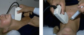
Breast ultrasound
If the woman is under 30 years old, the doctor may recommend an ultrasound instead of a mammogram because dense breast tissue in younger women is more difficult to examine with a mammograph.
If the woman is over 30 years old, the doctor will recommend a mammogram of both breasts followed by an ultrasound to evaluate the tumor.
Using an ultrasound, your doctor can determine whether a breast lump is solid or filled with fluid. When the mass is hard, there is a high probability that it is a fibroadenoma. A mass full of fluid indicates a cyst.