Women's health comes first for any woman, regardless of age. It so happens that sometimes we are given unpleasant diagnoses, one of which is breast fibroadenoma. This is the name for a benign lump in the mammary gland, which in some cases turns into cancer.
What is fibroadenoma
The content of the article
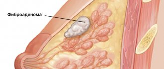
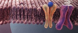
Fibroadenoma of the mammary gland is a mobile benign neoplasm of glandular and connective tissue. The pathology affects the chest, less often tendons, skin, and internal organs.
It is more often found in women under thirty years of age.
Glandular tissue is soft in nature, but overgrown with connective tissue, it takes on the appearance of a dense ball. In this form, the tumor is not dangerous - it does not extend beyond the frame and, unlike cancerous formations, does not metastasize. But if the capsule contains cysts (accumulations of purulent fluid) or inclusions of calcium deposits - calcifications, the neoplasm becomes dangerous as it can become malignant.
Why is fibrocystic mastopathy dangerous?
Mastopathy is always characterized by the formation of both cysts and fibrosis. When this process is balanced - equal parts of both, and it is distributed throughout the entire gland - then they speak of fibrocystic, diffuse or mixed mastopathy.
Fibrous mastopathy
Fibrous mastopathy is characterized by a predominance of the fibrous component over the cystic component in the development of fibrocystic mastopathy.
Increased formation of connective tissue at the site of former proliferation makes the breast gradually denser and more painful. It becomes difficult to evaluate her with mammography and even place her in a mammograph (due to pain). CESM and MRI are more suitable for diagnosis and screening .
Nodular mastopathy
Sometimes, with fibrocystic mastopathy, denser nodes appear in the breast tissue, often in the upper outer quadrants (where there is initially more glandular tissue). They often look suspicious for cancer upon examination and on mammograms and require MRI (or CESM ) and biopsy for definitive diagnosis.
Mastopathy compaction
Often such patients are even offered surgery—sectoral resection for localized fibroadenomotosis—as cancer is suspected. The operation can be combined with a breast lift, reduction or breast augmentation with implants.
Cystic mastopathy
Cystic mastopathy is characterized by the predominance of the process of cyst formation over fibrosis in the development of fibrocystic mastopathy.
These cysts are always multiple. At first they are small, but over time they can merge, reaching large sizes. puncture brings relief .
If the cysts recur, it is proposed to remove them, which can be combined with lifting, reducing or enlarging the mammary glands with implants.
Regarding mastopathy, monitoring for cysts and for preventive examinations of the mammary glands, we recommend that you consult a doctor:
Subbotina Olga Yurievna , AF-clinic, Spassky lane 11, M. Sadovaya, Sennaya, Spasskaya, tel. 310-00-33 and
Sokolova Valentina Ivanovna , Nova Vita Clinic, intersection of Engels and Thorez Ave., tel. 8(911)007-20-02
Dushina Irina Ilyinichna , SMT Clinic, Moskovsky Ave. 22, tel. 777-9-777
Raevskaya Natalya Aleksandrovna , Polyclinic No. 83, M. Sportivnaya, tel. 498-09-22 and 8(921)944-10-45.
Why do fibroadenomas form?
This type of pathology belongs to the category of little-studied. There are only assumptions about the causes of fibroadenomas, the most recognized among them being hormonal disorders.
It has been proven that any factors associated with hormonal fluctuations contribute to the occurrence of fibroadenoma:
- Independent, uncontrolled use of oral contraceptives, emergency contraception;
- Pregnancy, frequent abortions, in which the body, which is in the gestation stage, is forced to sharply adjust to a new rhythm;
- Intermittent lactation, breast refusal or inability to feed;
- The presence of pathologies, stressful situations, overload;
- Menopause.
Often fibroadenoma forms during the childbearing period, and after entering menopause it resolves. This indicates the important role of female sex hormones in this process.
Types and form of breast tumors
Kinds:
- Nodular
- a change in the shape of connecting tissues together with the ducts of the gland, ingrowth of connecting tissues into the ducts. - Leaf-shaped
. This is a dangerous, rare tumor. It actively increases in size and carries the risk of degeneration into sarcoma. This fibroadenoma gets its name from its structure: it consists of layers that look like leaves. - Mixed form
, which combines the symptoms of both cases.
Fibroids are divided into forms: immature and mature. In the first case, the tumor appears at an earlier age; there is no external capsule, which makes it possible to do without surgery. In the second case, the tumor is diagnosed after 20 years. The formation has a dense shell, so it cannot be treated with drugs.
Breast cancer support
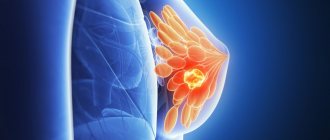
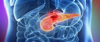
The diagnosis of breast cancer will not leave anyone indifferent. As a woman tries to cope with shock and fear for her future, she is asked to make important decisions about treatment.
A psychological attitude in the fight against a cancerous tumor will help the body and doctors together defeat the malignant neoplasm.
Everyone finds their own way to cope with a cancer diagnosis. The following tips may help:
- It's important to know everything you need to know about your form of cancer. Ask your doctor for details - type of cancer, degree of disease, hormonal receptor status. Find reliable sources of information about treatment options and don't trust publicist ploys. If the patient has high-quality and truthful information, it will be easier for her to make the right decision about treatment. However, some people do not want to know the details of their illness.
- Talk to other women with the same diagnosis or visit breast cancer forums. You are not alone.
- Keep your friends and family close, don't isolate yourself. When a woman tells her loved ones about the diagnosis, she will most likely receive many offers of help. Think of things you could use help with and don't be shy to ask. This communication makes us stronger.
- Maintain intimacy with your partner. Talk to him about your feelings and insecurities.
- Take care of your body. Get enough sleep every day. Eat plenty of fruits and vegetables. Make time for light exercise. Take more walks in the fresh air.
- Avoid bad habits. Remove cigarettes and alcohol from your life. Avoid foods high in fat.
Symptoms of the disease
First, a strange lump appears in the mammary gland, it is called a node. The nodule has a dense structure, can be easily felt and does not hurt. If the tumor is small, then it is difficult to notice at home; it is detected only by ultrasound of the mammary glands. There may be several fibroadenomas.
It is possible to recognize fibroadenoma more quickly if it appears near the nipples. In this case, in addition to compaction, the following may appear:
- Sores;
- Cracked nipples;
- Leakage of light fluid from the nipples;
- Pain on contact with the chest.
When a benign tumor becomes malignant, additional symptoms appear:
- The skin of the chest changes color to pale bluish or red;
- The tumor can grow quickly.
In any case, if you notice the slightest lump, you should immediately visit a gynecologist or mammologist. If necessary, after the examination he will redirect you to an oncologist.
Prevention
To prevent the appearance of seals, you should give up bad habits, be physically active, and avoid nervous overload. Visit your mammologist periodically and have your breasts examined in the first week after bleeding stops. Alarming symptoms:
- the shape of the gland has changed;
- upon palpation, nodules and tubercles are felt; they appear in the armpit area;
- the presence of thickenings in the tissues, bulges, dimples and folds;
- obvious swelling.
Carry out the examination while standing in front of a mirror. Raise your opposite hand and slowly feel your breast with your fingertips. Move in a spiral from the armpit to the nipple, then from top to bottom.
Take a lying position, put your hand behind your head, palpate the base of the gland with your fingers, moving towards the nipple, squeeze it to make sure there is no discharge.
If lumps are detected, contact your doctor to treat the pathology at an early stage.
Ultrasound table with norms and deviations of fibroadenoma
| Glandular tissue thickness | Fibroadenoma size | ||
| Normal up to 40 years – up to 14 mm | Deviations - more than 14 mm | Normally does not exceed 2-3 cm | Deviations – up to 9 cm |
| The norm after 40 years is up to 20 mm | Deviations - more than 20 mm | ||
If cancer is suspected, a fine-needle biopsy is prescribed. This is quite unpleasant, but the only adequate procedure that allows you to accurately determine whether there is cancer in the mammary gland.
The mammologist, using a thin hollow needle, inserts it into the tumor, capturing a small amount of tissue. To avoid mistakes, the doctor monitors the process using ultrasound. The cells obtained in this way are sent to the laboratory for cytological (cellular) examination. Under a microscope, the structure of cells is clearly visible: it is different for benign and malignant ones.
After the tests obtained, the doctor will prescribe further treatment, and if necessary, surgery.
Breast cancer - diagnosis
To diagnose cancer:
- An anamnesis is collected, and by interviewing the patient, the symptoms and duration of their action are found out. The doctor asks about the existence of possible risk factors, such as a family history of breast cancer, year of first and last menstruation, time of first birth and number of births, use of oral contraceptives or hormonal medications.
- The breast and lymph nodes (supraclavicular and axillary) are examined by inspection and palpation. During the examination, attention is paid to the size, shape and symmetry of the breasts, as well as possible changes in pigmentation, indentations in the skin or nipples. You should also pay attention to possible discharge from the nipple. Palpation determines the location of any changes, size, consistency (hard or soft), sensitivity, mobility to the surface and skin, as well as possible pain with suspicious changes in the breast.
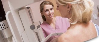
- Mammography is a diagnostic X-ray examination of the breast. It is still the most accurate and widely used non-invasive method. Mammography is used for early detection and confirmation of diagnosis. In addition, mammography identifies possible multicentric tumors and clusters of microcalcifications specific to breast cancer. Today it is recommended that a woman have her first mammogram at age 40 (with a positive family history at age 35). Then mammograms are done every two years.
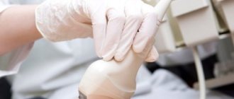
- Breast ultrasound is very often used to diagnose cancer. It is less sensitive and specific than mammography, especially in postmenopausal women. This method is indicated for young women in whom glandular tissue predominates in the breast and is very helpful in distinguishing breast cysts from solid tumors. The results of breast ultrasound depend on the phase of the menstrual cycle.
- Any suspicious breast changes require a biopsy. A breast biopsy is the removal of a tissue sample for pathological analysis, which means viewing under a microscope. The pathohistological result is the only reliable evidence of a malignant disease, such as breast cancer.
- In addition to a breast biopsy, it is necessary to determine the involvement of the lymph node draining the breast area (sentinel node). Determination of lymph node involvement is necessary to assess the stage of the disease.
How is fibroadenoma treated?
Having received the results, the mammologist resorts to the following types of treatment:
Monitoring the dynamics of tumor growth is suitable in several cases:
- The dimensions of the seal do not exceed two centimeters.
- Fibroadenoma is hormone-dependent and responds well to hormonal treatment.
- The tumor is not growing.
If drug treatment does not bring visible results, the breast hurts or cancer cells are found, surgery is required.
One type of surgery may be enucleation, where the tumor is excised from the breast. And the second type is sectoral resection. Excision is carried out locally, in a specific area. But low-traumatic interventions are especially popular:
- Radiofrequency ablation, in which the node is destroyed by radio waves. The operation is carried out under ultrasound control.
- Cryotherapy is the treatment of a tumor with liquid nitrogen delivered to the tumor through a thin probe.
After the operation, you need to take very good care of yourself so as not to miss the formation of new nodes.
Treatment of seals - adequate therapy
You should not experiment and self-medicate. If seals are detected, you must immediately contact a specialist who will prescribe therapy:
- Lactostasis - compresses using Vishnevsky ointment, camphor oil, etc.
- Mastitis - taking antibiotics, surgically opening the abscess.
- Mastopathy – the disease is monitored over time, it is recommended to come for an appointment every 6 months. An x-ray or ultrasound is prescribed, and hormonal or non-hormonal therapy is used. The goal is to eliminate the cause, which may lie in the malfunction of the reproductive organs, liver or nervous system.
- Nodular mastopathy requires special immunotherapy. The patient is given an allergenic vaccine, the dosage is increased, achieving stable remission. The therapy is ineffective - the nodes are excised using surgical intervention. The woman will be prescribed a special diet, vitamins, diuretics, and anti-inflammatory drugs. It is forbidden to wear a tight bra, visit saunas and solariums, or sunbathe for a long time.
- Small cysts - formation no more than 0.5 mm are subject to conservative treatment. The goal is to normalize hormonal levels. Single-chamber cysts are subject to resorption using puncture. Excess fluid is pumped out through a puncture on the chest, and a drug is injected that will destroy the capsule. Atypical cyst - tissue is excised during surgery. Aspiration method - a cannula will be inserted into the cavity of the cyst, through which the liquid will be pumped out. If traces of blood are detected, additional research is necessary. Immunostimulating drugs are prescribed.
- Thrombophlebitis. Conservative methods are used - bed rest, physiotherapy, compresses, wearing an elastic bandage. For lipoma, only surgical intervention is indicated.
Has it been established that the lump is a malignant formation? Assign:
- radical treatment - tumor tissue will be removed;
- auxiliary therapy - prescribed before or after surgery, less often instead. These are chemotherapy, radiotherapy, molecular targeted therapy, immunotherapy.
- palliative treatment – aimed at improving the quality of life of hopeless patients (presence of metastases).
Treatment of pathologies should not be delayed; there is a high risk of tumors degenerating into malignant formations.
Is it possible to prevent the formation of fibroadenoma and its degeneration into cancer?
Any female pathology can be prevented by observing the principles of careful hygiene, a healthy lifestyle, and timely diagnosis. Regular visits to a gynecologist-mammologist should be the norm in the life of any woman, starting from adolescence.
In our clinic you will meet qualified specialists who are ready to carefully examine each patient and help her get rid of any pathologies. An accurate diagnosis and targeted treatment using modern diagnostic equipment will help you forget about breast fibroadenoma.
CLICK TO MAKE AN APPOINTMENT, TEST OR ULTRASOUND
Diagnostic techniques
Each woman should palpate her breasts independently at different phases of the cycle. The lump can be detected at any age, but if you consult a doctor in time, you can defeat the disease, even oncology.
To establish an accurate diagnosis, you will be offered examinations:
- Ultrasound;
- x-ray of glands;
- biopsy - puncture will allow you to obtain a sample of breast tissue for an accurate diagnosis. The technique is highly accurate and accurately determines the nature of the tumor;
- galactography is a type of mammography. To examine the tissues and compaction, a special contrast will be injected into the milk ducts, which is clearly visible on x-rays. The result is that compacted areas in the ducts can be easily identified.
The decision on the choice of technique is made by the attending physician, who evaluates the clinical picture of the disease.