Damage to small vessels in diabetic angiopathy
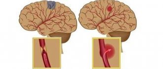
When small vessels are damaged, changes occur in their walls, blood clotting is disrupted and blood flow slows down. All this creates conditions for the formation of blood clots. Mainly small vessels of the kidneys, retina, heart muscles, and skin are affected. The earliest manifestation of diabetic angiopathy is damage to the lower extremities.
The processes occurring in blood vessels are of two types: thickening of the walls of arterioles and veins or thickening of capillaries. Initially, under the influence of toxic products that are formed during incomplete utilization of glucose, the inner layer of blood vessels swells, after which they narrow. The first manifestations of diabetic angiopathy are small hemorrhages under the nail plate of the big toe. The patient feels pain in the limbs, notices that the skin becomes pale, spots appear on it, nails become brittle, and the muscles of the legs “shrink out.” The pulse in the main arteries of the lower extremities does not change, but in the foot it may be weak.
Changes in the arteries of the retina may be detected and protein may appear in the urine. A specific painless bubble filled with bloody fluid appears on the skin of the feet. It heals on its own without forming a scar, however, microorganisms can enter the tissue and cause inflammation.
The following research methods are used to diagnose diabetic angiopathy:
- capillaroscopy;
- infrared thermography;
- introduction of radioactive isotopes;
- laser flowmetry;
- polarography or oxyhemography.
Damage to large vessels in diabetic angiopathy
In diabetic angiopathy, medium and large vessels can be affected. The inner membrane thickens in them, calcium salts are deposited and atherosclerotic plaques form.
The manifestations of the disease in this case are similar to those that occur with damage to small arteries. Pain in the feet bothers you, they become cold and pale, the nutrition of the tissues is disrupted, and they die over time. Gangrene of the fingers and then the feet develops. Diabetic angiopathy of internal organs
In diabetes mellitus, the pathological process most often affects the vessels of the retina and internal organs. This occurs due to the formation of toxic products due to incomplete “burning” of glucose. Almost all patients with high blood glucose levels have a retinal disease called retinopathy. With this disease, visual acuity first decreases, and then blood flows into the retina, and it peels off. This leads to complete loss of vision.
The second target organ whose vessels are affected in diabetes mellitus is the kidneys - nephropathy develops. In the initial stages, the disease does not manifest itself in any way; changes can only be detected during examination of the patient. Five years later, kidney function is impaired and protein appears in the urine. If changes are identified at this stage, they may still be reversible. But in the case when treatment is not carried out, the pathological process in the vessels of the kidneys progresses, and after ten years visible signs of the disease appear. First of all, a large amount of protein begins to be excreted in the urine. There is less of it in the blood, and this leads to the accumulation of fluid in the tissues and swelling. Initially, swelling is visible under the eyes and on the lower extremities, and then fluid accumulates in the chest and abdominal cavities of the body.
The body begins to use its own protein substances for life, and patients lose weight very quickly. They feel weak and have a headache. Also at this time, blood pressure rises, which stubbornly remains at high levels and does not decrease under the influence of medications.
The final stage of diabetic kidney angiopathy is the final stage of renal failure. The kidneys almost completely fail, they do not perform their function, and urine is not excreted. The body is poisoned by products of protein metabolism.
Diabetic macroangiopathy: treatment and diagnosis
Diabetic macroangiopathy is a concomitant disease of diabetes mellitus that affects large vessels of the cardiovascular system.
Diabetic macroangiopathy provokes thickening of arterial walls, resulting in the formation of plaques, impaired blood circulation, and a risk of thrombosis.
Causes of pathology
Factors that cause macroangiopathy include:
- obesity;
- arterial hypertension;
- increased blood clotting;
- hyperglycepia;
- insulin resistance;
- systemic inflammation, etc.
At risk are men over 45 years of age and women over 55 years of age, smokers, people exposed to occupational intoxication, as well as those who have diabetic macroangiopathy in previous generations of the family.
Symptoms of diabetic macroangiopathy
Tests will be required to identify symptoms of diabetic macroangiopathy. Among these symptoms:
- cardiac ischemia;
- thromboembolism;
- aneurysms;
- cardiogenic shock;
- heart failure;
- atherosclerosis.
The clinical picture for this pathology may include the following symptoms:
- cold feet;
- occasional lameness;
- swelling of the limbs;
- muscle pain from the buttocks and below;
- necrosis of leg tissue;
- trophic ulcers.
Diagnostics
To make a diagnosis and draw up a treatment plan, the patient will need to visit several specialists at once: an endocrinologist, a cardiologist, a vascular surgeon, a neurologist. Diagnosis of diabetic macroangiopathy is primarily aimed at assessing vascular damage. To do this, it is necessary to undergo a complete examination of the cardiovascular system. For effective diagnosis, a biochemical blood test is required. Neurological indicators of the disease are examined using ultrasound and duplex scanning of the head, neck and cerebral vessels. Additionally, ultrasound examination of the vessels of the extremities may be necessary.
Treatment of diabetic macroangiopathy
During treatment, it is important to slow down the development of the disease and prevent complications. First of all, therapy is aimed at restoring the normal functioning of body systems in case of hyperglycemia, hypercoagulation and other syndromes. Patients undergo insulin therapy with constant glucose monitoring. In addition, to correct carbohydrate metabolism, a diet and lipid-lowering medications are prescribed.
Diabetic angiopathy. Treatment at different stages of the disease
Successful treatment of diabetic angiopathy is possible only when it is possible to normalize blood glucose levels. This is what endocrinologists do.
In order to prevent irreversible processes in tissues and organs, it is necessary:
- control blood and urine sugar levels;
- ensure that blood pressure does not exceed 135/85 mm. Hg Art. in patients who do not have protein in the urine, and 120/75 mm. Hg Art. in patients in whom protein is detected;
- control fat metabolism processes.
In order to maintain blood pressure at the desired level, patients with diabetes need to change their lifestyle, limit their dietary salt intake, increase physical activity, maintain a normal body weight, limit their intake of carbohydrates and fats, and avoid stress.
When choosing drugs that lower blood pressure, you need to pay attention to whether they affect the metabolism of fats and carbohydrates and whether they have a protective effect on the kidneys and liver. The best drugs for these patients are captopril, verapamil, valsartan. You should not take beta blockers as they may contribute to the progression of diabetes. Patients with diabetic angiopathy are advised to take statins, fibrates, and medications that improve fat metabolism. In order to maintain a normal level of glucose in the blood, it is necessary to take gliquidone, repaglimide. If diabetes mellitus progresses, patients should be switched to insulin.
Diabetic angiopathy requires constant monitoring of glucose levels, fat metabolism and vascular condition. When the tissues of the limb become necrosis, operations are performed to remove them. In case of chronic renal failure, the only way to prolong the patient’s life is an “artificial” kidney. Retinal detachment as a result of diabetic angiopathy may require surgical intervention.
Diabetic macroangiopathy and self-control of glycemia
O.S. FEDOROVA
,
Federal State Budgetary Institution "Federal Bureau of Medical and Social Expertise" of the Ministry of Labor of Russia
Diabetic macroangiopathy is a generalized atherosclerotic lesion of large and medium-sized arteries in diabetes mellitus (DM). Macrovascular complications in diabetes mellitus, leading to cardiovascular diseases (CVD), are the main cause of death in patients with diabetes. CVD is less dependent on hyperglycemia or the intensity of hypoglycemia than microvascular complications. At the same time, there are large works confirming this relationship. Diabetic macroangiopathies include: coronary heart disease (CHD), cerebrovascular disease (CVD), chronic occlusive diseases of peripheral arteries [1].
The DCCT (Diabetes Control and Complications Trial) study demonstrated a trend toward a reduction in the risk of cardiovascular events with intensive insulin therapy. During 9-year follow-up after DCCT, the Epidemiology of Diabetes Interventions and Complications (EDIC) study showed that patients initially randomized to intensive control experienced a 57% reduction in the risk of nonfatal myocardial infarction, stroke, or cardiovascular death. diseases, compared with the group receiving standard therapy [2]. The beneficial effects of intensive glycemic control in this group of patients with type 1 diabetes have recently been shown to persist over several decades [3].
It has been proven that intensive glucose-lowering therapy in cases of newly diagnosed type 2 diabetes can reduce the risk of cardiovascular complications. In the UKPDS (United Kingdom Prospective Diabetes Study), there was a 16% reduction in the risk of cardiovascular events (fatal or nonfatal myocardial infarction and sudden death) in the intensive treatment group, but the reduction did not reach a statistically significant level (p = 0.052) [4] .
At the same time, during the subsequent 10-year follow-up period, patients initially randomized to the intensive treatment group had a statistically significant reduction in the incidence of myocardial infarction and death from all causes (by 13 and 27%, respectively) [5].
However, the ACCORD (Action to Control Cardiovascular Risk in Diabetes), ADVANCE (Action in Diabetes and Vascular disease: PreterAx and DiamicroN Controlled Evaluation) and VADT (Veterans Affairs Diabetes Trial) studies, which included patients with longer duration of type 2 diabetes, did not reveal a statistically significant reduction in the risk of adverse cardiovascular outcomes. All three studies included patients with an average duration of diabetes of 8–11 years, as well as with known cardiovascular risk factors or CVD.
Patients enrolled in the ACCORD trial were randomly assigned to either an intensive treatment group (target HbA1c < 6%) or a standard care group (target HbA1c 7–8%). However, the study was terminated early due to an increase in all-cause and cardiovascular mortality in the intensive control group (RR 1.22 [95% confidence interval 1.01–1.46]). The initial analysis of the data obtained could not fully explain the cause of the incident [6], but it was later revealed that the greatest risk of mortality was observed in those patients from the intensive control group who initially had a higher level of HbA1c [7].
The primary endpoint of the ADVANCE study was the composite of microvascular complications (nephropathy or retinopathy) and major cardiovascular events (myocardial infarction, stroke, death due to CVD). There was a reduction in the incidence of microvascular complications in the intensive treatment group, but no change in the risk of macrovascular outcomes.
The primary endpoint of the VADT study was a composite of cardiovascular events. Patients with type 2 diabetes who did not achieve compensation on insulin therapy or maximum doses of oral glucose-lowering drugs were randomly divided into 2 groups: the first aimed at a target glycated hemoglobin < 6.0%, the second received standard therapy. It was shown that the total incidence of cardiovascular events was lower in the intensive treatment group, but the difference was not statistically significant [8]. Additional analysis demonstrated a reduction in the likelihood of cardiovascular events with intensive treatment only in patients with lower prevalence of atherosclerosis. The effect of type of therapy on mortality depended on the duration of the disease before enrollment in the study. Patients with diabetes for less than 15 years had a reduced risk of mortality, while patients with diabetes for more than 20 years had a higher mortality rate with intensive treatment [9]. A meta-analysis of these studies showed that lowering glycemia leads to a moderate (9%) but statistically significant reduction in the risk of major cardiovascular events, primarily nonfatal myocardial infarction, but has no effect on mortality. Using a specially selected subgroup, it was found that this risk reduction was observed in patients without a history of CVD (HR 0.84 (95% confidence interval 0.74-0.94)) [10]. In addition, mortality data from the ACCORD and VADT studies suggest that the potential risks of intensive glycemic control may outweigh the benefits in some patients. Patients with a long history of diabetes, a history of severe hypoglycemia, advanced atherosclerosis, or low life expectancy will benefit more from less aggressive treatment strategies. Severe hypoglycemia should be avoided in patients with complicated diabetes. Severe or frequent hypoglycemia is an absolute indication for changes in therapy, including increasing the target values of glycemia and glycated hemoglobin HbA1c [4].
Various factors, as well as patient motivation and adherence to therapy, should be taken into account when choosing individual treatment goals [11] (Table 1).
Table 1. Algorithm for individualized selection of treatment goals based on the level of glycated hemoglobin HbA1c in type 1 and type 2 diabetes [1]
| Age | |||
| Young | Average | Elderly and/or life expectancy < 5 years | |
| No severe complications and/or risk of severe hypoglycemia | < 6,5% | < 7,0% | < 7,5% |
| There are severe complications and/or risk of severe hypoglycemia | < 7,0% | < 7,5% | < 8,0% |
Life expectancy - life expectancy
An increase in plasma glucose at 2 hours during an oral glucose tolerance test in nondiabetic individuals is associated with an increase in cardiovascular risk independent of fasting plasma glucose in some epidemiological studies. In patients with diabetes, postprandial hyperglycemia has a negative effect on surrogate markers of vascular pathology, such as endothelial dysfunction [12]. It is clear that both postprandial and preprandial hyperglycemia contribute to an increase in the level of glycated hemoglobin HbA1c (Table 2).
Table 2. Correspondence of target values of pre- and postprandial plasma glucose levels to target HbA1c levels for type 1 and type 2 diabetes [1]
| HbA1c,% | Plasma glucose on an empty stomach, before meals, mmol/l | Plasma glucose 2 hours after meals, mmol/l |
| <6,5 | <6,5 | <8,0 |
| <7,0 | <7,0 | <9,0 |
| <7,5 | <7,5 | <10,0 |
| <8,0 | <8,0 | <11,0 |
Note.
These targets do not apply to children, adolescents and pregnant women. Therefore, achieving therapeutic goals implies normalization of fasting glucose, 2 hours after a meal (postprandial glycemia) and glycated hemoglobin HbA1c - the main criterion for long-term compensation of diabetes. According to numerous observations, the HbA1c level begins to decrease as soon as the patient himself increases the frequency of glucose monitoring, regardless of the type of diabetes or the type of glucose-lowering therapy [13, 14].
It is especially important to control postprandial glycemia in patients who have achieved target fasting blood glucose but do not have target glycated hemoglobin HbA1c [4].
The American Diabetes Association currently offers the following recommendations for self-monitoring of glycemia:
- patients receiving insulin therapy in the regimen of multiple injections or using an insulin pump should self-monitor glycemia before main meals, snacks and at night, periodically monitor postprandial glycemia, before playing sports, if hypoglycemia is suspected, after relieving hypoglycemia until normoglycemia is achieved, before performing activities that require high concentration (for example, driving); — the results of self-monitoring may be useful for adjusting therapy in individuals receiving fewer insulin injections or therapy without insulin; — when prescribing glycemic self-monitoring, it is necessary to make sure that patients can use the results obtained to adjust therapy; - When used correctly, continuous glycemic monitoring (CGM) in combination with intensified insulin therapy is a useful tool for reducing glycated hemoglobin HbA1c in a certain category (aged 25 years and older) of people with type 1 diabetes; — CGM can also be used in children, adolescents, and young adults, despite the weak evidence base regarding the reduction of glycated hemoglobin levels; — CGM can be used as an additional tool to self-monitoring of glycemia for unrecognized hypoglycemia and/or frequent episodes of hypoglycemia.
Major clinical trials involving patients on insulin therapy have shown the benefits of intensive glycemic control and included self-monitoring of glycemia as part of multifactorial diabetes management, suggesting that self-management is a component of effective therapy. Self-monitoring allows patients to evaluate their own results of therapy and understand whether target glycemic values have been achieved. The results of self-monitoring help prevent hypoglycemia and are used to adjust therapy (in particular, prandial doses of insulin) and physical activity. The frequency and timing of self-monitoring is dictated by the patient's individual needs and goals. Self-monitoring of glycemia is especially necessary for patients receiving insulin therapy to prevent asymptomatic hypoglycemia and hyperglycemia [4].
A recent meta-analysis showed that self-glycemic control reduced HbA1c by 0.25% over 6 months. [15], however, a Cochrane review showed a gradual decline in the effect of self-control after 12 months. [16]. Thus, self-monitoring does not reduce glycemia in itself, but its results should be used by the patient and physician to adjust therapy.
One study in non-insulin-treated patients with suboptimal diabetes control showed that the use of structured self-glycemic monitoring (7 measurements over 3 days) resulted in a 0.3% reduction in HbA1c [17]. Patients should consider how to use the results of self-monitoring to change diet, exercise, or drug therapy to achieve individual glycemic goals. The appropriateness and frequency of self-monitoring of glycemia should be discussed and, if necessary, modified at each visit to the doctor [4]. The results of self-monitoring depend on the accuracy of the device and the accuracy of the actions of the person performing the measurement [18].
Modern glucometers must be accurate, convenient and reliable. The Bayer TC Circuit blood glucose monitoring system meets all of these requirements.
The Kontur TC glucose meter uses flavin adenine dinucleotide/glucose dehydrogenase without interaction with maltose and galactose, which allows it to be used in patients on peritoneal dialysis receiving icodextrin, as well as maltose-containing immunoglobulins. In addition, it suppresses interference with agents such as oxygen, uric acid, vitamin C, paracetamol, without distorting the accuracy of the results obtained. The TC circuit requires only 0.6 µl of blood. In addition, the test strips have a separate electrode that measures hematocrit, so the blood glucose value is determined by the corrected hematocrit. Measurement data appears on the meter screen within 8 seconds.
The TC Contour glucometer does not require entering a digital code or installing an encoded chip; it is automatically programmed using test strips, which makes its operation easier and more reliable. The TC circuit shows accurate and reproducible results, as confirmed by the International Organization for Standardization (ISO 15197:2003) [19].
Regular self-monitoring of blood glucose levels in patients with diabetes is an integral part of effective and adequate diabetes management, allowing patients to prevent the development of macrovascular complications, maintain a high quality of life and reduce the risk of mortality from cardiovascular diseases. The use of accurate, convenient and reliable glucometers that meet the criteria of the International Organization for Standardization makes it possible to successfully control diabetes by timely adjusting therapy, changing diet and intensity of physical activity, which increases adherence to treatment and helps compensate for diabetes.
Literature
1. Algorithms for specialized medical care for patients with diabetes mellitus. Ed. I.I. Dedova, M.V. Shestakova. Vol. 6. Ministry of Health and Social Development of the Russian Federation. M., 2013. 2. Nathan DM, Cleary PA, Backlund JY, et al. Diabetes Control and Complications Trial/Epidemiology of Diabetes Interventions and Complications (DCCT/EDIC) Study Research Group. Intensive diabetes treatment and cardiovascular disease in patients with type 1 diabetes. N Engl J Med 2005, 353: 2643-2653. 3. Nathan DM, Zinman B, Cleary PA, et al.; Diabetes Control and Complications Trial/Epidemiology of Diabetes Interventions and Complications (DCCT/EDIC) Research Group. Modern-day clinical course of type 1 diabetes mellitus after 30 years'duration: the Diabetes Control and Complications Trial/Epidemiology of Diabetes Interventions and Complications and Pittsburgh Epidemiology of Diabetes Complications experience (1983–2005). Arch Intern Med 2009, 169: 1307-1316. 4. Standards of Medical Care in Diabetes – 2014. American Diabetes Association. Diabetes Care 2014 Jan 37: S14-S80; doi:10.2337/dc14-S014. 5. Holman RR, Paul SK, Bethel MA, Matthews DR, Neil HA. 10-year follow-up of intensive glucose control in type 2 diabetes. N Engl J Med 2008, 359: 1577-1589. 6. Gerstein HC, Miller ME, Byington RP, et al. Action to Control Cardiovascular Risk in Diabetes Study Group. Effects of intensive glucose lowering in type 2 diabetes. N Engl J Med 2008, 358:2545-2559. 7. Riddle MC, Ambrosius WT, Brillon DJ, et al. Action to Control Cardiovascular Risk in Diabetes Investigators. Epidemiologic relationships between A1C and all-cause mortality during a median 3.4-year follow-up of glycemic treatment in the ACCORD trial. Diabetes Care, 2010, 33: 983-990. 8. Duckworth W, Abraira C, Moritz T, et al. VADT Investigators. Glucose control and vascular complications in veterans with type 2 diabetes. N Engl J Med 2009, 360: 129-139. 9. DuckworthWC, Abraira C, Moritz TE, et al. Investigators of the VADT. The duration of diabetes affects the response to intensive glucose control in type 2 subjects: the VA Diabetes Trial. J Diabetes Complications, 2011, 25: 355-361. 10. Turnbull FM, Abraira C, Anderson RJ, et al. Control Group. Intensive glucose control and macrovascular outcomes in type 2 diabetes. Diabetologia, 2009, 52: 2288-2298. 11. Ismail-Beigi F, Moghissi E, Tiktin M, Hirsch IB, Inzucchi SE, Genuth S. Individualizing glycemic targets in type 2 diabetes mellitus: implications of recent clinical trials. Ann Intern Med 2011, 154:554-559. 12. Ceriello A, Taboga C, Tonutti L, et al. Evidence for an independent and cumulative effect of postprandial hypertriglyceridemia and hyperglycemia on endothelial dysfunction and oxidative stress generation: effects of short- and long-term simvastatin treatment. Circulation, 2002, 106: 1211-1218. 13. Chernikova N.A. Diabetes for beginners. Diabetes. Lifestyle, 2012, July-August, 4: 57-59. 14. Chernikova N.A. Tell me, doctor... Diabetes. Lifestyle, 2012, May-June, 3: 7. 15. Willett LR. ACP Journal Club. Metaanalysis: self-monitoring in non-insulintreated type 2 diabetes improved HbA1cby 0.25%. Ann Intern Med 2012, 156: JC6-JC12. 16. Malanda UL, Welschen LMC, Riphagen II, Dekker JM, Nijpels G, Bot SD. Selfmonitoring of blood glucose in patients with type 2 diabetes mellitus who are not using insulin. Cochrane Database Syst Rev, 2012, 1. CD005060. 17. Polonsky WH, Fisher L, Schikman CH, et al. Structured self-monitoring of blood glucose significantly reduces A1C levels in poorly controlled, noninsulin-treated type 2 diabetes: results from the Structured Testing Program study. Diabetes Care, 2011, 34: 262-267. 18. Sacks DB, Arnold M, Bakris GL, et al.; National Academy of Clinical Biochemistry. Position statement executive summary: guidelines and recommendations for laboratory analysis in the diagnosis and management of diabetes mellitus. Diabetes Care, 2011, 34: 1419-1423. 19. Frank J1, Wallace JF, Pardo S, Parkes JL. Performance of the CONTOUR® TS Blood Glucose Monitoring System. J Diabetes Sci Technol., 2011, Jan., 1, 5(1): 198-205.


