Lipid metabolism disorders (dyslipidemia) affect the processes of absorption, transformation and metabolism of fats in the body. In addition to their energy function, fats are an important component of cell membranes, participate in the synthesis of hormones, transmit nerve impulses, and perform a host of other vital tasks. Therefore, a violation of lipid metabolism significantly affects the condition of the entire organism as a whole and can lead to the development of severe consequences.
Causes of dyslipidemia
- Nutritional – eating large amounts of animal and vegetable fats. The norm is set individually for each person, the average is from 0.8 to 1 g. fat per kilogram of body weight per day.
- Congenital disorders of lipid metabolism. They arise as a result of mutations in genes that are responsible for the processes of synthesis, breakdown, and transportation of fats. These mutations are passed on from generation to generation, so the disease can occur in young children for no apparent reason. Examples of genetic disorders of lipid metabolism are Gaucher disease, Tay-Sachs disease, and Niemann-Pick disease.
- Secondary disorders of lipid metabolism. Caused by other diseases that can affect the gastrointestinal tract, endocrine and enzyme systems, various internal organs (liver, kidneys).
Among the provoking factors that increase the risk of developing lipid metabolism disorders are bad habits (smoking, alcohol abuse), a sedentary lifestyle, excess weight, chronic stress, and taking hormonal medications.
General information
Lipids are organic substances in the body. Blood lipids are represented by cholesterol, fatty acids, triglycerides and phospholipids. Triglycerides accumulate in adipose tissue and provide energy. Cholesterol is produced in the liver and is necessary for the synthesis of hormones, membrane formation and bile acid synthesis. Phospholipids are part of cell membranes. Cholesterol and triglycerides are present in the blood in the form of a complex with proteins and are called lipoproteins (fat + protein). Lipoproteins transport lipids to organs and tissues, where they are used for their intended purpose. Liproteins are divided into:
- chylomicrons;
- very low density lipoproteins;
- low density;
- intermediate density;
- high density.
They all have different densities and sizes. About 75% of cholesterol is in LDL (the main atherogenic lipoprotein), and triglycerides are in chylomicrons. Catabolism of low-density lipoproteins occurs in peripheral tissues. 70-80% of low-density lipoproteins are removed from plasma every day, and the rest is taken up by cells. With normal lipid metabolism in the blood, the physiological level of essential lipids is determined. Violations of lipid metabolism lead to the development of atherosclerosis , against which cardiovascular diseases develop (coronary artery disease , hypertension , myocardial infarction and stroke ). A direct connection has been established between cholesterol levels and cardiovascular diseases and their complications. A manifestation of lipid metabolism disorders is dyslipidemia or hyperlipidemia .
What is dyslipidemia? This is a violation of the normal ratio of blood lipids (fats). Hyperlipidemia is a congenital (primary) or acquired (secondary) increase in the level of certain blood lipids, and since lipids in the blood are associated with transport proteins (lipoproteins), the term hyperlipoproteinemia is used. Typically, dyslipidemia, hyperlipidemia and hyperlipoproteinemia are identified. One way or another, these terms reflect the essence of one pathological process - increased levels of cholesterol, triglycerides and decreased levels of high-density lipoprotein cholesterol. The diagnosis of dyslipidemia is not independent, but is included in the clinical diagnosis of the underlying cardiovascular disease.
Hyperlipidemia occurs in 50-60% of adults and is an important factor in the development of cardiovascular diseases and a cause of increased mortality. Among patients with high levels of low-density lipoprotein, mortality is significantly higher. LDL contains the protein apolipoprotein B. Considering the importance of apoB-containing lipoproteins in the development of atherosclerosis, it is important to determine these particular lipoproteins to assess the risk of cardiovascular diseases. Elevated triglyceride levels also pose a risk of developing cardiovascular disease. The increasing prevalence of cardiovascular diseases necessitates the establishment of risk groups for patients, early detection and treatment of lipid metabolism disorders. Normalization of lipid parameters is the main task, the implementation of which reduces cardiovascular risk. The code for dyslipidemia according to ICD-10 is extensive - from E78.0 to E78.9 (the type of disorders is specified in the subheadings).
Fat metabolism disorder
General information
Fat metabolism is a set of processes of digestion and absorption of neutral fats, as well as their breakdown products in the gastrointestinal tract. Also responsible for the intermediate metabolism of fats and fatty acids and their removal from the body. Lipids that are part of plants and animal tissues are an important component of lipid metabolism in the body. Every day, about 70 g of fat enters the adult body with food. Lipid metabolism disorders are a factor in the development of many diseases. Partial breakdown of fats occurs in the stomach, but the main processes take place in the upper parts of the small intestine with the help of pancreatic lipase. Regulation of fat metabolism is carried out by the hypothalamus, which comes into effect already at the stage of breakdown of fats in the stomach. Neurohormonal influences on fat metabolism are associated with the process of mobilization of fatty acids from fat depots. During emotional stress, the level of free fatty acids in the blood increases, which leads to an increase in the release of catecholamines into the blood and activation of lipolysis. By activating or inhibiting lipolysis, the effect on fat metabolism is achieved.
Causes of fat metabolism disorders
Possible causes of impaired fat metabolism and incomplete breakdown of fats:
- lack of pancreatic lipase;
- decreased bile secretion;
- dysfunction of the intestinal epithelium;
- pancreatitis;
- jaundice;
- vagotomy;
- Whipple's disease;
- radiation sickness.
As a result of malabsorption, a large amount of fatty acids appears in the feces, which leads to steatorrhea. The body also stops receiving the required amount of fat-soluble vitamins. Blood plasma has a milky color due to the high content of chylomicrons. Treatment of pathology involves replacing natural fats with synthetic ones. Synthetic fatty acids are absorbed from the intestines directly into the blood. With reduced lipoprotein lipase activity, low-density lipoproteins accumulate in the blood. In this case, patients are given heparin intravenously and are also prescribed a diet low in fat and carbohydrates.
Causes of excess weight
In middle-aged and elderly people, excess fat accumulation may occur in adipose tissue. The reason is high-calorie foods combined with low energy costs. Overeating leads to obesity.
Excessive fat deposition can occur during the transition from an active to a sedentary lifestyle. At the same time, the outrageousness of the food center remains at the same level. The same appetite appears, but energy consumption is correspondingly reduced.
Morbid obesity is associated with the following factors:
- decreased activity of adipose tissue;
- increased conversion of carbohydrates to fats;
- increased excitability of the food center;
- decreased muscle activity.
Reduced fat mobilization also occurs when the functions of the thyroid and pituitary glands are weakened, since their hormones activate lipolysis. Decreased function of the gonads also leads to excess fat deposition. The main factor in the development of hereditary constitutional obesity is insulin hypersecretion.
The most noticeable sign of obesity is an increase in the volume of subcutaneous fat. With a high intensity of this process, obesity can be considered an independent disease. It is important to know that overweight people:
- do not tolerate physical activity well;
- suffer from shortness of breath;
- increased appetite;
- have high blood pressure.
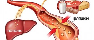
Obesity also increases the risk of developing diabetes. Also, with an increase in the content of lipids in the body, their content in the blood also increases. This leads to hyperlidemia. “Blood obesity” is very dangerous, since over time atherosclerosis plaques form, which clog blood vessels. As a result, the patient will suffer a heart attack or stroke.
Exhaustion and ketosis
Exhaustion with insufficient deposition of fat in tissues develops when the excitability of the food center is inhibited, with reduced absorption of fats and carbohydrates (enteritis), and with prolonged fasting. Disturbances in the formation of fat from carbohydrates can be observed with lesions of the autonomic centers of the hypothalamic-pituitary system, as well as the adrenal cortex. Such disorders underlie progressive wasting in pituitary cachexia and Addison's disease. Cachexia is extreme exhaustion of the body, which is characterized by:
- general weakness;
- sudden weight loss;
- activity of physiological processes;
- mental disorders.
Symptoms of cachexia include severe weakness, loss of ability to work, and sudden weight loss, often accompanied by signs of dehydration. Reduction of body weight up to 50% during the development of the disease.
The accumulation of fats in liver cells is a reaction to diseases and damage to the organ. Fatty infiltration of the liver is also observed in diabetes mellitus, protein deficiency, alcohol poisoning, and obesity. Ketosis is an increased formation and accumulation of ketone bodies in tissues and blood. In a state of ketosis, the body can function for a long time. For example, residents of the north eat animal food and fish throughout their lives without harming their health. But for the inhabitants of other parts of the world, the predominance of animal food in the diet is not historically typical. Therefore, a low-carbohydrate diet and introducing the body into a state of ketosis can negatively affect health, causing pathological changes in metabolism.
Prevention and treatment
Treatment of fat metabolism disorders involves completely eliminating the physical causes of the disorder and adjusting the body to proper metabolism. is also a must . If you are obese, you need to limit yourself to fatty and sweet foods, and if you are exhausted, you should gradually introduce foods high in fats and carbohydrates into your body. A nutritionist will be able to prescribe an adequate diet and also recommend additional prevention. For obesity, sports training helps, physical activity restores the balance of lipids in the blood. Sport also has a positive effect on metabolism.
It is possible to cope with fat metabolism disorders in three to four months. In case of serious disorders, it is better to seek help from an endocrinologist who can select a long-term form of treatment. The best prevention of fat metabolism disorders is diet and exercise training. A preventive examination by a nutritionist will help identify possible problems with metabolism in the body. The specialist will prescribe the necessary tests and select an individual diet to restore fat metabolism in the body. The patient can only monitor the diet and listen to all the doctor’s recommendations.
Pathogenesis
Lipoproteins whose diameter is less than 70 nm pass through the vascular endothelium. ApoB-containing lipoproteins in the arterial wall become trapped and trigger a process whereby lipids are deposited in the wall. Low-density lipoproteins are oxidized and, together with monocytes, form the core of an atherosclerotic plaque. The active substances released during this process are involved in the proliferation of smooth muscle cells in blood vessels and the breakdown of collagen. In patients with high levels of apoB-containing lipoproteins, more particles are deposited, and the atherosclerotic plaque progresses faster. Over time, other particles are deposited in the vessel wall, and the atherosclerotic plaque, reaching a critical point, ruptures with the formation of a blood clot on the surface. It becomes the cause of blockage of the vessel with the development of angina or myocardial infarction .
Dyslipidemia develops with
insulin resistance .
Under conditions of increased insulin secretion with decreased tissue sensitivity to insulin, increased breakdown of fats into fatty acids occurs. The latter are delivered to the liver, where they produce low-density lipoprotein cholesterol. Dyslipidemia causes increased blood pressure. The role of very low-density and low-density lipoproteins in the development of vascular endothelial dysfunction has been proven, which causes disruption of the synthesis of nitric oxide and an increase in the production of the vasoconstrictor peptide endothelin-1. As a result, vasoconstriction occurs and systemic blood pressure increases. Primary hyperlipidemia type 1 is associated with a mutation in the lipoprotein lipase gene. A defect in this enzyme blocks the metabolism of chylomicrons and they accumulate in large quantities in the plasma. With reduced lipoprotein lipase activity, triglycerides are not broken down and severe triglyceridemia .
Classification
Hyperlipidemia is divided into:
- Primary (genetically predisposed and hereditary).
- Secondary (symptom of diseases associated with metabolic disorders).
Secondary ones are divided into:
- Basal (detected during fasting examination and indicate serious disorders of fat metabolism).
- Induced (detected after taking excess amounts of fats or carbohydrates, hormonal drugs or after physical activity). For the most part, these are nutritional hyperlipidemias . Secondary dyslipoproteinemia is characterized by elevated levels of several classes of lipoproteins.
According to the classification of D. Fredrickson, there are several types of lipid metabolism disorders, depending on the ratio of different lipoproteins.
- Type I This is a fairly rare type that develops when there is a deficiency of the enzyme lipoprotein lipase or the protein that activates lipoprotein lipase . This type is manifested by an increase in the level of chylomicrons.
- II type . Hyperlipoproteinemia type 2 is the most common type of lipid metabolism disorder. This type is characterized by an increase in LDL cholesterol. This type is subdivided into types IIa and IIb. Type IIa is characterized by increased total cholesterol and LDL cholesterol. In type IIb, all fractions are elevated, including triglycerides. The frequency of occurrence of the latter type is 10%, found in metabolic syndrome). Statins, fibrates (for very high triglyceride levels), as well as a combination of these two groups of drugs are prescribed for treatment. ω3-fatty acids are also used.
- III type . This type is manifested by an increase in the level of chylomicrons and intermediate density lipids. The occurrence does not exceed 0.02%.
- IV type . This type is characterized by an increase in triglycerides. The occurrence reaches 1%.
- V type . Manifested by high levels of chylomicrons and VLDL.
In clinical practice, lipid metabolism disorders of types II and IV are important, the rest are very rare. In the prevention of atherosclerosis, the main thing is the correction of disorders that can be influenced and are associated with nutrition, lifestyle and obesity .
Treatment of dyslipidemia
Treatment of dyslipidemia is complex and includes:
- Drug therapy - fibrates, vitamins, statins and other drugs that correct lipid metabolism disorders;
- Non-drug treatment - weight normalization through fractional meals, dosed physical activity, limiting alcohol and smoking, and stressful situations.
- Diet therapy - foods rich in dietary fiber and vitamins are recommended (vegetables, cereals, fruits, beans, low-fat lactic acid products); fatty and fried meats are not allowed.
If lipid imbalance is a secondary pathology resulting from exposure to negative factors or any disease, NEARMEDIC cardiologists prescribe therapy aimed at timely detection and treatment of the underlying disease.
Dyslipidemia develops over years and requires equally long-term treatment. You can prevent further disturbances in lipid metabolism by strictly following the recommendations of doctors: move more, watch your weight, quit bad habits.
Contact your doctors on time!
In the early stages, stopping the pathological process is much easier. Timely therapy, elimination of risk factors and disciplined implementation of doctors’ recommendations significantly prolong and improve the lives of patients
Contact our clinics, do not delay your visit to the doctor. You will be consulted by experienced doctors, and you will undergo an expert examination using high-tech diagnostic equipment. Based on the results obtained, the cardiologist will prescribe competent treatment for dyslipidemia and recommend preventive measures.
To make an appointment with a cardiologist, call or fill out a request on the website.
Causes of lipid metabolism disorders
The cause of primary (hereditary) hyperlipidemia is mutations of certain genes (encoding apoprotein E, apoprotein B100, lipoprotein lipase). A defect in one gene is partially or completely compensated by various mechanisms, but under unfavorable conditions or with multiple mutations, lipid metabolism disorders develop.
The main causes of secondary dyslipidemias include:
- Eating disorders. With a high content of saturated fats in the diet, the content of low-density and very low-density lipoproteins, which have increased atherogenicity, increases in the blood. Consumption of trans fats leads to an increase in the synthesis of endogenous cholesterol and apoB lipoprotein. Eating easily digestible carbohydrates causes an increase in triglyceride levels. Alcohol consumption negatively affects triglyceride levels, disrupts the oxidation of fatty acids, which leads to the accumulation of lipids in the liver, and also affects the removal of VLDL from the body.
- Sedentary lifestyle.
- Psycho-emotional stress. Due to increased activity of the sympathetic nervous system, lipid metabolism is disrupted.
- Taking certain medications. Drug-induced dyslipidemias develop with long-term use of thiazide diuretics, retinoids, β-blockers, antiretroviral drugs, cyclosporine, antipsychotics, estrogen, tacrolimus, anabolic steroids, progestins, antidepressants and glucocorticoids. It should be noted that treatment with these drugs does not always cause lipid metabolism disorders (this depends on the dose and duration of use).
- Hormonal factor. During pregnancy , all indicators of the lipid spectrum increase moderately and gradually return to normal after the birth of the child. In women taking oral contraceptives, the levels of cholesterol, TG and low- and very low-density lipoproteins also increase. In women after menopause, the risk of lipid metabolic disorders increases, which is associated with a decrease in estrogen production.
Risk factors for dyslipoproteinemia are:
- Diabetes . The relationship between lipid and carbohydrate metabolism disorders is shown.
- Insulin resistance and metabolic syndrome .
- Hypothyroidism.
- Paraproteinemia.
- Polycystic ovary syndrome.
- Arterial hypertension (in hypertensive patients, hypercholesterolemia and low levels of high-density lipoproteins are detected).
- Chronic kidney disease.
- Fatty liver disease and cirrhosis .
- Obesity.
Nutrition, excess weight and a sedentary lifestyle directly or indirectly affect lipid metabolism; correction of exogenous factors reduces the severity of hyperlipidemia and the risk of coronary artery disease.
Diagnosis and treatment
Some specific tests are used to detect violations. They are necessary to figure out how to normalize lipid metabolism in the body:
- Blood biochemistry, measuring lipid metabolism.
- Determination of the atherogenic coefficient - determination of the ratio of high and low density lipoproteins.
- Ultrasound duplex scanning of color vessels using an ultrasound machine. With its help, the doctor identifies areas of poor circulation and compares blood flow in paired organs.
- Magnetic resonance angiography - allows you to evaluate the anatomical and functional features of blood flow.
- Computed angiography is used to visualize large blood vessels and identify their pathological changes.
Not all tests and methods are required. They are prescribed at the discretion of the doctor and there are no contraindications.
Specialists to contact with final results:
- cardiologist;
- neurologist;
- vascular surgeon..
After reviewing the patient’s tests, collecting anamnesis and examination, the doctor develops an individual treatment regimen. Therapy includes treatment with medications (eg, statins). These include a diet with a limited content of animal fats and feasible physical activity.
Sometimes extracorporeal hemocorrection is required, the purpose of which is to cleanse the blood of pathogenic elements (excess cholesterol, metabolic breakdown products, toxic substances and antibodies).
How methods of extracorporeal hemocorrection help with hypercholesterolemia
To correct lipid metabolism disorders, a number of techniques are used, such as:
- cryoapheresis (HELP-apheresis);
- plasmafiltration (cascade plasmapheresis);
- immunosorption.
The techniques help restore adequate cholesterol levels. After the procedure, the volume of cholesterol plaques on the walls of blood vessels decreases and blood viscosity decreases.
Other positive effects are also observed:
- Cell susceptibility to drugs increases.
- The work of internal organs becomes more efficient due to increased blood supply.
- Memory, concentration, and mood improve.
- Sleep is normalized.
- The likelihood of developing a stroke is reduced.
- The blood supply to the myocardium increases, which reduces the number of angina attacks and the risk of developing myocardial infarction.
- Physical activity is easier to bear.
- Blood circulation in the lower extremities improves, allowing trophic ulcers to heal faster. Reduces the likelihood of developing gangrene.
- Regeneration processes in the skin are stimulated by increasing blood circulation.
- A visible rejuvenating effect is achieved.
Signs of lipid metabolism disorders
In most cases, there are no signs of dyslipidemia, and it is detected by examining the blood when a patient visits for high blood pressure , heart pain , dizziness , etc. That is, the patient is examined already when the first manifestations of vascular atherosclerosis . With atherosclerosis of the vessels of the legs, pain in the legs appears when walking, and with atherosclerosis of the vessels of the brain - memory impairment, headaches and dizziness.
pancreatitis appear - bloating, mushy stools, heaviness and pain in the left hypochondrium or girdle pain. This is due to the fact that toxic products arising from the hydrolysis of large amounts of triglycerides affect the pancreas. An excess of chylomicrons causes microthrombosis in any organ. Triglycerides are deposited in the cells of the reticulohistiocytic system, so the spleen and liver enlarge. Ultrasound reveals fatty hepatosis (fatty liver), which is also a consequence of lipid metabolism disorders and often occurs without symptoms.
Characteristic skin changes are observed in hereditary hyperlipidemias.
- Flat xanthomas . These are yellowish stripes in the folds of the fingers and palms.
- A significant increase in triglyceride levels leads to the appearance of eruptive xanthomas . These are multiple nodular or spherical formations of yellow color, sometimes with a pink corolla. These elements are found throughout the body, but are more often detected on the back, buttocks, chest, elbows and knees.
- Most often, this skin pathology occurs in individuals with decompensated diabetes mellitus and severe triglyceridemia .
- Tendon xanthomas are yellowish nodules that form in the area of the tendons on the hands, in the area of the elbow joint and the Achilles tendon. Tendon xanthomas are a sign of familial hypercholesterolemia.
- Xanthelasma is a deposit of cholesterol under the skin of the eyelids in the form of flat yellow plaques. Xanthelasmas occur in biliary cirrhosis with normal lipid levels.
- The lipoid arch of the cornea appears as a white rim along the edges of the cornea. Its appearance at a young age indicates hereditary dyslipidemia.
Eruptive xanthomas
Xanthelasmas
With severe hypertriglyceridemia, the arteries and veins of the retina are creamy white.
Lipoid arch
Hypercholesterolemia: causes, symptoms, diagnosis and consequences.
Normally, cholesterol ensures the stability of cell membranes and participates in the synthesis of vitamins, bile acids, and hormones. If its level is high, especially for low-density lipoproteins, it is deposited in the walls of blood vessels, which entails an increased risk of developing severe cardiovascular accidents.
Why do lipid metabolism disorders occur?
The reasons that contribute to the development of hypercholesterolemia (a disorder of lipid metabolism in the human body, which leads to increased cholesterol levels in the blood) can be divided into primary and secondary. In the first case, we are talking about hereditary (family) forms.
Andrey Pristrom, Head of the Department of Cardiology and Rheumatology of BelMAPO, Doctor of Medical Sciences, Professor.Andrey Pristrom , Head of the Department of Cardiology and Rheumatology of BelMAPO, Doctor of Medical Sciences, Professor:
In the familial form of hypercholesterolemia, the metabolic processes of cholesterol synthesis are disrupted at the genetic level. Such forms are quite rare. However, they can lead to the development of cardiovascular accidents already at a young and even childhood age. There are often cases when the diagnosis of a hereditary form is established after one or another cardiovascular event has already occurred.
There are two forms of familial hypercholesterolemia: homozygous and heterozygous.
The homozygous form is rare but very severe. The prevalence rate is 1 case per 200-300 thousand people. With this form, the risk of developing some kind of vascular accident and even death is high already in adolescence (on average 12 years).
The heterozygous form is more common, but the prognosis is more favorable compared to homozygous dyslipidemia.
Andrey Pristrom:
If we talk about heterozygous dyslipidemia, then the average age when the first cardiovascular catastrophe develops is on average 35 years. These are young and able-bodied people who may outwardly appear absolutely healthy. For comparison: usually an atherosclerotic complication develops on average by the age of 55, that is, the difference in the timing of the development of adverse events is 20 years.
Among the secondary reasons are the following:
- the presence of an underlying disease that leads to increased cholesterol levels (hypothyroidism, diabetes mellitus, other endocrine disorders). When the underlying disease is corrected, cholesterol levels stabilize;
- pregnancy (after childbirth, as a rule, cholesterol returns to normal);
- taking certain medications (for example, glucocorticosteroids, hormonal contraceptives, beta-blockers).
In addition, there are a number of risk factors that can contribute to the development of hypercholesterolemia. This is smoking (the earlier a person starts smoking, the faster pathological changes occur), excessive consumption of foods rich in animals and refined fats, overweight and obesity, and low physical activity.
Diagnosis of hypercholesterolemia
As such, there are no specific complaints with elevated cholesterol levels. The main diagnostic method is a lipid profile (total cholesterol, high- and low-density lipoprotein cholesterol, triglycerides are determined). The emphasis is on the level of low-density cholesterol (LDL-C).
In a practically healthy person, this figure should not exceed 3 mmol/l. To confirm the diagnosis, a lipid profile is performed at least twice every one to several weeks.
In the case of a hereditary form, the LDL-C level is usually above 5 mmol/l.
Clinical manifestations are important:
- xanthomas (dense yellow formations) on the upper or lower eyelid or near the nose;
- xanthomas on the tendons (in the form of growths), can form on the Achilles tendon;
- the so-called lipoid arc of the cornea of the eyes - a rim of cholesterol (white or grayish-white).
Andrey Pristrom:
If the LDL-C level is above 5 mmol/l, there are clinical manifestations - there is a high probability of a hereditary form. In this case, family ties are important, in particular, identifying cardiovascular problems in first-degree relatives: father, mother, brother, sister. Hereditary forms are the most potentially dangerous. If a familial form of hypercholesterolemia is established, such patients automatically belong to a group of high cardiovascular risk. And if the patient already has a history of a cardiovascular accident, then the risk increases to very high.
In addition to the lipid profile, it is also recommended to perform an ultrasound of the brachiocephalic arteries in people with severe lipid profile disorders. This study is carried out to identify atherosclerotic plaques (deposits in the walls of blood vessels that are caused by elevated levels of LDL cholesterol), assess its location and condition, changes in the diameter and lumen of the vessel. Atherosclerosis, developing against the background of lipid metabolism disorders, is a systemic process. Therefore, the resulting changes in the brachiocephalic arteries will also occur in other vessels.
Prevention of cardiovascular diseases
With hypercholesterolemia, the issue of preventing the development of cardiovascular accidents is acute. Low-density cholesterol levels are crucial.
Andrey Pristrom:
There is a clear connection between the level of LDL cholesterol and cardiovascular complications: the lower this indicator, the lower the number of cardiovascular accidents. The key value for most patients is LDL-C – 1.4 mmol/l. Therefore, it is necessary to achieve indicators below this figure. Why is that? Studies have shown that at this level of cholesterol, processes in the atherosclerotic plaque are stabilized. The most important thing is that it stops expanding and growing and becomes stable, which, in turn, prevents the development of acute vascular adverse events.
The likelihood of developing cardiovascular accidents is determined by the SCORE scale (cardiovascular risk scale). With 5 or more points it is considered high.
The LDL-C target for patients directly depends on the level of risk:
- with low risk – less than 3 mmol/l;
- with moderate risk – less than 2.6 mmol/l;
- with a high level of risk – less than 1.8 mmol/l;
- with a very high risk level – less than 1.4 mmol/l.
The idea that too low LDL-C levels can have negative consequences for a person's health is erroneous. Although it is believed that the physiological level of LDL cholesterol is above 0.65 mmol/l.
Andrey Pristrom:
Today, thanks to the use of many modern drugs, it has been proven that even with very low LDL-C levels, no additional adverse events occur. Therefore, there is no limit as such below which risks increase. However, the literature has described cases of genetic disorders in individuals whose systems involved in cholesterol synthesis are damaged. Because of this, their LDL-C levels were very low. Observation of them showed that such people practically do not have vascular accidents or other complications.
Treatment of hypercholesterolemia
The main basic class of drugs used to treat lipid metabolism disorders are statins. Their action is aimed at reducing the formation of cholesterol in the liver, thereby reducing its level in the blood.
The algorithm of actions for choosing drug therapy consists of three steps. The first step is to use statins and increase the dose to the maximum (so-called high-intensity statin therapy). If such treatment is effective, therapy remains.
If the response to statins is insufficient, the next step is to add the drug Ezetimibe.
If the two previous prescriptions are ineffective, the regimen is replenished with drugs that block the synthesis of the PCSK-9 protein.
Andrey Pristrom:
If we are talking about those patients who develop an adverse reaction to statins, the algorithm of actions is different. In such cases, Ezetimibe is prescribed first. However, since its intensity in monotherapy is low, a second step is usually needed - drugs that block PCSK-9 protein synthesis.
Side effects of statins include a small risk of developing diabetes, muscle symptoms (weakness and muscle pain), and increased levels of liver enzymes. The true prevalence of such complications is low and often exaggerated (for example, the incidence of significant liver damage is 1 case per 100 thousand).
However, they cannot be ignored, as this can significantly worsen the patient’s quality of life. Therefore, lipid-lowering therapy requires annual laboratory monitoring of muscle and liver parameters. If statins are contraindicated, the question of prescribing other medications is raised.
If none of the drugs has an effect, it is possible to use a method such as plasma apheresis. Its effect is temporary. As a rule, the procedure should be carried out once every 10-14 days, in severe forms - more often.
This information is intended for medical and pharmaceutical professionals only. This information is subject to distribution at the venues of medical or pharmaceutical exhibitions, seminars, conferences and other similar events or direct transmission to medical and pharmaceutical workers. Dissemination of information by any other means that opens access to it to an indefinite number of persons is prohibited. The images used are not of actual patients.
Source
Tests and diagnostics
- Blood chemistry. Lipoproteins are studied fractionally (6 indicators). Traditionally, blood sampling is recommended on an empty stomach. However, studies have shown that the differences in blood parameters taken on an empty stomach and after a meal are very small. The result of the biochemical study is a statement of increased levels of lipids and protein. The analysis includes determining the atherogenicity coefficient, which reflects the ratio of atherogenic to antiatherogenic lipoproteins. This coefficient accurately reflects the favorable/unfavorable combination of lipoproteins to determine the risk of developing coronary artery disease. Sometimes the levels of apolipoproteins are additionally determined: apolipoprotein A1 , which is included in high-density lipoproteins and apolipoprotein B , which is included in low-density lipoproteins. ApoB is a marker of atherogenic lipoproteins and its increase indicates a high risk of cardiovascular diseases.
- Depending on the underlying disease, additional examinations are prescribed, which may include a blood test for sugar, insulin levels, Noma index, coagulogram, ECG, MRI of the brain, ultrasound of the vessels of the lower extremities, etc.
Diagnostic measures
Diagnostic procedures include a consultation with a physician, physical examination, medical history, and blood chemistry tests.
During a physical examination, the doctor notes the possible presence of xanthelasma, xanthomas, and lipoid arch of the cornea. Patients often experience high blood pressure. Auscultation (listening) and percussion (tapping) are not informative, since they are not accompanied by changes in dyslipidemia.
To identify inflammation and concomitant diseases, a laboratory test of blood and urine is prescribed. Genetic analysis identifies genes that carry hereditary information and are responsible for the development of type 2 dyslipidemia.
A biochemical blood test allows you to determine the level of total blood protein and sugar, uric acid and creatinine to detect concomitant organ damage. Immunological analysis determines the content of antibodies to cytomegalovirus and chlamydia, as well as the level of C-reactive protein.
But the main method for diagnosing dyslipidemia is a lipidogram - a blood test for fat-like substances, lipids. When measuring the lipid spectrum
The levels of triglycerides, total cholesterol, LDL and HDL are determined.
The studies are carried out on an empty stomach, in NEARMEDIC's own laboratory.
Treatment of dyslipidemia is prescribed after a complete diagnosis, accurate diagnosis and determination of the cause of the pathology.
Procedures and operations
Patients in whom drug treatment is ineffective undergo extracorporeal removal of low-density lipids. Most often, this method is used for familial hypercholesterolemia (homozygous and heterozygous). These are hardware methods based on plasma filtration and plasma sorption. Other types of techniques are also used: immunosorption, heparin precipitation, plasmapheresis. During the procedure, lipoproteins are precipitated and removed by membrane filtration, and the plasma is returned to the patient. There are diseases for which plasmapheresis is used as the first line of treatment, while for others it is used as the second line or in combination with other treatments.
Diet
Diet for vascular atherosclerosis
- Efficacy: therapeutic effect after 2 months
- Dates: no data
- Cost of products: 1700-1800 rubles. in Week
DASH Diet
- Efficacy: therapeutic effect after 21 days
- Timing: constantly
- Cost of products: 1700-1800 rubles. in Week
Patients are prescribed a diet low in fat, simple carbohydrates and an increased intake of dietary fiber up to 40 g. In the diet of patients, the amount of unsweetened fruits and vegetables with a low starch content, vegetable oils, beans, nuts, chickpeas, soybeans, fish, whole grain products, low-fat yoghurts At the same time, red meat, processed meats and salt are reduced. The traditional diet for hypercholesterolemia is the DASH and Mediterranean . These diets are effective in reducing cardiovascular disease factors. The Mediterranean diet includes olive oil or nuts.
When following this diet, there is a 30% decrease in cardiovascular diseases. Canola, flax, corn, olive and soybean oils reduce LDL cholesterol levels.
The main points of nutrition for patients are:
- Avoiding consumption of trans fats.
- Limiting saturated fats (only 7% of them are allowed in the diet).
- Limiting the intake of cholesterol from food (less than 300 mg/day - one chicken egg covers the need for cholesterol).
- Reducing the amount of carbohydrates, it is advisable to completely eliminate simple carbohydrates from the diet. Carbohydrates have a neutral effect on low-density lipoproteins, but excessive consumption of highly refined carbohydrates indirectly (via insulin) adversely affects triglycerides and low-density lipoproteins.
- Reduce consumption of foods with cholesterol.
- Increase in dietary fiber, which has a hypolipidemic effect. Soluble dietary fiber, which is found in fruits, vegetables, legumes, and whole grain cereals, is effective in this regard. Coarse plant fiber (cabbage, lettuce, carrots, grapefruits, apples, pears) partially adsorbs cholesterol and also prevents the absorption of fats.
- Eating foods rich in phytosterols . Regular consumption of such foods reduces cholesterol levels by 10%. Phytosterols are found in corn, soybeans, lentils, peas, vegetable oils, beans, whole grains, pumpkin seeds, sesame seeds, pistachios, and walnuts.
- Consuming omega-3 PUFAs.
Publications in the media
The term atherosclerosis (A) comes from the Greek words athere - gruel, skleros - hard. Atherosclerosis is a chronic disease characterized by fatty infiltration in the intima (inner lining of the arteries) and accompanied by changes in the media (middle layer of the vascular wall).
In this case, the arterial wall thickens: due to hyperplasia of smooth muscle cells (an increase in their number), the appearance of fibrous plaques, the formation of a parietal thrombus and platelet adhesion, and the elasticity of the arterial wall decreases and turbulent blood flow occurs in this area of the arteries. Atheromatous plaques protruding into the lumen of blood vessels consist of 25% cholesterol and have a lipid base. Vascular disease, with its effects on the brain, heart, kidneys, and other vital organs, remains a leading cause of morbidity and mortality in many industrialized countries.
The main risk factors causing the occurrence of atherosclerosis are: 1. arterial hypertension; 2. abnormal levels of lipids in the blood serum, as a rule, these are increased levels of low-density lipoproteins (LDL) and low levels of high-density lipoproteins (HDL); 3.diabetes mellitus; 4. burdened heredity, genetically determined, for example, by a deficiency of necessary enzymes - lipoprotein lipase (LPL), lecithin cholesterol acyltransferase and other disorders; 5.age, gender (male predisposition), lifestyle and bad habits: smoking; alcohol consumption; eating fatty foods rich in cholesterol; obesity (due to physical inactivity and excess nutrition).
Atherosclerosis is characterized by an unsystematic course until the development of a critical degree - when the cap of the atheromatous plaque ruptures and a thrombus forms in the damaged area, and occlusion of the lumen of the arteries by the thrombus leads to ischemia, infarction or embolism. The danger of arteriosclerosis increases significantly when the level of cholesterol in the blood plasma is about 7.8 mmol/l (300 mg/100 ml). When the concentration of cholesterol in plasma increases, it is necessary to take into account which transport protein (lipoprotein) it is associated with. Its connection with LDL is considered critical (atherogenic), because LDL transports cholesterol from the liver to the periphery. It is considered harmless when cholesterol binds to HDL, because the latter, on the contrary, transport cholesterol from the periphery to the liver and thereby contribute to its removal not only from the walls of the arteries, but also from the entire body. The regulation of the content of most lipoproteins in the blood plasma is largely regulated by LP receptors, especially LDL receptors of the liver and other tissues.
Cholesterol in the form of esters is deposited in tissues; the majority of esterified cholesterol is found in “foamy” - endothelial cells and macrophages (“foamy” - a cell overloaded with cholesterol and embedded under the endothelial surface of the arteries). A high concentration of non-esterified cholesterol leads to its crystallization; in the crystalline state, cholesterol is found outside the cells. The main task of prevention, treatment of atherosclerosis and prevention of its complications is to reduce the elevated level of atherogenic lipoproteins in the blood plasma - LDL, VLDL, LDLP and increase anti-atherogenic HDL. Treatment begins with the prescription of diet therapy and if it turns out to be ineffective, then they switch to medication.
To achieve the most complete pharmacotherapeutic effect, as part of treatment, drug therapy should be combined with a diet that limits the intake of cholesterol and saturated fatty acids.
Antiatherosclerotic drugs (drugs) are divided into 7 groups.
1. Sequestrants of bile acids (sequestrate – remove). Medicines that increase the excretion of bile acids from the body and reduce the absorption of cholesterol in the intestine. Anion exchange resins - Cholestyramine (Questran), Colestipol (Choletide) and the drug Ezitimib (Ezetrol).
2. Statins are inhibitors of cholesterol synthesis. Drugs that inhibit 3-hydroxy-3-methylglutaryl coenzyme A reductase (HMG-CoA). Lovastatin (Mevacor), Simvastatin (Zocor), Pravastatin (Pravachol), Fluvastatin (Lescol), Atorvastatin (Liprimar), Rosuvastatin (Crestor).
3. Fibrates. Drugs that lower the level of VLDL in the blood and increase the breakdown of cholesterol - Bezafibrate (Holestenorm), Fenofibrate (Grofibrate, Lipantil-200M), Gemfibrozil (Gevilon), Ciprofibrate (Lipanor), Etofibrate (Lipo-Merz).
4. Nicotinic acid and its preparations. Niacin (nicotinic acid, vitamin PP or B3). Preparations – Nicotinic acid, Xanthinol nicotinate, Enduracin, Nicobid.
5. Antioxidants. Drugs that inhibit the early stage of cholesterol biosynthesis and facilitate its transport, reducing the effects of lipid peroxides and free radicals. Medicines – Probucol (Alcolex, Fenbutol).
6. Angioprotectors. Endotheliotropic drugs that reduce cholesterol in the intima of arteries and prevent platelet aggregation. Drugs – Pericarbate (Parmidin, Anginin).
7. Additional drugs. This group includes drugs containing unsaturated fatty acids - Linetol, Lipostabil; plant drugs containing a sum of saponins - Tribusponin, Polysponin; containing vitamins - rosehip and sea buckthorn syrup; garlic preparations - Alisat, Allitera, Allilchep, Allicor and dietary supplements (dietary supplements) - Cholestade, Lysivit S.
Anion exchange resins - cholestyramine, colestipol from the gastrointestinal tract (GIT) are not absorbed and are not destroyed by digestive enzymes. The mechanism of action is the binding of bile acids (necessary for the absorption of dietary cholesterol) in the intestinal lumen with the formation of insoluble compounds due to quaternary ammonium groups. The resulting complex is excreted in excrement. As a result, the enterohepatic recirculation of bile acids is disrupted and enteral cholesterol resorption is reduced.
In the liver, the synthesis of the missing fatty acids necessary for the formation of cholesterol is activated, the number of surface LDL receptors increases and, thereby, the rate of removal of LDL from the plasma increases. Anion exchange resins are started at 1.0 g x 6 times a day and, if well tolerated, the dose is increased to 8.0 g - 24.0 g per day. The level of cholesterol in the blood decreases by 15-20%. The development of serious systemic complications is unlikely, because they are not absorbed. The most common gastrointestinal disorders are: nausea, vomiting - due to unpleasant taste; dyspeptic disorders; constipation With long-term use, it must be remembered that drugs impede the absorption of fat-soluble vitamins and iron, and when used together, they adsorb many drugs.
Ezetrol, when entering the small intestine, prevents the absorption of cholesterol from the intestine by 50%, reduces its content in the liver and increases the removal of cholesterol from the blood. It does not affect the excretion of bile acids and the synthesis of cholesterol in the liver. The drug is prescribed in combination therapy with statins. Side effects are minor: headache, dyspepsia, fatigue.
Statins - these drugs act through competitive inhibition of the enzyme HMG-CoA reductase, thus blocking the conversion of HMG-CoA to mevalonic acid. The ability of drugs to inhibit HMG-CoA reductase for a long time is distributed: lovastatin < simvastatin < fluvastatin < pravastatin < atorvastatin. The potency of the last two drugs increases ~70% due to active metabolites. As a result, the process of formation of cholesterol, necessary for the synthesis of fatty acids in the liver, is inhibited. To compensate for cholesterol deficiency, hepatocytes increase the number of LDL receptors and stimulate the flow of cholesterol from plasma into the liver, reducing its plasma concentration by 30-40% and increasing anti-atherogenic HDL. Lovastatin was isolated from various strains of the fungus; simvastatin, fluvastatin, pravastatin, etc. were obtained semi-synthetically and synthetically. Lovastatin and simvastatin are “prodrugs”, highly lipophilic, in the body the lactone ring opens and they are converted into a β-hydroxy acid, which penetrates well through the blood-brain and placental barriers. In contrast, pravastatin and fluvastatin are hydroxy acids; they are less lipophilic and do not penetrate these barriers. The drug is prescribed once a day in the evening, because Cholesterol is synthesized mainly at night. Doses of drugs - Lovastatin from 20 to 80 mg, simvastatin, fluvastatin, pravastatin 10-40 mg and atorvastatin from 10 mg/day-80 mg. Side effects are typical for all drugs, the most serious are myopathy, hepatotoxicity, allergic manifestations; alopecia, impotence.
Drugs that lower triglyceride levels (VLDL) are divided into 3 groups: fibrates, nicotinic acid and preparations containing fish oil.
Fibrates – increase the activity of endothelial lipoprotein lipase; increase the number of LDL receptors and stimulate the catabolism of LDL and VLDL by the liver, which significantly contributes to their removal from the body; The excretion of bile acids increases and the amount of neutral sterols in the feces increases. Fibrates have a slight effect on cholesterol synthesis. The level of total serum cholesterol in the blood decreases by 15-20% and triglycerides (VLDL) by ~ 30-40%. The main side effects of this group of drugs are gastrointestinal dysfunction - nausea, vomiting, diarrhea, allergic manifestations - itching, urticaria. When using clofibrate, the appearance of gallstones of cholesterol origin was observed; at present, it is used much less frequently. New drugs from the group of fibrates (fenofibrate, gemfibrozil) are less likely to cause side effects and are less toxic.
Nicotinic acid is found in animal organs: liver, kidneys, muscles, etc.; in fish, milk, yeast, vegetables and fruits, buckwheat and other products. The taken dose of nicotinic acid 3.0 g/day provides a drop in cholesterol levels by 10%, triglycerides by 28%, which is due to a decrease in the activity of intracellular lipase, inhibition of lipolysis in adipose tissue, this affects the supply of fatty acids to the liver and a decrease in biosynthesis VLDL, LDL. The level of HDL, which transports atherogenic lipoproteins to the liver, increases and lipoprotein lipase, which destroys them, is activated. Nicotinic acid causes a number of side effects - skin hyperemia, itching, diarrhea, vomiting, liver dysfunction, etc. This drug is undesirable for use in the secondary form of hyperlipidemia caused by diabetes mellitus, because a side effect is hyperglycemia.
Antioxidant - Probucol has moderate activity in reducing serum cholesterol (15%) and has a negligible effect on VLDL levels. However, its mechanism of action is not yet clear enough. It is believed to reduce the absorption of dietary cholesterol, inhibit the early stage of cholesterol biosynthesis, and increase LDL catabolism. Side effects: irritates the gastrointestinal tract; dizziness; angioedema; eosinophilia, thrombocytopenia. Use 0.5 g in the morning and evening with meals.
Endotheliotropic drugs include the angioprotector pericarbate (parmidine), which reduces the permeability of the endothelium to atherogenic lipoproteins. In places where cholesterol is deposited, it promotes the regeneration of elastic and muscle fibers and prevents platelet aggregation. Adverse reactions: minor gastrointestinal disorders, allergic reactions, palpitations. Among the drugs that promote catabolism and cholesterol excretion are polyunsaturated fatty acids (PUFAs) and saponins. PFAs convert cholesterol from esters of insoluble fatty acids into soluble, easily excreted ones, thereby increasing the cholesterol content in bile and excrement and reducing the level of cholesterol in the blood plasma. Side effects: dyspepsia, pain in the gallbladder area. Medicines of plant origin containing saponins delay the absorption of cholesterol in the gastrointestinal tract; a side effect is considered to be dysfunction of the gastrointestinal tract.
In many cases, in the treatment of hyperlipoproteinemia, the most pronounced effect is observed not with monotherapy, but with the combined use of drugs with different mechanisms of action and always in combination with diet therapy.
Forecast
The prognosis for type I lipid metabolism disorders is relatively favorable, but there is a predisposition to pancreatitis . In type II, the prognosis depends on the severity of vascular damage. If early atherosclerosis develops and lipid metabolism disorders are not treated, complications are possible (coronary artery disease, stroke). Rational and timely treatment of hyperlipidemia can significantly reduce the risk of complications and improve the prognosis. For primary hyperlipidemia, lifelong therapy is carried out.
List of sources
- ESC/EAS 2021 recommendations for the treatment of dyslipidemia: modification of lipids to reduce cardiovascular risk/Russian Journal of Cardiology 2020; 25 (5), pp. 121-193.
- Ershova A.I., Al Rashi D.O., Ivanova A.A., Aksenova Yu.O., Meshkov A.N. Secondary hyperlipidemia: etiology and pathogenesis. Russian Journal of Cardiology. 2019; (5): 74-81.
- Diagnosis and correction of lipid metabolism disorders for the prevention and treatment of atherosclerosis. Russian recommendations VI revision. Atherosclerosis and dyslipidemia. 2017; 3 (28): 5-22.
- Cronenberg G. M. et al. Obesity and lipid metabolism disorders / Trans. from English edited by I. I. Dedova, G. F. Melnichenko. M.: Read Elsiver LLC, 2010. 264 p.
- Vizir V.A., Berezin A.E. Modern approaches to the treatment of hyperlipidemia/Zaporozhye Medical Journal. - 2011. - volume 13, no. 1, pp. 108-117.