What is type 1 diabetes and why is it dangerous?
The content of the article
Juvenile type 1 diabetes (T1DM) is a disease associated with metabolic disorders, namely a deficiency of the hormone insulin and increased concentrations of glucose in the blood. It is an autoimmune disease in which the immune system mistakenly destroys the body's own cells, making it difficult to treat. The disease affects both adults and children. A baby can become insulin dependent after suffering a virus or infection. If we compare statistics on type 1 and type 2 diabetes, type 1 diabetes occurs in approximately one in 10 cases.
Type 1 diabetes is dangerous due to severe complications - it gradually destroys the entire vascular system. For example, T1DM significantly increases the risk of developing cardiovascular diseases: people suffering from hyperglycemia are more likely to suffer strokes and heart attacks. The life expectancy of a woman suffering from type 1 diabetes is 15 years less than that of a healthy woman of the same age. Men with hyperglycemia live on average to 50-60 years and die 15-20 years earlier than their peers.
Diabetics must follow a diet and daily routine throughout their lives, take insulin and monitor their blood sugar levels. If you follow all the recommendations of the endocrinologist, and it is this doctor who treats type 1 and type 2 diabetes mellitus, you can avoid dangerous complications and live a normal life.
Diabetes mellitus is a disease that develops as a result of a relative or absolute deficiency of insulin or a disruption of its interaction with the receptor, resulting in hyperglycemia. It is characterized by a chronic course and a violation of all types of metabolism: carbohydrate, fat, protein, mineral and water-salt.
Risk factors for developing diabetes:
- Presence of diabetes in 1st degree relatives (parents, brothers, sisters)
- Impaired fasting glucose or glucose tolerance at previous examination
- Other clinical conditions that are associated with impaired glucose tolerance
- Presence of cardiovascular diseases
- Low physical activity
- In women - polycystic ovary syndrome, birth of a child weighing more than 4 kg, gestational diabetes
- BP > 140/90 or therapy for hypertension
- HDL cholesterol < 0.9 mM/l and/or triglycerides > 2.82 mM/l
- Testing for prediabetes and diabetes in asymptomatic adults is indicated if they are overweight (BMI greater than 25 kg/m2) and have one or more risk factors for diabetes. If there are no additional risk factors, the test should be performed starting at age 45 at intervals of 3 years.
There are two main types of diabetes mellitus:
- Type 1 diabetes mellitus (insulin-dependent, IDDM) occurs in 10-15% of all diabetic patients. Hyperglycemia develops as a result of insulin deficiency caused by autoimmune destruction of insulin-producing β-cells of the pancreas. The peak incidence was observed at the ages of 3-5 and 11-14 years.
- Type 2 diabetes mellitus (non-insulin dependent, NIDDM) is much more common (80-85% of cases). Hyperglycemia is caused by insulin resistance. The risk of morbidity is observed after 50 years of life.
Main differences between both forms of diabetes
| IDDM ( type 1) | index | NIDDM ( type 2) |
| Up to 30 years old | Patient's age | After 50 years |
| Shortage | Body mass | 80-90% obesity |
| Acute | Onset of the disease | Gradual |
| Spring-winter period | Seasonality of the disease | Absent |
| Labile | Course of diabetes | Stable |
| Tendency to ketoacidosis | Ketoacidosis | Not developing |
| High hyperglycemia | Blood analysis | Moderate hyperglycemia |
| Reduced | Insulin and C-peptide in the blood | Normal, often elevated |
| Detected in 80-90% in the first weeks of the disease | Autoantibodies to the islets of Langerhans Autoantibodies to insulin Autoantibodies to glutamic acid decarboxylase | None |
| HLA DR3-B8, DR4-B15, C2-1, C4, A3, Dw4, DQw8 | Immunogenetics | No different from healthy ones |
In addition to type 1 and type 2 diabetes, there are other specific types of diabetes. Diabetes mellitus in such patients is a consequence of a certain primary disease (genetic or acquired), and this form of diabetes is often called secondary diabetes mellitus. The main causes are the following diseases:
- Genetic defects in β-cells or insulin action;
- Acromegaly (gigantism): GH interferes with the uptake of glucose into fat and muscle tissue by suppressing the action of insulin;
- Pheochromocytoma: the action of catecholamines is aimed at mobilizing glucose from the depot and suppressing the effects of insulin;
- Itsenko-Cushing syndrome: excessive secretion of cortisol inhibits glucose uptake in many tissues;
- Chronic pancreatitis or surgery on the pancreas causes tissue damage to the pancreas (including β-cells);
- Toxic effects on the pancreas of drugs and chemicals.
A normal pregnancy is accompanied by numerous hormonal changes that predispose to the development of hyperglycemia and, accordingly, to the development of diabetes. Up to 14% of pregnant women suffer from transient diabetes.
DIAGNOSIS OF DIABETES MELLITUS The task of a laboratory examination if diabetes is suspected is to identify or confirm the presence of absolute or relative insulin deficiency in the patient. The main biochemical signs of insulin deficiency are: fasting hyperglycemia or an abnormal increase in glucose levels after meals, glycosuria and ketonuria. In the absence of symptoms, laboratory results alone provide an accurate diagnosis. The WHO Expert Committee recommends screening for the presence of diabetes in the following categories of citizens: 1) All patients over the age of 45 years (if the test result is negative, it is repeated every three years). 2) Younger patients with:
- Obesity;
- Hereditary burden of diabetes;
- High-risk ethnic/racial background;
- History of gestational diabetes mellitus;
- The birth of a child weighing more than 4.5 kg;
- Arterial hypertension;
- Previously identified impaired glucose tolerance or high fasting glycemia;
Blood glucose testing Testing the concentration of glucose in the blood is the most common method for diagnosing diabetes. Glucose is evenly distributed between plasma and blood cells, with some excess of its concentration in plasma. The glucose content in arterial blood is higher than in venous blood, which is explained by the continuous use of glucose by the cells of tissues and organs. This explains the advantage of studying glucose in plasma or serum of venous blood over its determination in capillary blood for diagnosing diabetes. Venous blood additionally reflects such an important point as the use of glucose by cells of tissues and organs. Therefore, for the diagnosis of diabetes, it is preferable to determine glucose in venous blood.
Reference values for blood glucose concentrations
| Age | Glucose concentration in blood plasma, mmol/l | mg/dl |
| newborns | 2,8 — 4,4 | 50 — 115 |
| children | 3,9 — 5,8 | 70 — 105 |
| adults | 3,9 — 6,1 | 70 — 110 |
Glycosylated hemoglobin Glycosylated hemoglobin is formed as a result of a non-enzymatic reaction between hemoglobin A contained in red blood cells and serum glucose. The degree of glycosylation of hemoglobin depends on the concentration of glucose in the blood and on the duration of contact of glucose with hemoglobin (the lifespan of a red blood cell). Red blood cells circulating in the blood have different ages, therefore, to average the level of glucose associated with it, we focus on the half-life of red blood cells (60 days). There are at least three variants of glycosylated hemoglobins: HbA1a, HbA1b, HbA1c, but only the latter variant quantitatively predominates and gives a closer correlation with the severity of diabetes mellitus. The results of the study of glycosylated hemoglobin are assessed as follows:
- 4-6% indicate good compensation of diabetes in the last 1-2 months,
- 6.2-7.5% – satisfactory level,
- above 7.5% – unsatisfactory.
Unlike glucose, the level of glycosylated hemoglobin does not depend on physical activity, food intake, medications, or the patient’s emotions.
Relationship between blood glucose concentrations and glycosylated hemoglobin levels
| Glycosylated hemoglobin level | Blood glucose concentration | |
| mmol/l | mg/dl | |
| 4,0 | 2,6 | 50 |
| 5,0 | 4,7 | 80 |
| 6,0 | 6,3 | 115 |
| 7,0 | 8,2 | 150 |
| 8,0 | 10,0 | 180 |
| 9,0 | 11,9 | 215 |
| 10,0 | 13,7 | 250 |
| 11,0 | 15,6 | 280 |
| 12,0 | 17,4 | 315 |
| 13,0 | 19,3 | 350 |
| 14,0 | 21,1 | 380 |
Glucose tolerance test The most informative method for diagnosing diabetes is considered to be the dynamics of changes in the level of glucose in the blood of patients in response to a sugar load - the glucose tolerance test (glucose tolerance test). A glucose tolerance test should be performed in patients if the fasting plasma glucose level is from 6.1 to 7.0 nmol/l, as well as in persons with identified risk factors for the development of diabetes (diabetes mellitus in close relatives, birth of a large fetus, impaired tolerance history of glucose deficiency, obesity, hypertension). To carry out the test, the patient must receive a diet containing at least 125 g of carbohydrates for 3 days (all hospital food tables meet this requirement). The test is carried out in the morning after 10-14 hours of fasting. An initial blood sample is taken on an empty stomach, then the patient takes 75 g of glucose dissolved in 200 ml of water, and the child takes 1.75 g of glucose per 1 kg of weight, but not more than 75 g. The blood is taken again after 120 minutes and the samples are examined for glucose content (interpretation of test results, see below).
Examination of urine for glucosuria In healthy people, glucose entering the primary urine is almost completely reabsorbed in the renal tubules and is not determined in urine by conventional methods. When the glucose concentration increases above the renal threshold (8.88-9.99 mmol/l), glucose begins to enter the urine and glucosuria occurs. Glucose can be detected in the urine in two cases: with a significant increase in glycemia and with a decrease in the renal threshold for glucose - renal diabetes. In patients with diabetes mellitus, glucosuria is studied to assess the effectiveness of treatment and as an additional criterion for compensation of diabetes:
- In type 1 diabetes mellitus, urinary loss of 20-30 g of glucose per day is allowed.
- The criterion for compensation of type 2 diabetes mellitus is the achievement of aglucosuria.
Determination of glucose in urine is not usually used to diagnose diabetes mellitus. Moreover, with age, the renal threshold for glucose increases and in older people can be above 16.6 mmol/l before glucosuria appears.
Autoantibodies Currently, IDDM is considered an autoimmune disease with destruction of β-cells of the pancreatic islets. Inducers of the autoimmune process in type 1 diabetes mellitus include Coxsackie B viruses, mumps, encephalomyelitis, rubella, etc. The detection of autoantibodies to islet cell antigens has the greatest prognostic significance in the development of type 1 diabetes mellitus. They appear 1-8 years before the clinical manifestation of diabetes. Their identification allows the clinician to diagnose prediabetes, select a diet and carry out immunocorrective therapy. Carrying out such therapy plays an extremely important role, since clinical symptoms of insulin deficiency in the form of hyperglycemia and associated complaints appear when 80-90% of insulin-producing β-cells are affected, and the possibilities of immunocorrective therapy during this period are limited. The high level of autoantibodies in the preclinical period and at the beginning of the clinical period gradually decreases over several years, until complete disappearance. The use of immunosuppressants in the treatment of type 1 diabetes also leads to a decrease in the level of autoantibodies.
Antibodies to insulin Antibodies to insulin belong to the IgG class. Antibody levels can be elevated in 35-40% of patients with newly diagnosed type 1 diabetes (i.e. not treated with insulin) and in almost 100% of children within 5 years from the time of onset of IDDM. The appearance of autoantibodies to insulin is based on hyperinsulinemia, which occurs in the initial stage of the disease, and the reaction of the immune system. Therefore, the determination of autoantibodies can be used to diagnose the initial stages of diabetes, its debut, erased and atypical forms (sensitivity - 40-95%, specificity - 99%). After 15 years from the onset of diabetes, autoantibodies to insulin are found in only 20% of patients. Long-term insulin therapy usually causes an increase in the number of circulating antibodies to the administered insulin drug. Autoantibodies are the cause of insulin resistance. However, there is not always a direct relationship between the level of antibodies and the degree of insulin resistance. Autoantibodies can also be detected in patients treated with oral antidiabetic drugs (in particular bucarban, glibenclamide). Determination of the level of autoantibodies can be used to assess the risk of developing diabetes over 5 years in first-degree relatives. In the presence of autoantibodies more than 20 units. The risk increases almost 8 times and is 37%; when autoantibodies to islet cell antigens and insulin are combined, it reaches 50%. The main antigen representing the main target for autoantibodies associated with the development of IDDM is glutamic acid decarboxylase, a β-cell membrane enzyme. Antibodies to glutamic acid decarboxylase (anti-GAD) are a very informative marker for diagnosing prediabetes, as well as identifying individuals at high risk of developing IDDM (sensitivity - 70%, specificity - 99%). Elevated levels of anti-GAD can be detected in patients 7-14 years before clinical manifestations of the disease. Over time, the level of anti-GAD decreases, and after years, antibodies are detected in only 20% of patients. Anti-GAD is found in 60-80% of patients with type 1 diabetes; their detection in patients with type 2 diabetes indicates the involvement of autoimmune mechanisms in the pathogenesis of the disease and serves as an indication for immunocorrective therapy.
Criteria for diagnosing diabetes mellitus Diabetes mellitus can be diagnosed if one of the following criteria is present:
- Clinical symptoms of diabetes (polyuria, polydipsia, unexplained weight loss) and an incidentally detected increase in plasma glucose concentration ≥11.1 mmol/l (≥200 mg%).
- Fasting plasma glucose level (no food intake for at least 8 hours) – ≥ 7.1 mmol/l (≥ 126 mg%).
- 2 hours after an oral glucose load (75 g glucose) – ≥ 11.1 mmol/l (≥ 200 mg%).
Diagnostic criteria for diabetes mellitus and other categories of hyperglycemia
| Category | Glucose concentration, mmol/l | |||
| Whole blood | Plasma | |||
| venous | capillary | venous | capillary | |
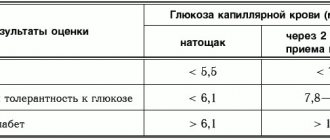
| Diabetes mellitus: on an empty stomach and/or 120 minutes after taking glucose | >6,1 >10,0 | >6,1 >11,1 | >7,0 >11,1 | >7,0 >12,2 |
| Impaired glucose tolerance: on an empty stomach and 120 minutes after ingestion of glucose | <6,1 6,7 — 10,0 | <6,1 7,8 — 11,1 | <7,0 7,8 — 11,1 | <7,0 8,9 — 12,2 |
| Impaired fasting glucose: fasting 120 min after glucose intake | 5,6 — 6,1 <6,7 | 5,6 — 6,1 <7,8 | 6,1 — 7,0 <7,8 | 6,1 — 7,0 <8,9 |
WHO recommends using only venous plasma test results to make a diagnosis of diabetes mellitus
- normal fasting plasma glucose levels are up to 6.1 mmol/l (<110 mg%);
- fasting plasma glucose levels from 6.1 mmol/l (≥110 mg%) to <7.0 mmol/l (<128 mg%) are defined as impaired fasting glucose;
- fasting plasma glucose level > 7 mmol/l (> 128 mg%) is regarded as a preliminary diagnosis of diabetes mellitus, which must be confirmed according to the above criteria.
When conducting a glucose tolerance test, the following indicators are important:
- Normal glucose tolerance is characterized by a plasma glucose level 2 hours after a glucose load of <7.8 mmol/L (<140 mg%);
- An increase in plasma glucose concentration 2 hours after a glucose load ≥ 7.8 mmol/L (≥ 140 mg%), but below 11.1 mmol/L (<200 mg%) indicates impaired glucose tolerance;
- A plasma glucose level 2 hours after an oral glucose load > 11.1 mmol/l (> 200 mg%) indicates a preliminary diagnosis of diabetes mellitus, which must be confirmed according to the criteria given above.
To evaluate the results of the glucose tolerance test, 2 indicators are calculated: hyperglycemic and hypoglycemic coefficients:
- Hyperglycemic – the ratio of glucose content after 30 or 60 minutes (the largest value is taken) to its level on an empty stomach. Normally, this coefficient should not be higher than 1.7.
- Hypoglycemic – the ratio of glucose levels after 2 hours to its fasting level. Normally, this coefficient should be less than 1.3.
If the patient does not have impaired glucose tolerance, but the value of both or one of the coefficients exceeds normal values, the glucose load curve is interpreted as “doubtful”. Such a patient should be advised to abstain from carbohydrate abuse and repeat the test after 1 year.
Recommended laboratory tests to establish the diagnosis of DIABETES MELLITUS
First level tests Blood
- The amount of glucose in venous blood
- Amount of glucose after exercise (vein)
- Glycosylated hemoglobin
- Fructosamine
Urine
- Amount of glucose
- Acetone in urine
Second level tests (differential diagnosis of diabetes)
- Blood insulin level
- Blood C-peptide level (fasting)
- Autoantibodies to cells of the islets of Langerhans
- Autoantibodies to insulin
- Autoantibodies to glutamic acid decarboxylase
- Immunogenetic typing (HLA)
- DR3-B8
- DR4-B15
- B15
- DR4
- Dw4
Third level tests (diagnosis of diabetes complications) Hypoglycemic coma
- Blood insulin level
- Blood glucose level
Diabetic nephropathy (tests at least once every 4-6 months)
- Microalbuminuria
- Protein in urine (proteinuria)
- Study of 24-hour urine protein (rate of increase in proteinuria)
- Rehberg test (creatinine clearance)
- Hypercreatininemia
- Hypoalbuminemia
- Apolipoprotein A1
- Apolipoprotein B (Apo B)
- Triglycerides
- Cholesterol
- HDL cholesterol
- LDL cholesterol
Diabetic macroangiopathy
- Hyperglycemia
- Hyperinsulinemia
- Total cholesterol (increased levels)
- Triglycerides (increased levels)
- High density lipoproteins
- Accelerated thrombus formation (see “diagnosis of thrombophilia”)
- Microalbuminuria
- Proteinuria
Plan for laboratory examination of a patient diagnosed with DIABETES MELLITUS
Laboratory tests at the first visit and at annual follow-up Blood
- The amount of glucose in plasma
- Amount of glucose after exercise
- Glycosylated hemoglobin
- Fructosamine
- Apolipoprotein A1
- Apolipoprotein B (Apo B)
- Triglycerides
- Cholesterol
- HDL cholesterol
- LDL cholesterol
- Creatinine
- Electrolytes (K/Na/Cl)
- Blood insulin level
- Blood C-peptide level
Urine
- Glucosuria
- Acetone in urine
- Microalbuminuria
- Ketonuria
- Bacteriuria
- Urine sediment
Laboratory tests at follow-up visit (every 3 months) Blood
- The amount of glucose in plasma
- Amount of glucose after meals
- Glycosylated hemoglobin
- Triglycerides
- Cholesterol
- HDL cholesterol
- LDL cholesterol
Urine
- Glucosuria
- Proteinuria
Assessing the effectiveness of diabetes treatment (diabetes compensation criteria)
| Laboratory indicators | Criteria for compensation of diabetes mellitus | ||
| DIABETES MELLITUS, TYPE 1 | optimal | satisfactorily | unsatisfactory |
| Glycemic profile | |||
| Fasting glycemia, mmol/l | 4,0 — 5,0 | 5,1 — 6,5 | >6,5 |
| Glycemia after meals (peak), mmol/l | 4,0 — 7,5 | 7,6 — 9,0 | >9,0 |
| Glycemia before bedtime, mmol/l | 4,0 — 5,0 | 6,0 — 7,5 | >7,5 |
| Glycosylated hemoglobin,% | <6,1 | 6,2 — 7,5 | >7,5 |
| Lipid profile | low risk | moderate risk | high risk |
| Total cholesterol, mmol/l | <4,8 | 4,8 — 6,0 | >6,0 |
| LDL-C, mmol/l | <3,0 | 3,0 — 4,0 | >4,0 |
| HDL-C, mmol/l | >1,2 | 1,0 — 1,2 | <1,0 |
| Triglycerides, mmol/l | <1,7 | 1,7 — 2,2 | >2,2 |
| DIABETES MELLITUS, TYPE 2 | optimal | satisfactorily | unsatisfactory |
| Glycemic profile | |||
| Fasting blood glucose (capillary blood), mmol/l | <5,5 | >5,5 | >6,0 |
| Glycemia after meals (peak, capillary blood), mmol/l | <7,5 | >7,5 | >9,0 |
| Fasting glycemia (venous blood plasma), mmol/l | <6,0 | >6,0 | >7,0 |
| Glycosylated hemoglobin,% | <6,5 | >6,5 | >7,5 |
| Lipid profile | low risk | moderate risk | high risk |
| Total cholesterol, mmol/l | <4,8 | 4,8 — 6,0 | >6,0 |
| LDL-C, mmol/l | <3,0 | 3,0 — 4,0 | >4,0 |
| HDL-C, mmol/l | >1,2 | 1,0 — 1,2 | <1,0 |
| Triglycerides, mmol/l | <1,7 | 1,7 — 2,2 | >2,2 |
Types of blood glucose curves during the glucose tolerance test. Changes in glucose concentration during hyperinsulinism (1); in healthy individuals (2); with thyrotoxicosis (3); with mild form (4) and severe form (5) of diabetes mellitus.
Laboratory diagnosis of complications of diabetes mellitus Diabetic nephropathy (DN) is currently the leading cause of high morbidity and mortality in patients with diabetes. The frequency of its development ranges from 40 to 50% in patients with insulin-dependent diabetes (IDDM) and from 15 to 30% in patients with non-insulin-dependent diabetes (NIDDM). The danger of this complication is that developing quite slowly and gradually, DN goes unnoticed for a long time, since clinically it does not cause a feeling of discomfort in the patient. And only at a pronounced (often terminal) stage of kidney pathology does the patient develop complaints related to intoxication of the body with nitrogenous wastes; however, at this stage it is not always possible to radically help the patient. Therefore, the main task of a general practitioner, endocrinologist or nephrologist is to timely diagnose DN and carry out adequate pathogenetic therapy as early as possible. In the pathogenesis of DN, changes that develop in the structure of the glomerular basement membrane and capillaries are important, which leads to disruption of hemodynamic parameters of renal function. They manifest themselves in an increase in glomerular filtration rate during the period of manifestation and the first years of diabetes. Due to relaxation of the afferent and narrowing of the efferent arterioles, intraglomerular pressure increases, which has a damaging effect on the surface of endothelial cells, both through increased mechanical stress and through an increase in the permeability of the basement membranes of the glomerular capillaries for lipids and various protein components of the plasma. Disruption of the normal structure of the glomerular barrier contributes to the proliferation of mesothelial cells with the deposition of lipids and proteins, thickening of the basement membrane, increased formation of the extracellular matrix and sclerosis of the renal tissue. As sclerotic changes progress, glomerular occlusion and renal tubular atrophy develop. Hyperfiltration (more than 140 ml/min/1.73 m2), observed in the initial stages of DN, is replaced by hypofiltration, which is accompanied by an increase in creatinine and urea in the blood serum and the appearance of clinical symptoms of renal failure. DN in the first years after the manifestation of diabetes occurs latently. This period lasts about 10-15 years, and only then does proteinuria, clinical signs of renal failure appear, and at the end of the disease - symptoms of uremia, combined with anemia, acidosis, and arterial hypertension.
Classification of stages of development of diabetic nephropathy (according to Mogensen S. E.)
| Stage of diabetic nephropathy | Clinical and laboratory characteristics | Development timeframe |
| Hyperfunction of the kidneys | increased GFR (> 140 ml/min); PC increase; kidney hypertrophy; normoalbuminuria (<30 mg/day). | In debut |
| Stage of initial structural changes in kidney tissue | thickening of the basement membranes and capillaries of the glomeruli; expansion of the mesangium; high GFR remains; normoalbuminuria. | After 2 - 5 years |
| Beginning nephropathy | microalbuminuria (from 30 to 300 mg/day); GFR is high or normal; unstable increase in blood pressure; | In 5 - 15 years |
| Severe nephropathy | proteinuria (more than 500 mg/day); GFR is normal or moderately reduced; arterial hypertension. | In 10 - 25 years |
| Uremia | decreased GFR < 10 ml/min; arterial hypertension; symptoms of intoxication. | After 20 or more years or after 5 - 7 years from the onset of proteinuria |
Algorithm for screening and management of diabetic nephropathy (from the moment of diagnosis)
| Test | Frequency | Acceptable Next steps | |
| Microalbuminuria | 1 time per year | < 20 mg/l | 20 mg/l Rule out other diseases (infections, heart failure) Determine daily albumin excretion |
| Rate of daily albumin excretion | 1 time per year | <15 µg/min | 15 – 200 mcg/min – if blood pressure is elevated, start treatment > 200 mcg/min – determine serum creatinine |
| Serum creatinine | Age norm | <120 – control once every 6 months 120 – 200 – control once every 3 months 200 – 450 – management together with a nephrologist >450 – management by a nephrologist | |
How does type 1 diabetes develop?
What is the trigger for the development of type 1 diabetes, like all autoimmune diseases, there is still no exact answer. But the main cause of the disease is known - a lack of insulin occurs due to the death of cells in the islets of Langerhans. The islets of Langerhans are areas on the tail of the pancreas that produce endocrine cells that participate in various life processes.
The role of endocrine cells is extensive, to be convinced of this, it is enough to consider a few examples:
- Alpha cells produce glycogen, which stores energy in the liver. This polysaccharide is the main form of glucose storage: glycogen reserves in the liver in a healthy person can reach 6% of the total body weight. Glycogen from the liver is available to all organs and can quickly replenish glucose deficiency in the body.
- Beta cells produce insulin, which converts glucose from the blood into energy. If the number of beta cells is insufficient or their work is poor, there is not enough insulin, so glucose remains unchanged in the blood.
- Delta cells are responsible for the production of somatostatin, which is involved in the functioning of the glands. Somatostatin limits the secretion of somatotropin - growth hormone.
- PP cells stimulate the production of gastric juice, without which complete digestion of food is impossible.
- Epsilon cells secrete a secretion that stimulates appetite.
The islets of Langerhans are equipped with capillaries, innervated by the vagus and peripheral nerves, and have a mosaic structure. The islets that produce certain cells are interconnected. Beta cells that produce insulin inhibit the production of glucogen. Alpha cells suppress the production of beta cells. Both islets reduce the amount of somatostatin produced.
Failure of immune mechanisms leads to the body's immune cells attacking the cells of the islets of Langerhans. Due to the fact that 80% of the surface of the islets is occupied by beta cells, they are the ones that are destroyed the most.
Dead cells cannot be restored; the remaining cells produce too little insulin. It is not enough to process the glucose entering the body. All that remains is to take insulin artificially in the form of injections. Diabetes mellitus becomes a lifelong death sentence; it cannot be cured and leads to the development of concomitant diseases.
Causes of type 1 diabetes mellitus
The development of type 1 diabetes is provoked by the following diseases:
- Severe viral infections
(rubella, chickenpox, cytomegalovirus, hepatitis, mumps). In response to infection, the body produces antibodies that, simultaneously with the virus cells, destroy beta cells, which are in many ways similar to the infection cells. In 25% of cases, after suffering from rubella, a person is diagnosed with diabetes mellitus. - Autoimmune diseases of the thyroid gland and adrenal glands
that produce hormones: autoimmune thyroiditis, chronic adrenal insufficiency. - Hormonal diseases
: Itsenko-Cushing syndrome, diffuse toxic goiter, pheochromocytoma. - Long-term use of a number of medications
. Antibiotics, tablets against rheumatism, dietary supplements with selenium are dangerous - they all provoke hyperglycemia - increased levels of glucose in the blood. - Pregnancy
. Hormones produced by the placenta increase blood sugar levels. The pancreas is under increased stress and cannot cope with the production of insulin. This is how gestational diabetes develops. This disease requires observation and may disappear without a trace after childbirth. - Stress.
When a person is very nervous, a large amount of adrenaline and glucocorticosteroids are released into the blood, which destroy beta cells. In patients with a genetic predisposition, it is after stress that a diagnosis of type 1 diabetes is made.
2.What is insulin and why is it needed?
Typically, the hormone insulin is produced in small quantities in the pancreas. When you eat, the sugar (glucose) in the food stimulates the pancreas to produce insulin. Its amount is proportional to the amount of food taken. Insulin's main role is to help move certain nutrients, especially sugar, into the body's tissues and cells. Cells use sugar and other nutrients as an energy source to function.
The amount of sugar in the blood decreases when it enters the cells. This usually signals the beta cells to reduce the amount of insulin they secrete to avoid hypoglycemia (low blood sugar), and the amount of insulin they secrete also decreases. But the destruction of beta cells disrupts the functioning of the entire system.
Visit our Endocrinology page
Causes of type 1 diabetes in children and adolescents
Many parents are mistaken in thinking that diabetics got sick because they ate a lot of chocolate and sugar. If you limit your child’s sweets, he can be protected from diathesis rather than from diabetes. Children get diabetes at an early age not because of poor diet. This is evidenced by the findings of scientists studying this problem.
The authoritative scientist Andreas Beyerlein from the Munich Helmholtz Center conducted a study, upon completion of which the following conclusions were made:
- A severe viral infection suffered at the age of 0-3 years leads to the development of type 1 diabetes mellitus in 84%, and the pathology is more often diagnosed when the child reaches the age of 8.
- Acute acute respiratory viral infection suffered by infants under 3 months causes diabetes in 97% of cases.
- In children with a hereditary predisposition to hyperglycemia, the risk of developing the disease increases depending on nutritional factors (nutrition): artificial feeding, early consumption of cow's milk, high birth weight (above 4.5 kg).
There are two peak ages for detecting diabetes in children - 5-8 years and adolescence (13-16 years). Unlike adults, childhood diabetes develops very quickly and rapidly. The disease manifests itself as an acute form of ketoacidosis (poisoning by ketone bodies formed in the liver) or diabetic coma.
As for heredity, the likelihood of transmitting T1DM is low. If the father has type 1 diabetes, the risk of transmission to children is 10%. If the mother, then the risks are reduced to 10%, and in late births (after 25 years) to 1%.
Identical twins have different risks of getting the disease. If one child is sick, the disease occurs in the second no more than 30-50%.
Complications of type 1 diabetes
In addition to diabetes itself, its complications are no less dangerous. Even with a slight deviation from the norm (5.5 mmol/liter on an empty stomach), the blood thickens and becomes viscous. Vessels lose their elasticity, and deposits in the form of blood clots (atherosclerosis) form on their walls. The internal lumen of arteries and vessels narrows, organs do not receive sufficient nutrition, and the removal of toxins from cells is slowed down. For this reason, places of necrosis and suppuration appear on the human body. Gangrene, inflammation, rash occurs, and blood supply to the extremities deteriorates.
High blood sugar disrupts the functioning of all organs:
- Kidneys
. The purpose of the paired organs is to filter the blood from harmful substances and toxins. When the sugar level is more than 10 mmol/liter, the kidneys stop doing their job efficiently and pass sugar into the urine. A sweet environment becomes an excellent base for the development of pathogenic microflora. Therefore, hyperglycemia is usually accompanied by inflammatory diseases of the genitourinary system - cystitis (inflammation of the bladder) and nephritis (inflammation of the kidneys). - The cardiovascular system.
Atherosclerotic plaques, formed due to increased blood viscosity, line the walls of blood vessels and reduce their capacity. The cardiac muscle myocardium ceases to receive adequate nutrition. This is how a heart attack occurs - necrosis of the heart muscle. If a sick person does not suffer from diabetes, he will feel discomfort and a burning sensation in the chest during a heart attack. A diabetic suffers from decreased sensitivity of the heart muscle and may die unexpectedly. The same goes for blood vessels. They become brittle, increasing the risk of stroke. - Eyes
. Diabetes damages small vessels and capillaries. If a blood clot blocks a large vessel of the eye, partial death of the retina occurs, and detachment or glaucoma develops. These pathologies are incurable and lead to blindness. - Nervous system.
Poor nutrition, associated with serious restrictions in type 1 diabetes, leads to the death of nerve endings. A person stops reacting to external stimuli, he does not notice the cold and freezes his skin, does not feel the heat and burns his hands. - Teeth and gums.
Diabetes is accompanied by diseases of the oral cavity. The gums soften, tooth mobility increases, gingivitis (inflammation of the gums) or periodontitis (inflammation of the inner surface of the gums) develops, which leads to tooth loss. The effect of insulin-dependent diabetes on the teeth of children and adolescents is especially noticeable - they rarely have a beautiful smile: even their front teeth deteriorate. - Gastrointestinal tract
. In diabetes, beta cells are destroyed, and along with them the PP cells responsible for the production of gastric juice. Patients with diabetes often complain of gastritis (inflammation of the stomach lining), diarrhea (diarrhea due to poor digestion of food), and the formation of gallstones. - Problems with bones and joints
. Frequent urination leads to the leaching of calcium, resulting in damage to the joints and skeletal system, and an increased risk of fractures. - Leather
_ Increased blood sugar leads to the loss of protective functions of the skin. Small capillaries become clogged with sugar crystals, causing itching. Dehydration makes the skin wrinkled and very dry. In some cases, patients develop vitiligo - the breakdown of skin cells that produce pigment. In this case, the body becomes covered with white spots. - Female reproductive system
. A sweet environment creates favorable soil for the development of opportunistic microflora. In type 1 diabetes, frequent relapses of thrush are typical. Women have poor vaginal lubrication, which makes sexual intercourse difficult. Hyperglycemia negatively affects fetal development in the first 6 weeks of pregnancy. Diabetes also leads to premature menopause. Early menopause occurs at 42-43 years.
Symptoms of type 1 diabetes
External signs help determine diabetes, because the disease affects the functioning of the entire body. In young people under 18 years of age, diabetes develops very quickly and rapidly. It often happens that 2-3 months after a stressful event (ARVI, moving to another country), a diabetic coma occurs. In adults, symptoms may be milder and gradually worsen.
The following signs are cause for concern:
- Frequent urination, a person goes to the toilet several times a night.
- Weight loss (dieting and the desire to lose weight in adolescence is fraught with the rapid development of hyperglycemia).
- The appearance of age-appropriate wrinkles, dry skin.
- Increased feeling of hunger with lack of weight.
- Lethargy, apathy, the teenager quickly gets tired, he begins to have painful thoughts.
- Fainting, severe headache, vision problems.
- Constant thirst, dry mouth.
- A specific smell of acetone from the mouth, and in severe cases from the body.
- Night sweats.
If at least a few symptoms are noticed, the patient should be immediately sent to an endocrinologist.
The younger the body, the faster the coma sets in.
Diagnosis of diabetes mellitus
The endocrinologist will definitely prescribe the following tests for diabetes:
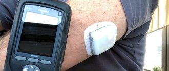
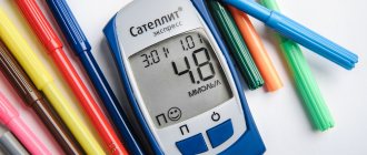
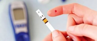
- Test for blood glucose levels
. Blood is taken on an empty stomach; the last meal should be no earlier than 8 hours before. A reading below 5.5 mmol/liter is considered normal. An indicator of up to 7 mmol/liter indicates a high predisposition; 10 mmol/liter and above indicates hyperglycemia. - Oral glucose tolerance test
. This test is done for those who are at risk of developing diabetes. The patient takes a glucose solution on an empty stomach. Then, 2 hours later, blood is taken for sugar. Normally, the reading should be below 140 mg/dL. A blood sugar level above 200 mg/dL confirms diabetes mellitus. - Test for glycosylated hemoglobin A1C
. Excess sugar in the blood reacts with hemoglobin, so the A1C test shows how long the body's sugar levels have been above normal. Monitoring is carried out every 3 months, the level of glycosylated hemoglobin should not exceed 7%. - Blood test for antibodies
. Type 1 diabetes is characterized by an abundance of antibodies to the cells of the islets of Langerhans. They destroy body cells, which is why they are called autoimmune. By identifying these cells, the presence and type of diabetes is determined. - Urinalysis - microalbuminuria
. Detects protein in urine. It appears not only with kidney problems, but also with vascular damage. High levels of albumin protein lead to heart attack or stroke. - Screening for retinopathy
. High glucose levels lead to blockage of small vessels and capillaries. The retina of the eye does not receive nourishment; it peels off over time and leads to blindness. Special digital equipment allows you to take pictures of the back surface of the eye and see the location of damage. - Test for thyroid hormones.
An overactive thyroid gland leads to hyperthyroidism—excessive production of hormones. Hyperthyroidism is dangerous because the breakdown products of thyroid hormones increase the level of glucose in the blood; diabetes is accompanied by acidosis (high levels of acetone in the urine), osteoporosis (leaching of calcium from the bones), and arrhythmia (failure of heart rhythm).
Gestational diabetes
Gestational diabetes is manifested by hyperglycemia with blood glucose levels that exceed normal values, but do not reach diagnostic values for making a diagnosis of diabetes. Gestational diabetes is diagnosed during pregnancy. Women with this diagnosis have an increased risk of complications during pregnancy and childbirth. They also have an increased risk of developing type 2 diabetes in the future. Most often, the diagnosis of gestational diabetes is made during prenatal screening; patients do not complain.
Decreased glucose tolerance and impaired fasting glycemia
Reduced glucose tolerance (PTH) and impaired fasting glucose (IFG) are intermediate conditions between normal and diabetes. People with PTH and IFN are at high risk of developing type 2 diabetes, but this may not happen.
Treatment of type 1 diabetes
Type 1 diabetes is not curable because beta cells cannot be restored. The only way to maintain normal blood sugar levels in a sick person is to take insulin, a hormone produced by the beta cells of the islets of Langerhans.
Based on the speed of action and duration of effect, medications containing insulin are divided into categories:
- Short-acting (Insuman Rapid, Actrapid)
. They begin to act 30 minutes after administration, so they should be taken half an hour before meals. When the drug is administered intravenously, it is activated after a minute. The duration of the effect is 6-7 hours. - Ultra-short-acting (Lispro, Aspart).
They begin to work 15 minutes after the injection. The action lasts only 4 hours, so the drug is used for pump administration. - Medium duration (Insuman Bazal, Protafan).
The effect occurs an hour after administration and lasts 8-12 hours. - Long-term exposure (Tresiba).
The drug is administered once a day, it does not have a peak effect.
Medicines are selected individually for the patient in combination with other drugs that prevent the negative effects of high blood glucose.
References
- Clinical recommendations. Diabetes mellitus type 1 in adults, 2019. - 167 p.
- Guide to laboratory diagnostic methods. - M.: GEOTAR Media, 2007. - 800 p.
- Guyton, A.K., Hall, J.E. Medical physiology / ed. IN AND. Kobrina. - M.: Logosphere, 2008. - 1296 p.
- Regnell, S., Lernmark, Å. Early prediction of autoimmune (type 1) diabetes. Diabetologia, 2021. - Vol. 60(8). - P. 1370-1381.
- Nazarenko, G.I., Kishkun, A.A. Clinical evaluation of laboratory results. - M.: Medicine, 2000. - 544 p.
- Kishkun, A.A. Guide to laboratory diagnostic methods. - M.: GEOTAR-Media, 2007. - 800 p.
Exercise for diabetes
Physical activity for type 1 diabetes is simply necessary, although there are restrictions regarding the type of sport. Exercise normalizes blood pressure, improves well-being, and normalizes weight. But in some cases, physical activity causes spikes in blood glucose levels.
If you have type 1 diabetes, you cannot overload yourself, so training should not exceed 40 minutes a day. The following sports are acceptable:
- walking, cycling;
- swimming, aerobics, yoga;
- table tennis, football;
- exercises in the gym.
Any exercise is contraindicated if ketones, protein breakdown products, are found in the urine, and if blood pressure has increased or problems with blood vessels have appeared.
Detailed description of the study
Diabetes mellitus type 1 (DM 1) is an autoimmune pathology, which is based on the destruction of pancreatic cells and insulin deficiency. These cells, also called islet cells, produce the hormone insulin, which transports glucose into tissues.
Features of this type of diabetes are the following:
- The appearance of the disease in the vast majority of cases in childhood or adolescence;
- Acute onset;
- Insufficiency of insulin in the body, in which the only treatment is the administration of insulin from outside.
In the clinical picture of the disease, the following symptoms can be observed:
- Thirst and dry mouth;
- Increased volume of urine excreted;
- General weakness;
- Loss of body weight;
- Increased appetite;
- Ketoacidosis, diabetic coma (a life-threatening condition requiring immediate medical attention, in which the blood glucose level is noticeably elevated and can reach critical levels).
Differentiation of T1DM is carried out with type 2 diabetes and other forms of diabetes. Typically, type 2 diabetes develops gradually in adults with underlying medical conditions, such as hypertension.
As stated earlier, T1DM most often develops in young adulthood or childhood. It is usually preceded by an acute viral infection or a stressful condition. Risk factors for the occurrence of pathology are genetic predisposition and the influence of environmental factors (such as viruses, psychosocial factors, diet). Often, a viral illness can occur several weeks before the first symptoms of T1DM. First-degree relatives of patients who have been diagnosed with T1DM are at particular risk.
Early diagnosis of type 1 diabetes allows timely treatment to be prescribed to prevent severe complications. This laboratory examination includes six tests that are highly likely to help identify T1DM or a predisposition to it. The study includes the determination of antibodies to insulin, pancreatic beta cells, blood glucose concentration, glycated hemoglobin, and antibodies to GAD.
Glucose is a molecule that belongs to the group of saccharides and is the main source of human energy. Its concentration in the blood is normally within certain limits; an increase in level is most often observed in diabetes mellitus. However, it should be noted that an increase in the concentration of this molecule in some cases (during acute infection, injury or stress) may be temporary (transient) and does not apply to diabetes mellitus.
Glycated hemoglobin is a type of hemoglobin (one of the blood proteins) that is bound to glucose. This form is referred to as HbA1c. Determining HbA1c in a patient plays an important role in the diagnosis of T1DM, because The degree of glycosylation (addition of glucose to Hb) of hemoglobin depends on the level of glucose in a person’s blood and on the duration of contact with it.
Insulin synthesis is a complex multi-step process, which results in the formation of an inactive substance - proinsulin. Its breakdown leads to the formation of the hormone insulin, which enters the blood and reaches organs, where it is involved in the transport of glucose into tissues.
C-peptide is involved in the synthesis of insulin and is secreted by beta cells of the pancreas. After being released from the pancreas, it travels through the bloodstream to the liver and kidneys, then is excreted from the body.
Normally, immunoglobulins (antibodies, antibodies) serve to protect against microorganisms and foreign substances. In T1DM, the body produces abnormal immunoglobulins called autoantibodies. They trigger a cascade of reactions aimed at destroying one’s own beta cells, and can also be produced against insulin.
Autoantibodies to glutamate decarboxylase (GAD65). GAD65 belongs to the beta cell molecules of the pancreas. These antibodies affect beta cells and can be detected even before the onset of the disease.
Thus, the study includes a number of important indicators that are necessary for timely and accurate diagnosis of type 1 diabetes. You need to donate venous blood for the test.
A detailed description of the studies and reference values are presented on the pages with descriptions of individual studies.