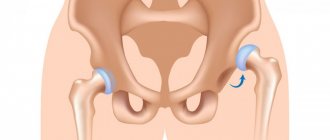
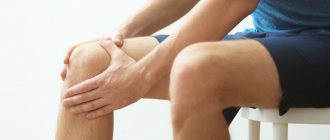
The hip joint is one of the most important in the musculoskeletal system. By supporting the weight of the entire body, the joint can withstand enormous overloads.
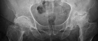
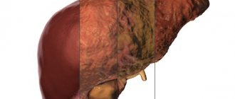
This is what the disease looks like.
It is presented in a spherical shape and allows the use of all axes of movement: adduction and abduction (sagittal axis), flexion and extension (frontal axis), pronation and supination (vertical axis). Due to its mobility, the joint is often damaged.
Structure of the hip joint
One of the most dangerous injuries is avascular necrosis of the femoral head (AFH). It is a consequence of a gross disruption of local microcirculation with the development of ischemia and necrotization of the bone marrow components of the femoral head. DDI is a consequence of previous operations on the hip joint, a history of trauma, previous osteomyelitis of the femur, and the development of epiphyseal and/or spondyloepiphyseal dysplasia.
We list several causes of vascular disorder leading to necrosis:
- repeated heart attacks due to arterial thrombosis;
- long-term insufficiency of arterial blood supply to the joint;
- venous stasis;
- combined disorder of the arterial-venous network.
The factors that provoke this situation are: congenital hypoplasia of the vessels of the hip joint, disruption of neurohumoral regulatory mechanisms, insufficient vascularization of the femoral head associated with the anatomical and functional immaturity of the vascular network.
Blood supply to the hip joint
Malnutrition of the bone components of the hip joint is the cause of necrotic changes.
Content
- Causes and risk factors
- Diagnostic methods
- Treatment methods
- Treatment programs
- Who carries out the treatment?
- Why do they contact us?
- Reviews from our patients
- Sign up for treatment
The mechanism of development of the disease is associated with deterioration of the blood supply to the femoral head for various reasons. ANFH is a polyetiological disease, that is, the appearance of the disease is caused by several reasons simultaneously. Deterioration of bone nutrition leads to disruption of the structure, macro- and microarchitecture of bone tissue, with the formation of foci of ischemia, cystic restructuring and death. As a result, deformation of the head develops, the bone becomes brittle, and microfractures occur.
Treatment begins with conservative therapy. Surgical assistance is required in desperate cases, for example, when other methods of therapy have not brought the desired result. Endoprosthesis replacement is recommended at stage 4 of the disease.
Symptoms and differences from coxarthrosis
The symptoms of ANFH are determined by the stages of the pathology. The onset is marked by asymptomatic symptoms or minor pain for no apparent reason. At the same time, the normal range of motion in the joint is maintained, pain is noted when the hip rotates inward.
The progression of the disease is expressed by paroxysmal, severe pain. Such a symptom may indicate the presence of collapse or fracture of the femoral head, inherent in the final stage of degenerative changes. They lead to a decrease in range of motion and the presence of constant pain, crepitus and instability of the femoral head.
The clinical symptoms of ANFH are similar to the classic picture of coxarthrosis:
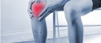
- pain in the groin that runs along the anterolateral surface of the thigh and radiates to the knee joint. The load only intensifies these sensations, with the pain moving to the lumbar region. They always bother a person, even at night;
- the range of motion in the affected joint is noticeably limited. These people need help with self-care;
- severe lameness on the affected leg when walking;
- rapidly progressive hypotrophy of the muscular frame of the thigh on the affected side;
- shortening of the thigh.
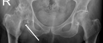
Necrosis of the right hip joint
Risk of developing avascular necrosis due to trauma
One of the main reasons for the development of ANFH is fractures of the head or neck of the femur. The greatest risk is a transepiphyseal fracture (impaired blood supply to the proximal fragment up to 97.6%), subepiphyseal fracture (impaired blood supply to the proximal parts of the head up to 97.4%), subcapital fracture (impaired blood supply up to 92%).
There are several types of fractures:
Type 1: abduction, subcapital, incomplete fracture without displacement. It occurs due to the action of diverting tension. During a fracture, the neck and diaphysis of the femur are directed towards the head. The bone fragments are firmly wedged, and the fracture surface line is close to horizontal. The disturbance of blood supply is minimal (up to 10.2%). The prognosis for recovery is good.
From left to right: subcapital, transcervical, basicervical fractures.
Type 2: adduction, subcapital, complete fracture without displacement. The fracture is located in a vertical plane, passing through the femoral neck. Trabeculae and lower cortex are torn, without displacement. Microcirculation is impaired by 23.6%. The prognosis is favorable.
Type 3: adduction fracture with incomplete displacement of bone structures. It is characterized by varus deformity of the femoral neck, but the fragments remain connected due to the posteroinferior block. The distal bone fragment is in a position of external rotation and abduction with an angle open anteriorly. There is a comminuted fracture of the posterior surface of the neck. The trabeculae and lower cortex are torn. Vascularization disorders reach 42.8%. Risk of developing avascular necrosis.
Type 4: adduction fracture with complete displacement. The head is deprived of all connections with the synovial membrane of the capsule, as a result of which the fragment becomes free. The disturbance of blood supply is maximum (54.4%). The risk of aseptic necrosis is increased.
To summarize: in the event of a potential injury to the hip joint, you must go to the nearest emergency room and have an x-ray taken. Early diagnosis can save the joint from destruction and maintain a high quality of life!
Minimally invasive endoprosthetics in the Czech Republic: doctors, rehabilitation, terms and prices.
Find out more
Reasons for development
There are many factors that can lead to the development of ANFH. These include:
- dislocations of the hip joint, fractures of the femoral neck and the head itself, as well as surgical interventions;
- long-term use of corticosteroids, especially those intended for systemic use (tablets);
- alcohol abuse, which leads to metabolic disorders;
- long-term use of painkillers and NSAIDs;
- autoimmune diseases, including scleroderma, hemorrhagic vasculitis, systemic lupus erythematosus;
- decompression sickness, characteristic of divers, miners and representatives of other professions, which require them to work in conditions of rapid decrease in the pressure of the inhaled gas mixture;
- radiation sickness or radiation therapy performed to suppress a malignant tumor;
- cauda equina syndrome, caused by entrapment of the spinal roots at the level of the lumbosacral vertebrae.
However, in approximately 30% of patients, it is not possible to determine exactly what exactly caused the development of ANFH.
ANGKB stages according to ARCO
However, in Russia the classification into five stages is more common:
- There are no radiological signs. The histological specimen shows signs of necrosis of the spongy substance of the head and bone marrow structures. Clinically expressed by aching pain and stiffness in the joint, increasing muscle weakness.
- multiple impression fractures. Against the background of necrosis, many microscopic fractures occur. The radiograph shows a homogeneous darkening of the femur, its height is reduced, the surfaces of the head in places are in the form of compacted facets, the joint space is widened. MRI data indicate a necrotic defect in the head.
- sequestration. The articular head is flattened and looks like structureless isolated fragments with different shapes and sizes. The neck of the bone shortens and thickens, and the joint space widens even more.
- reparative. The spongy substance of the femoral head is restored. On X-ray, sequestration-like zones are not noticeable; the shadow of the head is outlined, but with rounded cyst-like clearings.
- secondary deforming arthrosis. The bone structure of the femur begins to be traced, significantly changed, the congruence of the articular surfaces is disrupted.
Disease in dynamics.
Important: collapse of the femoral head occurs in an incredibly short period of time - 5 months.
Diagnostics
If you experience pain in the hip joint, it is important to contact an orthopedic traumatologist as soon as possible. This symptom may indicate the development of various joint pathologies, only one of which is ANKB. Therefore, the doctor must prescribe instrumental research methods:
- X-ray of the hip joint in 3 projections. The method makes it possible to establish not only the presence of ANKB, but also the stage of development of the disease. But it must be taken into account that at stage 1 there are no radiological signs of necrosis of the femoral head. This may cause a diagnostic error. Therefore, it is important to repeat the X-ray of the hip joint after six months.
- CT. It is prescribed, if necessary, to obtain layer-by-layer images of the hip joint and examine the characteristics of blood flow.
- MRI. The method allows you to detect the slightest changes in the condition of the hip joint, especially hyaline cartilage and other soft tissue structures.
- Densitometry. This diagnostic method makes it possible to measure bone density and detect signs of osteoporosis in the hip joint.
Additionally, biochemical blood and urine tests may be prescribed. The greatest attention is paid to the level of calcium, phosphorus, magnesium, DPID, pyridonine, osteocalcin.
Study of the blood circulation of the femoral head
Laser Doppler flowmetry and microsensory intraosseous pressure transducers are used. Histological examination makes it possible to diagnose the disease, differentiate it from other pathologies and determine the pathogenetic paths of development of the process.
As the disease progresses, the bone structure undergoes changes. In the subchondrial zone and the zone of necrosis, the activity of osteoclasts increases, and in the zone of sclerosis, on the contrary, the activity of osteoblasts increases.
With ANFH, laboratory parameters such as prothrombin time, aPTT (activated partial thromboplastin time) are normal. However, in a comparison of patients with non-traumatic necrosis and healthy volunteers, significant deviations in the concentration of coagulation factors in the blood plasma were revealed.
In people with ANFH, the level of ghrelin is reduced, the level of von Willebrand factor, plasminogen activator inhibitor-1 (PAI-1), and C-reactive protein is increased, which indicates the involvement of these factors in the mechanism of pathogenesis.
The use of certain laboratory parameters as markers for the diagnosis of ANFH is a promising method.
Treatment
For each stage according to ARCO, the duration of each of them is determined. For stage I and stage II - up to 6 months, stage III - 3-6 months and an immediate transition to stage IV.
Early initiation of treatment, while symptoms reveal only minor discomfort in the periarticular area with irradiation to the groin area and knee joint, is extremely important.
Tasks during the treatment period:
- For patients with a history of trauma, intoxication, and those at risk (taking glucocorticoids, cytostatics), use the diagnostic algorithm for ANFH in the early stages. It includes early clinical manifestations, information analysis of cardiac signals using the Screenfax system, MRI/CT, radiography, laboratory tests of markers of bone metabolism, scintigraphy, X-ray/ultrasound densitometry.
- Using a non-surgical treatment method, improve metabolic processes in the lesion, regenerate the elements of the femur and restore the function of the joint itself.
Taking into account the fact that ANFH is a multifactorial pathology, it is necessary to use an integrated approach in diagnosis, treatment and rehabilitation aimed at restoring:
- optimal functioning of all systems and organs;
- microvasculature and hemocoagulation parameters;
- neuroregulatory factors;
- immune reactions;
- myodiscoordinate processes and biomechanics of joints.
Treatment of stages 1-2
Seems most effective for a favorable prognosis. The most important condition for successful treatment is strict adherence to the regimen. You should give up bad habits (alcohol and smoking), avoid overloading and hypothermia of the affected joint.
Therapy is directed in several areas: medication, physiotherapy and exercise therapy.
Drugs . For aseptic necrosis, the following groups of drugs are prescribed:
- Bone resorption inhibitors (bisphosphonates). They slow down the destruction of bone tissue and prevent compression and deformation of the femoral head.
- Calcium and vitamin D preparations. In the form of active forms, they facilitate the formation of bone structures and their mineralization. The dose is selected individually.
- Additional sources of minerals and phosphates (ossein-hydroxyapatite).
- Since blood coagulation factors change in ANFH, antiplatelet drugs (chimes, dipyridamole, xanthinol) are necessarily prescribed.
- Non-steroidal anti-inflammatory drugs (NSAIDs). They have an analgesic and anti-inflammatory effect. The most commonly prescribed drugs are ibuprofen, ketoprofen, diclofenac and their analogues.
- Muscle relaxants to relieve muscle tension around the joint.
- Multivitamin complexes, chondroprotectors.
Physiotherapy. The main task is to stimulate blood flow in tissues and start/accelerate regenerative processes.
- a) hyperbaric oxygenation. The patient is placed in a pressure chamber, where he is exposed to air enriched with oxygen under conditions of high pressure. This procedure improves oxygen supply to the damaged area.
- b) shock wave therapy (SWT). Represents the action of a sound wave. It is carried out pointwise, certainly at the site of damage. It effectively affects pathological tissues that interfere with the healing of diseased joints (calcium crystals or adhesions). UVT improves blood supply to the treated area and thereby enhances the reparative effect.
- c) myostimulation. Allows you to restore muscle tone during developing hypotrophy, which is often found in ANFH. Stimulation relieves spasm of the muscle fibers surrounding the joint and accelerates local blood flow.
Physiotherapy
The initial stage is unloading the affected joint. In this case, the patient is fitted with crutches, which can take up to a year to walk on.
If after therapy the pain has decreased and bone turnover markers have returned to normal, crutches should be discontinued after 2-3 months. But don't rush. Unloading the joint will relieve pain and prevent further destruction of the femoral head. The gentle regime accelerates healing, it is faster and easier.
On the other hand, the patient needs exercise therapy. A special set of exercises will reduce pain and swelling of the joint and increase its mobility. Physical exercise will develop not only the joint, but also the muscles surrounding it, preventing the development of malnutrition.
Obese patients are advised to combine exercise with a specially selected diet. This combination will allow you to achieve better results.
The importance of physical therapy at the rehabilitation stage should be especially emphasized. It will help not only to develop the operated/replaced joint, but also to prevent its instability.
Department of Traumatology and Orthopedics
article in PDF format
QUOTE LINK:
A. V. ANTONOV, V. E. VOLOVIK
KGBOU DPO "Institute for Advanced Training of Health Care Specialists" MHHC, Khabarovsk
Information about the authors:
Antonov Alexander Vadimovich – Postgraduate student of the Department of Traumatology and Orthopedics of the KGBOU DPO "Institute for Advanced Training of Healthcare Specialists" of the Ministry of Health of the Khabarovsk Territory, e-mail
Volovik Valery Evgenievich - Head of the Department of Traumatology and Orthopedics of the KGBOU DPO "Institute for Advanced Training of Healthcare Specialists" of the Ministry of Health of the Khabarovsk Territory, Honored Worker of Science and Education, Honored Health Worker of the Russian Federation, Doctor of Medical Sciences, Prof., Corresponding Member. RAE, e-mail
Aseptic necrosis of the femoral head is a common multifactorial, polyetiological disease, representing a complex medical and social problem, affecting people of working age and accounting for 1.2–4.7% of all degenerative-dystrophic diseases of the musculoskeletal system according to various authors. The lack of timely diagnosis and the inappropriateness of conservative treatment when citizens present late lead to their disability, and subsequently to a complete joint replacement. Endoprosthetics as a method of choice for surgical treatment is associated with both long-term rehabilitation of patients and the risks of developing instability of components, and with the onset of 2021, many economic aspects. This problem remains relevant and requires further study, improvement of methods of conservative and surgical treatment in order to achieve stable long-term remission, and possibly complete recovery of the patient.
Key words: aseptic; ANGBK; necrosis; idiopathic necrosis; necrosis of the femoral head.
Aseptic necrosis of the femoral head (AFH) is a severe chronic common pathology, which accounts for 1.2–4.7% of all degenerative diseases of the musculoskeletal system according to various authors. The problem of early diagnosis due to the absence of obvious symptoms during the onset of the disease, the similarity of clinical signs with other pathological conditions and the rapid development of the disease lead to characteristic degenerative processes. Changes in the usual rhythm of life and the inability to receive medical care due to the lack of effective treatment methods sooner or later lead to persistent impairment and disability of patients [1].
The risk group for the development of this pathology is predominantly the male population aged 20–50 years, exposed to various harmful factors (alcoholism, smoking, use of corticosteroids, radiation), as well as a number of serious diseases: sickle cell anemia, coagulopathies, vasculitis, hyperlipidemia, decompression sickness, Gaucher disease, bleeding disorders, chronic liver and kidney diseases, etc., requiring the use of aggressive treatment methods such as hemodialysis, chemotherapy, etc.
The nature of the changes occurring in the femoral head is not fully understood, but the traumatic etiology of the disease remains undoubted. According to S. Delaunay, depending on the severity of the injury, 10–50% of patients with various injuries to the hip joint (HJ) develop aseptic necrosis in the immediate or long term [2]. A number of foreign authors [3, 4], having studied modern methods of surgical treatment for fractures and their long-term consequences, believe that the development of ANFH is influenced by the patient’s age and delay of surgery, but not by the method of treatment [5]. Along with these conclusions, there are non-traumatic theories of origin associated with insufficient blood supply (including atypical variants of the development of the topographic-anatomical picture of the branches of the deep femoral artery) [6], increased intraosseous pressure, functional inferiority of the hip joint, the toxic effect of drugs (protein tyrosine kinase inhibitors, hormone-induced ANFH) [7, 8], with metabolic (lipid metabolism disorders) [9], neurogenic disorders [10]. Considering the many theories of the origin of the pathological process, today there are no suitable experimental models to find the cause of the disease [11].
The study of the pathogenesis of the disease over the last century has not yielded fruitful results of the same type, making this problem, from the point of view of many specialists, unsolvable. For example, M.G. Proves (1938) determined that the arteries of the femoral head do not end blindly, suggesting the possibility of a thromboembolic mechanism. Gratsiansky (1955) believed that ANFH is a secondary pathology that occurs after a fracture of bone beams, representing the trigger mechanism as microtraumatization or overload of the joint, activating the processes of fatigue. In this case, pathological impulses directed from the lesion to the cerebral cortex contribute to the formation of a reverse signal, causing vasospasm, stagnation of blood and lymph, metabolic disorders, accumulation of decay products, changes in the physicochemical properties of bone tissue, and subsequently destruction of bone beams with difficulty in local blood circulation and, accordingly, necrosis. For the first time, A.L. spoke about the possible participation of the parasympathetic nervous system in the development of the pathological process. Gimmelfarb, who argued that pathological impulses from the focus contribute to the development of reflex vascular spasm, disrupting the blood supply. Focusing on the presence of vascular disorders, a number of researchers examined separately the arterial, venous and mixed components (L. Solomon, N.F. Moroz); at the same time, Leriche, believing that the root cause may be a disorder of lipid metabolism, refers to the need to revise the ischemic theory of pathogenesis.
The historically recognized foundations of ischemic disorders were also confirmed and expanded by O.P. Bolshakov, who determined the possibility of atypical variants of development of the topographic-anatomical picture of the branches of the deep femoral artery in the form of an increase in the angle of departure of the circumflex arteries or the complete absence of the medial circumflex artery.
Over the past 10 years, thanks to many foreign experimental models, the number of assumptions regarding the pathogenesis of the disease has increased significantly. Changes occurring in the head of the femur are primarily due to local disturbances in blood flow, decreased perfusion due to the anatomical features of the feeding vessels [12], which leads to the development of ischemic phenomena. As a result of changes in blood supply and the hydrodynamic influence of elastic deformations, redistribution disorders, perimedullary venous congestion, disruption of tissue metabolism, increased osteoclastic resorption, thinning and sponging of the compact substance, an increase in bone marrow spaces and the development of necrosis occur. Lipid disorders, including those resulting from increased levels of steroids, play an important role in joint nutrition and are most likely one of the causes of vascular disorders [13]. H. Kantor believes that approximately 45% of patients have a triad of metabolic disorders - lipid metabolism disorders, liver diseases and chronic alcoholism. But despite the harmony of the alcohol-induced theory, according to D. Orlic, the incidence of pathology in alcohol abusers is 3.9%. This indicates the participation of other processes in pathogenesis, in addition to lipid metabolism disorders [2]. The study of the genetic aspects of the pathology has shown the important role of oxidative damage to the DNA of hematopoietic bone marrow cells [14], polymorphism of various genes and endothelial synthase [15, 16].
Proponents of molecular studies conclude that there is a causal relationship with osteoprotegerin synthesis [17], fibroblast growth factors [18], leptin levels in the bone marrow [19] and proteoglycan loss, compared with inflammatory changes in arthritis [20], considering that ANFH may be preceded by an inflammatory response [21]. In any case, an important pathomorphological link in the development of the disease is: violation of the conditions of transcapillary exchange, inhibition of mineralization, degeneration of the red bone marrow. A pathological type of blood circulation is formed with the development of ischemia, and subsequently the death of bone cells with the formation of a zone of bone tissue decay [11] and microcrystallization of cartilage [23]. If the load-bearing capacity of the necrotic bone decreases, the subsequent disruption of the distribution of contact stress leads to the collapse of the femoral head and its complete destruction [24].
Among all theories of pathogenesis, the concept of deposition of calcium-containing crystals in articular cartilage and para-articular tissues occupies a special position. In this case, certain types of crystals are detected, such as calcium pyrophosphate, hydroxyapatite, tri-octacalcium phosphates. This phenomenon is called “microcrystalline stress” and, according to A.I. Dubikov, occurs in all patients with aseptic necrosis, which indicates the need to revise existing theories of pathogenesis [23].
Changes that occur in the head of the femur are usually accompanied by changes in the color, structure and shape of the surface; tuberosity or roughness of the cartilaginous cover appears, while hyaline cartilage remains of dense consistency, gray, less often brown, with additional inclusions. Dystrophic changes in cartilage and bone tissue are accompanied by thinning of the cortical layer, expansion of the interbeam spaces, angiomatosis, as well as cicatricial degeneration of the joint capsule. There are signs of fatty degeneration of the bone marrow, fibrous tissue, resorption, necrobiosis, lymphoid-plasmacytic and histocytic infiltration of the stroma, and in some cases, an inflammatory reaction.
Classifications of this disease are quite rare and are represented mainly by the x-ray picture and symptoms, not counting the form of the process. There are 4 types of ANFH: peripheral, which consists of localizing the pathological focus in the outer part of the femoral head under the articular cartilage; central, with the formation of a necrosis zone in the center of the femoral head; segmental, in the form of a small area of necrosis in the shape of a cone in the upper or upper-outer part; complete defeat.
The expansion of radiological diagnostic capabilities in recent years has made it possible to make a significant step forward in the examination of patients in the early stages of the development of ANFH. The sensitivity and specificity of magnetic resonance imaging (MRI) [25] in the differential diagnosis of this disease is 98%, making it possible to identify the pathological process at the initial stage (grade I) [26], computed tomography (grade II), and radiographic methods (grade III). –IV century) according to the international classifications Ficat and Arlet [27]. Pre-radiological stages of the pathological process, not diagnosed by MRI, include circulatory disorders, manifested only on angiography and scintigraphy [28] in the form of increased intraosseous pressure and histological changes. V. Packialakshmi, conducting a series of experimental studies, isolated a number of low-molecular proteins specific for ANGBC, suggesting their use as biomarkers [29].
The effectiveness of conservative treatment depends primarily on the location and severity of the process, as well as on the age and general health of the patient, the outcome of which correlates with the stage of the disease and should take an integrated approach. Conservative therapy traditionally includes: compliance with the optimal orthopedic regimen (with dosed load on the affected joint), therapeutic exercises, medication and physical therapy (extracorporeal shock wave therapy, electropulse therapy, hyperbaric oxygenation) [30]. Pharmacotherapy includes the use of bisphosphonates [31], low molecular weight heparins [32], ossein-hydroxyapatite complex, vasodilators, antiplatelet agents, calcium and vitamin D preparations, using intra-articular administration of dimexide, chondroitin, perfluoran [33], hydrogen-enriched saline [34] , platelet-rich plasma [35] – with positive dynamics at stages I–II. Some authors note positive dynamics with oral use of imatinib with hydroxyurea [36]. J. Pak, isolating stem cells from adipose tissue and using platelet-rich plasma together with hyaluronic acid, reports complete recovery of a patient with stage I necrosis of the femoral head [37]. Despite the many proposed methods, treatment of ANFH is possible only in the early stages of the disease, in which, as a rule, standard research methods do not provide timely recognition of the pathological condition.
Existing surgical methods are used with varying success rates, and no one method can be considered the best. Classic surgical treatment options, such as tunnelization [38], have been repeatedly revised and are used together with transplantation of bone marrow mesenchymal cells [39], recombinant human bone morphogenetic proteins [40], replacement of the tunnel with an autograft of the ilium [41], fibula [ 42], a vascularized graft on a nutrient pedicle [43], and a porous tantalum rod. Multiple tunneling techniques have been developed to maximize revascularization of the femoral head [44], with replacement with calcium phosphate composite, decalcified bone matrix, bone cement, biphasic ceramic implants, and even in combination with partial synovectomy of the hip joint. But despite this, M. Olsen, according to his latest research, came to the conclusion that complete removal of the tantalum rod after tunneling poses a problem due to the destruction of the metal structure during use, and osteoplastic operations using fibula autograft are complicated in 23% cases (T. Fang), which does not exclude a similar percentage of complications with other methods of autotransplantation.
Various corrective osteotomies proposed recently: detorsion-variation, anterior rotation, transtrochanteric, valgus (Y. Nakashima), double subtrochanteric transverse osteotomies [45], changing the point of maximum load on the femoral head, do not allow to restore the configuration of the articular surface in case of advanced destructive process, creating technical difficulties for subsequent arthroplasty [46]. The above methods, used individually, provide only minor dynamics, making it possible to delay radical surgery for a short time.
In modern literature, there has recently been increasing evidence of positive results of arthroscopic methods of treating ANFH in the early stages, using arthroscopic curettage of the necrotic area with the introduction of autogenous bone cylinders. Endoscopic navigation, which allows for detailed assessment of the accuracy and effectiveness of surgical procedures, is comparable in its effectiveness only to decompressive treatment methods [47] and will have a number of advantages in the future [48]. One of the approaches to the possibilities of revascularization of the femoral head is considered by R.M. Rasulov, which consists of incomplete ligature occlusion of the femoral artery [49]. Total hip arthroplasty is the main method of modern treatment, which is used everywhere in stages III–IV of the disease and has relatively good medium-term clinical results, but is not recommended due to the high risk of revision interventions [22].
Effective prevention of aseptic necrosis of the femoral head is a difficult task, including early clinical examination of risk groups, a strict sequence of patient management with the earliest diagnosis of the corresponding stage of the process, compliance with the complexity of rehabilitation measures, and is practically not found in modern literature.
Aseptic necrosis of the femoral head is a common multifactorial polyetiological disease, affecting mainly men of working age, the initial link of the pathogenesis of which has not been precisely studied. Diagnosis of ANFH presents significant difficulties due to both the late presentation of patients for medical help and the lack of clear diagnostic signs, expressed only by bone marrow edema in the initial stage of the pathological process on MRI. Traditional conservative treatment is not effective enough and provides only short-term improvement only in the early stages of the process due to the use of drugs with low or unproven effectiveness, which make it possible to delay complete joint replacement only for a short period of time. Surgical methods, as a rule, are traumatic and require long-term rehabilitation, without providing stable long-term remission, and endoprosthetics are associated with high risks of component instability. This problem is relevant and requires further study of individual factors for each patient, health status, concomitant diseases (endocrine and immunological status) and the level of activity of patients, improving methods of conservative and surgical treatment in order to achieve stable long-term remission, and possibly complete recovery of the patient .
Conflict of interest
The authors of this article have confirmed that there are no conflicts of interest to disclose.
Bibliography
1. Bashkova, I.B. Multiple non-traumatic osteonecrosis of large joints in a young man, provoked by short-term treatment with glucocorticoids / I.B. Bashkova, I.V. Madyanov // Russian. honey. magazine Rheumatology. – 2021. – No. 2. – P. 125–128.
Bashkova, IB Mnozhestvennye netravmaticheskie osteonekrozy krupnykh sustavov u molodogo cheloveka, sprovotsirovannye neprodolzhitel'nym lecheniem glyukokortikoidami / IB Bashkova, IV Madyanov // Rus. med. zhurn. Revmatologiya. – 2021. – No. 2. – P. 125–128.
2. Blishch, O.Yu. Pain in the hip joint: modern ideas about the capabilities and role of various methods of radiation diagnostics in determining the causes of pain // Radiation diagnostics and therapy. – 2014. – No. 2 (5). – pp. 37–44.
Blishch, O. Yu. Bol' v tazobedrennom sustave: sovremennye predstavleniya o vozmozhnostyakh i roli razlichnykh metodov luchevoi diagnostiki v opredelenii prichin bolevogo sindroma // Luchevaya diagnostika i terapiya. – 2014. – No. 2 (5). – P. 37–44.
3. Bolshakov, O.P. The importance of functional and anatomical factors in the choice of treatment method for adult patients with aseptic necrosis of the femoral head and children with Legg-Calvé-Perthes disease / O.P. Bolshakov, N.V. Kornilov, R.M. Rasulov // Vestn. traumatology and orthopedics named after. N.N. Priorova. – 2007. – No. 2. – P. 27–31.
Bol'shakov, OP Znachenie funktsional'nykh i anatomicheskikh faktorov v vybore metoda lecheniya vzroslykh bol'nykh s asepticheskim nekrozom golovki bedrennoi kosti i detei s bolezn'yu Legga–Kal've–Pertesa / OP Bol'shakov, NV Kornilov, RM Rasulov // Vestn. travmatologii i ortopedii im. NN Priorova. – 2007. – No. 2. – P. 27–31.
4. Volkov, E.E. Microcirculation parameters in the area of the hip joints in healthy volunteers: orig. research / E.E. Volkov, A.M. Vasilenko, S.E. Mezhov // Thrombosis, hemostasis and rheology. – 2021. – No. 2 (66): June. – pp. 43–46.
Volkov, EE Parametry mikrotsirkulyatsii v zone tazobedrennykh sustavov u zdorovykh dobrovol'tsev : orig. issledovaniya / EE Volkov, AM Vasilenko, SE Mezhov // Tromboz, gemostaz i reologiya. – 2021. – No. 2 (66). – P. 43–46.
5. Guryev, V.V. Revascularization of the hip joint in persons with post-traumatic coxarthrosis of the initial stage / V.V. Guryev, V.I. Zorya, E.D. Sklyanchuk // Moscow. surgeon. magazine – 2011. – No. 2 (18). – pp. 44–48.
Gur'ev, VV Revaskulyarizatsiya tazobedrennogo sustava u lits s posttravmaticheskim koksartrozom nachal'noi stadii / VV Gur'ev, VI Zorya, ED Sklyanchuk // Mosk. khirurg. zhurn. –
2011. – No. 2 (18). – P. 44–48. 6. Dubikov, A.I. The phenomenon of microcrystallization of cartilage during
coxarthrosis and aseptic necrosis of the femoral head (original research) / A.I. Dubikov, M.A. Kabalyk, T.Yu. Petrikeeva // Scientific and practical rheumatology. – 2012. – No. 54 (5). – pp. 37–41.
Dubikov, AI Fenomen mikrokristallizatsii khryashcha pri koksartroze i asepticheskom nekroze golovki bedrennoi kosti (orig. issledovaniya) / AI Dubikov, MA Kabalyk, T.Yu. Petrikeeva // Nauchno-prakticheskaya revmatologiya. – 2012. – No. 54 (5). – P. 37–41.
7. Kovalenko, A.N. Etiology and pathogenesis of aseptic necrosis of the femoral head / A.N. Kovalenko, I.F. Akhtyamov // Genius of orthopedics. – 2010. – No. 2. – P. 138–144.
Kovalenko, AN Etiologiya i patogenez asepticheskogo nekroza golovki bedrennoi kosti / AN Kovalenko, IF Akhtyamov // Genii ortopedii. – 2010. – No. 2. – S. 138–144.
8. Mustafin, R.N. Aseptic necrosis of the femoral head // Lecheb. case. – 2015. – No. 4. – P. 7–20.
Musta n, RN Asepticheskii nekroz golovki bedrennoi kosti // Lecheb. business – 2015. – No. 4. – P. 7–20.
9. Prokhorenko, V.M. The use of perftoran in the conservative treatment of the initial stages of aseptic necrosis of the femoral head / V.M. Prokhorenko, A.G. Shusharin, M.P. Half // Vestn. traumatology and orthopedics named after. N.N. Priorova. – 2015. – No. 4. – P. 26–31.
Prokhorenko, VM Ispol'zovanie per orana pri konservativnom lechenii nachal'nykh stadii asepticheskogo nekroza golovki bedrennoi kosti / VM Prokhorenko, AG Shusharin, MP Polovinka // Vestn. travmatologii i ortopedii im. NN Priorova. – 2015. – No. 4. – P. 26–31.
10. Rasulov, R.M. The influence of redistribution of arterial blood flow in the femoral vessels on the restitution of the femoral head during its necrosis in an experiment // Clinical Medicine. Medical academic journal. – 2004. – T. 4, No. 4. – P. 89–95.
Rasulov, R.M. Meditsinskii akademicheskii zhurnal. – 2004. – T. 4, No. 4. – P. 89–95.
11. Rempel, D.P. Possibility of methods for radiological diagnosis of damage to a symmetrical joint with diagnosed aseptic necrosis of the femur / D.P. Rempel, A.V. Bryukhanov, Yu.M. Farmhand // RADIOLOGY – practice. – 2013. – No. 6. – P. 24–32.
Rempel', DP Vozmozhnost' metodov luchevoi diagnostiki porazheniya simmetrichnogo sustava pri diagnostirovannom asepticheskom nekroze bedrennoi kosti / DP Rempel', AV Bryukhanov, Yu.M. Batrak // RADIOLOGIYa – praktika. – 2013. – No. 6. – P. 24–32.
12. Fedorov, V.G. Medium-term monitoring of the results of bone grafting in aseptic necrosis of the femoral head // International. butt magazine and foundation. research. – 2015. – No. 11. – P. 689–693.
Fedorov, VG Srednesrochnyi monitoring rezul'tatov kostnoi plastiki pri asepticheskom nekroze golovki bedrennoi kosti // Mezhdunar. zhurnal priklad. i fundament. researched. – 2015. – No. 11. – P. 689–693.
13. Shusharin, A.G. Aseptic necrosis of the femoral head: options for conservative treatment and results / A.G. Shusharin, M.P. Half, V.M. Prokhorenko // Scienti c reviews. Fundamental research. – 2014. – No. 10. – P. 428–435.
Shusharin, AG Asepticheskii nekroz golovki bedrennoi kosti: varianty konservativnogo lecheniya i rezul'taty / AG Shusharin, MP Polovinka, VM Prokhorenko // Scienti c reviews. Fundamental research. – 2014. – No. 10. – P. 428–435.
14. A comparative study on the measurement of femoral head necrosis lesions using ultra-thin layer slicing and computer aided identification / SD Li, SB Xu, C. Xu et al. // Zhongguo Gu Shang. – 2021. – No. 29 (2) : Feb. – P. 131–135.
15.Arthroscopic management and platelet-rich plasma therapy for avascular necrosis of the hip / J. Guadilla, N. Fiz, I. Andia, M. Sánchez // Knee Surgery, Sports Traumatology, Arthroscopy: o c. J. of e ESSKA. – 2012. – Vol. 20 (2) : Feb. – P. 393–398. DOI: 10.1007/s00167-011-1587-9
16. Avascular necrosis of femoral head as the initial manifestation of CML. Informa Healthcare / S. Kumar, D. Bansal, M. Prakash, P. Sharma // J. of Pediatric, Hematology/Oncology. – 2014. – No. 31 (6): Sep. – P. 568–573. DOI: 10.3109/08880018.2013.831961
17. Bergh, C. Increased risk of revision in patients with non-traumatic femoral head necrosis // Acta Orthopaedica. – 2014. – Vol. 85 (1): Feb. – P. 11–17. DOI: 10.3109/17453674.2013.874927
18. Chang, T. Treatment of early avascular necrosis of femoral head by core decompression combined with autologous bone marrow mesenchymal stem cells transplantation // Chinese J. of Reparative and Reconstructive Surgery. – 2010. – Vol. 24 (6): Jun. – P. 739–743.
19. Daniel, M. Contact stress in hips with osteonecrosis of the femoral head / M. Daniel, S. Herman, D. Dolinar // Clinical Orthopedics and Related Research. – 2006. – Vol. 447 : Jun. – P. 92–99. DOI: 10.1097/01.blo.0000203472.88926.c8
20. Deleanu, B. Avascular necrosis of the femoral head at 2 years a er pertrochanteric fracture surgery / B. Deleanu, R. Prejbeanu, D. Vermesan et al. // Annals of Medicine and Surgery (Lond). – 2015. – No. 5: Dec. – P. 106–109. DOI: 10.1016/j.amsu.2015.12.053
21. Downregulation of basic broblast growth factor is associated with femoral head necrosis in broilers / PE Li, ZL Zhou, CY Shi, JF Hou // Poultry Science J. – 2015. – Vol. 94 (5) : May. – P. 1052–1059. DOI: 10.3382/ps/pev071
22. Drilling decompression for femoral head necrosis at collapse stage / M. Wei, ZG Wang, YJ Liu, ZL Li // Zhongguo Gu Shang. – 2015. – No. 28 (6): Jun. – P. 562–566.
23. E ects of leucocyte and platelet-rich plasma on osteogenic di erentiation of bone marrow mesenchymal stem cells in treating avascular necrosis of femoral head in rabbits / J. Fang, Z. Yin, W. Wang et al. // Zhongguo Xiu Fu Chong Jian Wai Ke Za Zhi. – 2015. – No. 29 (2): Feb. – P. 227–233. https://dx.doi. org/10.1155/2016/9458396
24.E ects of osteoprotegerin, RANK and RANKL on bone destruction and collapse in avascular necrosis femoral head / MY Xiong, LO Liu, SQ Liu SQ et al. // Amer. J. of Transl. Research. – 2021. – No. 8 (7): Jul. 15. – P. 3133–3140. PMCID: PMC4969450
25. F-18 uoride positron emission tomography/computed tomography in the diagnosis of avascular necrosis of the femoral head: Comparison with magnetic resonance imaging / S. Gayana, A. Bhattacharya, RK Sen et al. //Ind. J. of Nuclear Medicine. – 2016. – No. 31 (1) : Jan.-Mar. – P. 3–8. DOI: 10.4103/09723919.172337
26. Femoral Head Avascular Necrosis Is Not Caused by Arthroscopic Posterolateral Femoroplasty / R. Stoner, F. Strambi, I. Bohacek, T. Smoljanovic // Orthopedics. – 2021. – No. 39 (6): Nov. 1. – P. 330. DOI: 10.3928/01477447-20161020-01
27. Govaers, K. Endoscopically assisted core decompression in avascular necrosis of the femoral head // Acta Orthopaedica Belgica. – 2009. – Vol. 75 (5) : Oct. – P. 631–636.
28. Huang, SL Hydrogen-rich saline attenuates steroid-associated femoral head necrosis through inhibition of oxidative stress in a rabbit model / SL Huang, J. Jiao, HW Yan // Experimental and eraprutic Medicine. – 2021. – No. 11(1) : Jan. – P. 177–182. DOI: 10.3892/etm.2015.2883
29. Kawai, K. Steroid – induced accumulation of lipid in the osteocytes of the rabbit femoral head. A histochemical and electron microscopic study / K. Kawai, A. Tamaki, K. Hirohata. // Surg. – 1985. – Vol. 67-A, No. 5. – P. 755–763.
30.Li, D. Alterations of sympathetic nerve bers in avascular necrosis of the femoral head // Intern. J. of Clinical and Experimental Pathology. – 2015. – Vol. 8 (9): Sep, 01. – P. 10947–10952. PMCID: PMC4637627
31.Li, Z. BCB1 gene polymorphisms and glucocorticoid-induced avascular necrosis of the femoral head susceptibility: a meta-analysis // Intern. Medical J. of Experimental and Clinical Research. – 2014. – Vol. 20 : Dec. 29. – P. 2811–2816. DOI: 10.12659/MSM.891286
32.Liu, T. Correlation between local microenvironment leptin expression and avascular necrosis of the femoral head // Chinese J. of Reparative and Reconstructive Surgery. – 2012. – Vol. 26 (11): Nov. – P. 13–19.
33. Mohanty, SP Management of non-traumatic avascular necrosis of the femoral head-a comparative analysis of the outcome of multiple small diameter drilling and core decompression with bular gra ing // Musculoskeletal Surgery. – 2021. – Oct. DOI: 10.1007/s12306-016-0431-2
34. Packialakshmi, B. Poultry Femoral Head Separation and Necrosis: A Review // Avian Diseases. – 2015. – Vol. 59 (3): Ser. – P. 349–354. DOI: 10.1637/11082-040715-Review.1
35. Pak, J. Complete resolution of avascular necrosis of the human femoral head treated with adipose tissue-derived stem cells and platelet-rich plasma // e J. of Intern. Med. Research. – 2014. – Vol. 42 (6): Dec. – P. 1353–1362. DOI: 10.1177/0300060514546940
36. Pan, ZX E ect of recombinant human bone morphogenetic protein 2/poly-lactide-co-glycolic acid (rhBMP-2/PLGA) with core decompression on repair of rabbit femoral head necrosis // Asian Paci c J. of Tropical Medicine . – 2014. – Vol. 7 (11): Nov. –
P. 895–899. DOI: 10.1016/S1995-7645(14)60156-5 37. Proteomic Changes in the Plasma of Broiler Chickens with Femoral Head Necrosis / B. Packialakshmi, R. Liyanage, Jr. Lay JO et al. // Biomarker Insights. – 2021. – No. 11 : Apr. 27. –
P. 55–62. DOI: 10.4137/BMI.S38291 38. Roles of oxidative DNA damage of bone marrow hematopoietic
cells in steroid-induced avascular necrosis of the femoral head / ZQ Zhao, R. Bai, WL Liu et al. // Genetics and Molecular Research. – 2021. – No. 15 (1) : Mar. 24. DOI: 10.4238/gmr.15017706
39. S3 guideline. Part 2: Non-Traumatic Avascular Femoral Head Necrosis in Adults – Untreated Course and Conservative Treatment / A. Roth, J. Beckmann, U. Smolenski et al. // Zeitschri fur Orthopadie und Unfallchirurgie. – 2015. – No. 153(5) : Oct. – P. 488–497. DOI: 10.1055/s-0035-1545903
40. Should thorough Debridement be used in Fibular Allogra with impaction bone grafting to treat Femoral Head Necrosis: a biomechanical evaluation / G. Zhou, Y. Zhang, L. Zeng et al. // BMC Musculoskeletal Disorders. – 2015. – No. 16 : Jun 10. – P. 140. DOI: 10.1186/s12891-015-0593-3
41. Steroid-induced ischemic bone necrosis of femoral head: Treatment strategies. Professional Medical Publications / B. Wu, Z. Dong, S. Li, H. Song // Pakistan J. Med. Sciences. – 2015. – No. 31(2) : Mar.-Apr. – P. 471–476.
42. Total hip arthroplasty a er failed curved intertrochanteric varus osteotomy for avascular necrosis of the femoral head / Y. Takegami, D. Komatsu, T. Seki et al. ; Nagoya University School Of Medicine // Nagoya J. Med. Science. – 2021. – No. 78 (1): Feb. – P. 89–97. PMCID: PMC4767517
43. Treatment of bilateral avascular necrosis of femoral head by free vascularized bula gra ing with unilateral bula as donor. Publisher: China NLM ID: 9425194 Publication Model: Print Cited Medium: Print ISSN: 1002-1892 / X. Liu, J. Sheng, C. Zhang et al. // Zhongguo Xiu Fu Chong Jian Wai Ke Za Zhi. – 2011. – No. 25 (6): Jun. – P. 641–645.
44. Tribological changes in the articular cartilage of a human femoral head with avascular necrosis / EM Seo, SK Shrestha, CT Duong // Biointerphases. – 2015. – No. 10 (20): Jun. 29. – P. 021004. DOI: 10.1116/1.4919020
45. Wang, L. Study on e ect of sensory neuropeptide in steroid-induced avascular necrosis of femoral head // Chinese J. of Reparative and Reconstructive Surgery. – 2010. – Vol. 24 (9): Sep. – P. 1078–1081.
46. Wang, T. Analysis of risk factors for femoral head necrosis a er internal xation in femoral neck fractures // Orthopedics. – 2014. – Vol. 37 (12) : Dec. – P. e1117–23. DOI: 10.3928/0147744720141124-60
47.Wang, W. Study on the relationship between osteoporosis and mRNA expressions of vascular endothelial growth factor and bone morphogenetic protein 2 in nontraumatic avascular necrosis of femoral head // Chinese J. of Reparative and Reconstructive Surgery. – 2010. – Vol. 24 (9) : Sep. – P. 1072–1077. DOI: 10.3928/01477447-20141124-60
48. Yassin, MA Dasatinib Induced Avascular Necrosis of Femoral Head in Adult Patient with Chronic Myeloid Leukemia // Blood Disorders. – 2015. – Vol. 8:Jul. 23. – P. 19–23. DOI: 10.4137/CMBD.S24628
49. Zhang, GP Correlation between polymorphism of endothelial nitric oxide synthase and avascular necrosis of femoral head // Intern. J. of Clinical and Experimental Medicine. – 2015. – Vol. 8 (10) : Oct. 15. – P. 18849–18854. PMCID: PMC4694406
ASEPTIC NECROSIS OF THE FEMORAL HEAD (LITERATURE REVIEW)
AV ANTONOV, VE VOLOVIK
Postgraduate Institute for Public Health Workers, Khabarovsk
Degenerative diseases of a hip joint (for example aseptic necrosis of the femoral head), one of modern problems of the orthopedics which appears at working-age people. ANFH as multifactory pathology with not clear etiology and the beginning, high risk of development at young age and lack of e cient methods of treatment requires the greatest attention and studying.
Key words: necrosis, ANFH; aseptic necrosis; femoral necrosis; avascular necrosis.
Treatment of stages 3-4 necrosis
If the joint has already entered these stages, then the process of destruction of the femoral head has already begun. Exercise therapy and physiotherapy remain in the same vein as in the initial stages, but drug therapy undergoes changes. This is due to severe pain that patients can no longer tolerate. Stronger painkillers (diclofenac, tramadol) are prescribed.
The therapeutic effect is complemented by intra-articular injections of hyaluronic acid and the introduction of platelet-rich plasma.
Hyaluronic acid is an important component of synovial fluid. Such injections improve the shock absorption of congruent surfaces and facilitate their sliding relative to each other.
PRP.
PRP therapy (injection of platelet-rich plasma) activates the healing process. This is a young but promising direction. Under the influence of platelets, the production of collagen (the main “building material” of cartilage, connective and bone tissue) increases. Integration of osteoblasts occurs, helping to build new tissue, microcirculation improves due to the formation of new vessels.
Important: despite the breadth of possibilities of modern medicine, conservative treatment is effective only in the early stages. At the first signs of destruction of the head, the joint cannot be restored.
In this situation, a decision will be made regarding surgical intervention. Basic combinations of groups of surgical techniques [14]:
- intertrochanteric corrective osteotomy;
- modeling of the femoral head, including with intertrochanteric corrective osteotomy;
- modeling of the femoral head with autoplasty, including intertrochanteric corrective osteotomy;
- subchondral autoplasty of the femoral head, including intertrochanteric corrective osteotomy;
- segmental autoplasty of the femoral head, including intertrochanteric corrective osteotomy;
- reconstruction of the pelvic components, namely: pelvic osteotomy according to Chiari, including with intertrochanteric corrective osteotomy, supraacetabular acetabuloplasty, including with intertrochanteric corrective osteotomy.
No operation guarantees a full recovery. They reduce destructive processes inside the joint and reduce the risk of developing secondary coxarthrosis.
What happens in the bone?
As a result of circulatory disorders, bone cells die, because the blood vessels supplying the head remain blocked.
As a result, the supply of oxygen, minerals and nutrients to the bone of the hip joint is very limited.
The main function of bone cells is to maintain a balance between the decrease and increase of bone during the process of its adaptation to changing loads. The dead bone tissue of the hip joint can no longer support this natural process.
As a result, the bone beams lying inside the bones, which are responsible for the stability and shape of the femoral head, are no longer renewed: the bones affected by necrosis break due to insufficient strength. A hole forms in the bone under the cartilage! The underlying cartilage can be severely damaged and there is a risk of arthrosis of the hip joint! The result is irreversible damage to the joint and bone mass.
Comparing with the frozen surface of a lake, where the ice breaks where its layer is not thick enough, the bone also breaks in the thinnest place.
The biggest problem with femoral head necrosis is that the disease affects the regions adjacent to the joint and therefore leads to joint destruction. Bone infarcts, for example, which are often diagnosed incidentally on x-rays, lie within the bone away from the joint and therefore do not play a major role.
If left untreated
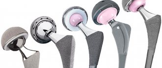
Aseptic necrosis of the head of the hip joint is a severe pathology that will not resolve on its own. If the situation is left without proper attention, the disease can lead to disability. In one case, secondary coxarthrosis may develop, requiring replacement of the joint with a prosthesis (hip arthroplasty). During the operation, the head of the hip joint is removed along with part of the femur and a prosthesis is placed in its place.
Joint replacement allows you to count on a favorable prognosis. The prosthesis allows you to completely restore lost functions.
Another outcome of the disease can be ankylosis - complete immobility in the joint. Complete degeneration of the cartilaginous cover of the articular surfaces occurs with the proliferation of connective fibrous or bone tissue.
Disability
ANGKB affects all areas of life: health, performance, socialization. With a long duration of the disease at different stages of conservative treatment, patients were forced to change activities with increased physical activity. They choose professions that do not require long periods of being on their feet, moving, or close to their place of residence.
The disability group of such people is determined during a medical and social examination in accordance with the Appendix to the Order of the Ministry of Health of the Russian Federation dated January 27, 1977 No. 33 to the Resolution of the Ministry of Labor and Social Development of the Russian Federation dated January 27, 1977 No. 1: “Classifications and temporary criteria used in the implementation of medical social expertise".
Disability is assigned based on the severity of the disease and decreased quality of life. Changes in the level of disability were carried out only in the case of surgical intervention.
In other words, surgical intervention is most effective in treating aseptic necrosis. Despite the impressiveness of conservative treatment methods, proven effectiveness is questionable.