Sometimes this is the result of inflammation in the scar area, sometimes it is a consequence of the formation of weak connective tissue that makes up the scar (a feature of the patient’s body). As a result, a defect (weak spot) appears at the site of the postoperative scar, through which a protrusion of internal organs is formed - a hernia is formed.
According to statistics, a hernia of this type occurs in every fifth of the operated patients. In half of them, a hernia occurs already in the first year after surgery. For the rest - over the next 5 years.
Causes of postoperative hernia formation
You can never name one single reason why a particular person developed a hernia. There are many factors that contribute to or influence the development of a ventral hernia. Below are the most common conditions that contribute to the formation of a hernia:
Heredity
Some hereditary diseases of connective tissue (for example, Marfan syndrome) are manifested by its weakness, including the weakness of formed scars at the site of damage (surgeries). People with such genetic abnormalities are predisposed to the formation of postoperative hernias.
Impaired regeneration (wound healing).
Inflammation of a postoperative wound can significantly affect the formation of the scar, thereby making it not strong enough, which also increases the risk of a hernia.
Intolerance of the patient's body to suture material.
In this case, an inflammatory reaction of the body to the threads occurs, which can lead to the appearance of defects in the postoperative scar.
Violation of the postoperative regimen by the patient.
Untimely and, most importantly, a sharp increase in physical activity, violation of diet and nutrition with the formation of constipation, during the formation of the scar, leads to a disruption of its structure and a decrease in strength.
Concomitant diseases
Any disease that leads to increased intra-abdominal pressure
- Bronchial asthma
- Bronchitis
- Intestinal disorders, chronic constipation
- Prostate adenoma, chronic urinary retention
- Obesity, etc.
With all these diseases, the edges of the wound experience constant tension, normal microcirculation and innervation are disrupted, which leads to a rapid disruption of the formation of a dense scar.
Technical errors during the operation
Errors in tissue joining techniques can also, over time, cause the formation of defective areas and recurrent hernias.
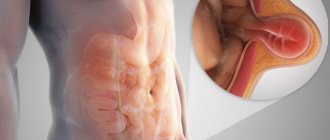
1.What is a ventral hernia
Ventral hernia is a pathology related to surgical diseases. A hernia is an abnormal protrusion of internal organs into the subcutaneous fat space. The organs protrude along with the peritoneal tissues if the hernia is located on the abdomen. Less common types of ventral hernia are inguinal-scrotal hernia and hernia of the white line of the abdomen.
The classification of ventral hernias is based not only on their location. Ventral hernias are divided into acquired (postoperative or post-traumatic) and congenital. There are uncomplicated and complicated hernias. The size of the hernia (from small to extensive and even gigantic) is also significant. There are also recurrent hernias and those that are eliminated for life after treatment.
A must read! Help with treatment and hospitalization!
Course of the disease
Postoperative hernias in the early stages are reducible and are not accompanied by pain. However, with sudden straining, falling, or lifting, pain appears and the protrusion increases. As the hernia progresses, the pain intensifies, sometimes becoming cramping in nature. At the same time, intestinal lethargy, constipation, flatulence, nausea, belching develop, the activity of patients sharply decreases, fecal stagnation, accompanied by intoxication, is periodically observed.
2.Why do ventral hernias appear?
There are two main factors that determine the predisposition to the occurrence of a ventral hernia:
- increased pressure inside the peritoneum;
- weakness of the abdominal wall.
This or that factor may have congenital causes or be acquired under the influence of certain negative conditions during life. These include:
- rapid weight loss or weight gain;
- surgical intervention;
- poor quality or improper suturing of the postoperative incision;
- an abscess localized in the abdominal cavity;
- prostate adenoma in men.
As for postoperative ventral hernias, responsibility for their occurrence may lie equally with the surgeon who sutured the surgical wound and with the patient himself. If the patient does not comply with the recommendations during the rehabilitation period, the likelihood of developing a ventral hernia increases significantly.
Also, the appearance of a hernia is more likely against the background of a long-term healing process or if surgery is performed repeatedly in the same area.
Visit our Traumatology and Orthopedics page
Causes of hernias
In the first year after gastroenterological surgery, ventral hernias form due to a number of complications - too traumatic surgery, errors in suturing, poor-quality suture material, stitching of heterogeneous tissues, wound infection, as well as everything that complicates the rehabilitation period - improper patient behavior, chronic cough or constipation, too early physical activity, spontaneously occurring concomitant diseases. Aggravating factors include the urgency of the previous operation, the patient's advanced age, excess weight, diabetes, and bad habits.
3. Symptoms and possible complications of ventral hernia
An uncomplicated ventral hernia may not bother you at all. It manifests itself only by some protrusion of the abdominal wall outward in a standing position.
A more severe course is accompanied by pain, general malaise, and periodic fever. If the ventral hernia is located along the white line of the abdomen, this leads to a divergence of the muscles of the abdominal wall. In this case, nausea and vomiting are quite common.
A hernia localized in the groin may not have pronounced manifestations, especially given the location, it is often not even visually detected by the patient himself. Only when large in size does an inguinal hernia become noticeable as an asymmetry in the scrotum. Only when you consult a doctor can he identify a small ventral hernia by palpation.
A ventral hernia is not as dangerous in itself as its possible complications are. Without treatment, unfortunately, the hernia becomes complicated quite often.
A complicated hernia cannot be repaired if it is strangulated. As a rule, the hernia is strangulated due to physical activity, heavy lifting, and sudden movements of the body. Also dangerous factors are straining during bowel movements or childbirth, and even a normal cough.
In case of complications, the infringement is accompanied by severe pain. The hernia itself becomes bluish or red in color, and the skin on it becomes shiny and glossy. This complication requires immediate treatment, as it threatens the development of necrosis of abdominal wall tissue and an abscess.
About our clinic Chistye Prudy metro station Medintercom page!
Prevention
To reduce the risk of developing a hernia after surgery, you should carefully follow the recommendations of your doctor:
- wearing a bandage of the appropriate size;
- if you are overweight, weight loss is desirable;
- limiting the load after abdominal surgery for a period established by the doctor;
- compliance with aseptic rules to prevent infection from entering the wound;
- a balanced diet that prevents flatulence and constipation.
Visiting a medical facility after discharge and contacting your doctor if inflammatory processes occur.
4. Treatment of ventral hernia
Ventral hernias cannot be treated conservatively. Of course, you can live with a hernia for years, but if it enlarges and, especially, if a complication develops, surgical help is inevitable.
It is possible to repair a strangulated hernia only within 6-8 hours after the development of this complication. For a longer period, surgical assistance is necessary.
The purpose of surgical treatment is to strengthen the abdominal wall. Whenever possible, this is done using the patient's own tissue. If the abdominal wall around the hernia does not have sufficient strength and prognostically does not exclude the possibility of relapses, mesh grafts are used.
Before installing a mesh prosthesis, excess fatty tissue is removed, and the hernia itself is excised. Such measures make it possible to strengthen the hernia gate and ensure better engraftment of the artificial material. After this treatment, the abdomen acquires a smooth surface.
If the hernia is localized in the groin area or on the white line of the abdomen, wearing a special bandage may be recommended as symptomatic treatment. However, this measure is not considered treatment; it only facilitates general well-being if the hernia causes nagging pain. The bandage also prevents strangulation and increase in size of the hernia.
Preparing for surgery
Before surgery, it is necessary to undergo an examination to determine the general condition of the patient and identify possible contraindications. The patient undergoes blood tests, ECG and other studies in advance. Familiarize yourself with the procedure for hospitalization and the list of necessary tests before hernioplasty.
Staying in hospital
The timing of postoperative rehabilitation is determined not so much by the type of operation as by the type and size of the hernia.
Rehabilitation period
To avoid the risk of relapse or minimize it, the patient will need to strictly follow all the doctor’s recommendations, limit physical activity and wear an abdominal band.
Modern methods of surgical treatment of hernias of the anterior abdominal wall
Surgery is an art, and, as such, it most of all requires creativity and least of all tolerates a pattern.
Vikenty Vikentievich Veresaev, doctor and writer
Modern approaches to surgical treatment of hernias of the anterior abdominal wall should implement the following principles as much as possible:
The reliability of the operation is to operate in such a way that the hernia does not occur again. It is based on the principle of stitching tissue without tension and using prosthetic techniques.
Less traumatic - the less tissue traumatization during surgery, the less surgical trauma - less pain, less need to stay in the hospital, less time to spend on recovery, faster you can get back to work and the normal rhythm of life. All this is achieved using modern minimally invasive, endoscopic techniques using modern low-traumatic methods of tissue fixation, reducing the time of the operation itself.
The safety of the operation is directly related to the use of modern approaches and techniques in the treatment of hernias, the use of biocompatible materials - modern composite implants, as well as the preparedness of surgeons and anesthesiologists performing this operation.
Functionality is the achievement of surgical restoration of the physiological function of the anterior abdominal wall, which can significantly improve the patient’s quality of life.
Aesthetics – restoration and improvement of the contours of the anterior abdominal wall, using modern approaches and principles of plastic and minimally invasive surgery.
All these requirements are met by the surgical techniques and modern materials I use in the treatment of hernias of varying locations and degrees of complexity.
Method of preperitoneal repair of inguinal and femoral hernia ONSTEP.
This method is a minimally invasive and tension-free version of surgical intervention, which is performed through a small incision (3-4 cm) remote from the main nerves, reducing the risk of long-term postoperative pain. Special “self-expanding” Onflex or PolySoft implants are used, installed preperitoneally and partially intramuscularly, which allows you to reliably close all potentially “weak” areas of the groin area and prevent the further development of other hernias in this area. In addition, fixation of the implant with sutures is not required, which reduces damage to the patient’s tissues and reduces the time of the operation itself. This technique is an alternative to laparoscopic hernioplasty.
The technique has proven itself well in the surgical treatment of recurrent inguinal hernias, including those arising after repair with mesh implants.
Photos of the stages of inguinal hernia repair using the ONSTEP method.
Advantages of the technique:
- Minimal, unnoticeable incision. (cosmetic effect).
- Possibility of quick recovery, shorter and less painful postoperative period.
- Possibility of use for surgical interventions for recurrent inguinal and femoral hernias.
Photos of patients after hernia repair using the ONSTEP method.
The use of self-fixing implants ADHESIX (Adgesix) in the treatment of inguinal hernias.
A special feature of the use of this type of implant is its ability to self-fix (glue) to tissues. In these implants, a biocompatible “self-adhesive” gel is applied to macroporous lightweight polypropylene, providing quick, easy and reliable fixation of the implant without the use of additional fixing sutures. This significantly reduces the amount of “surgical trauma” during surgery, which is very important for some groups of patients (for example, those taking anticoagulant therapy for pathologies of the cardiovascular system, patients who have had a heart attack and thrombophlebitis). Reducing tissue damage significantly reduces postoperative pain.
View of the Adhesix implant before installation
View of the posterior wall of the inguinal canal with the Adgesix implant installed)
Inguinal hernia repair using the Lichtenstein method using the self-fixing Adhesix implant from Bard.
About 25% of all operations for hernias of the anterior abdominal wall are operations for postoperative ventral hernias and primary ventral hernias with large hernial orifices. In all these cases, the edges of the hernia defect cannot be compared without tension.
Solutions to this problem exist today.
- Bridging
- Separation of the components of the anterior abdominal wall
- Temporary chemical denervation
Bridging (English: Bridge) is a device designed to combine segments.
Such a device, as a rule, is an implant that covers the defect and creates an additional supporting “frame” in the area of its installation. This refers to plastic surgery of the defect without covering it with aponeurotic tissue. These techniques are used mainly in cases where it is impossible or dangerous to reduce the edges of the hernial orifice due to the possibility of developing cardiopulmonary complications associated with a decrease in the volume of the abdominal cavity. The location of the implant can be different in relation to the layers of the anterior abdominal wall and depends on its quality characteristics, the method and complexity of its installation.
“inlay-bridging” - provides for the placement of the implant over the hernial orifice without suturing the edges of the aponeurosis.
“sublay-bridging” - the implant is located under the aponeurosis or under the muscles.
“intrapreritoneal (IPOM) - bridging” - the implant is located preperitoneally.
Intraperitoneal repair of ventral hernias using composite implants Ventralex, VentralexST, Ventrio, VentrioST.
Currently, taking into account the understanding of the mechanisms of integration of modern implantation systems in human tissue, special attention is paid to the spatial arrangement of the implant and the optimally simple, safe method of its installation.
The IPOM (intraperitoneal onlay mesh) hernia repair technique is an intraperitoneal position of the implant in the area of the hernial orifice directly on the peritoneum, that is, “inside the abdomen.”
With this installation method, the implant is pressed against the anterior abdominal wall due to intra-abdominal pressure. In this case, you can get by with its rare fixation in the area of unchanged tissues or even not use it. There is no contact of the implant with subcutaneous fat, which eliminates the negative impact on the course of the wound process and allows you to avoid complications associated with the development of inflammatory reactions.
Installation of the implant can be performed through an incision (usually minimal) - open IPOM technique, or through separate punctures on the abdominal wall using laparoscopic technologies - closed IPOM technique. One of the options for the closed IPOM technique is the IPOM plus , in which the defect is eliminated using special transfascial sutures, and then an implant is installed.
The Ventralex implant from Bard (USA), which is a self-expanding, non-absorbable sterile prosthesis, consisting of a top layer of polypropylene mesh and a layer of ePTFE (stretched polytetrafluoethylene) with microscopic pores, which helps to minimize tissue adhesion, has proven well in practical use. VentralexST implants have a resorbable anti-adhesion barrier based on Sepra® Technology hydrogel applied to the inner surface.
Straightening and fixation of the endoprosthesis is carried out using an elastic ring integrated into the implant with shape memory, thanks to which during the operation there is no need to straighten the device after insertion, as well as fix it (with a thread or herniostapler), which significantly reduces the duration of the operation. This also affects the patient’s feelings after surgery - discomfort and pain are minor and pass relatively quickly.
The “memory ring” keeps the implant in a tensioned position, which prevents it from shrinking over time, thereby eliminating the possibility of relapse. The implant does not migrate while in the body.
Photos of the stages of hernia repair using the Ventralex implant
The use of Ventralex composite implants is effective, safe and technically simple. It makes it possible to reliably perform hernioplasty for umbilical and small ventral hernias and to close postoperative defects with a diameter of up to 8 cm. Moreover, it is possible to install them “through a puncture” using endoscopic or minimally invasive technology.
Hernia repair using the open intraperitoneal (IPOM intraperitoneal onlay mesh) technique for an umbilical hernia using the Ventralex composite implant.
Ventrio and Ventrio ST implants are used to close large defects (more than 8 cm), which usually arise after abdominal surgery or after recurrence of previously operated hernias. They are also equipped with shape memory rings and special positioning “pockets” to facilitate their installation. All of them are created using modern composite biocompatible materials, which make it possible to solve serious reconstructive problems of the anatomical integrity of the anterior abdominal wall with the restoration of its function without the use of complex combined traumatic interventions that produce a large number of relapses.
Hernia repair using the open intraperitoneal (IPOM intraperitoneal onlay mesh) technique for a ventral hernia using the Ventrio composite implant.
I have accumulated extensive experience in using these operations and modern implants in the treatment of patients who are overweight, with diseases that reduce the reparative ability of tissues (diabetes mellitus, autoimmune diseases, oncopathology), the possibility of closing “complex” defects in lateral hernias, including hernias arising after placement of intestinal ostomies.
The practical application of low-traumatic technologies for installing these implants has proven to ensure reliable fixation of the endoprosthesis to the tissues of the anterior abdominal wall, avoids postoperative complications and relapses, and quickly restores daily activity and quality of life of patients.
Closed intraperitoneal repair (IPOM) of postoperative ventral hernias using implants with the ECHO PS positioning system.
The essence of this operation is that the intervention takes place without the use of traditional incisions. Everything is performed through small punctures on the anterior abdominal wall, and all adhesions caused by previous interventions are separated with minimal tissue damage. And most importantly: the hernia defect is closed from the inside with the Ventralight ST ECHO PS implant (Ventralite ST) from the American company Bard, coated with a special layer of hydrogel, which minimizes the formation of adhesions and adhesions. The implant is equipped with a special positioning system ECHO PS. This is a system of microtubules, which, when filled with air, unfolds the implant in the abdomen and allows it to be tightly aligned with the tissues from the inside. The implant is fixed using the SorbaFix hernia stapler, with special clips, which then completely dissolve. Upon completion of the installation and fastening of the implant, the “tubes” are deflated and then quickly and completely removed from the abdomen.
This technology helps maintain optimal implant deployment, eliminating some of the challenges of placement and positioning within the abdominal cavity, especially for larger defects. Simplifies the process of using an effective and high-tech method of treating hernias.
For the first time such an operation in the Samara region was performed by the author of this article on February 20, 2021.
Endoscopic surgery using the IPOM plus method using the Ventralight ST ECHO PS implant (Ventralite Sepra Technology ECHO PS) with a self-positioning system in the treatment of postoperative recurrent ventral hernia.
Separation plastic surgery of the anterior abdominal wall.
Based on the technique of separating the muscular-fascial components of the abdominal wall to increase the volume of the abdominal cavity. This allows suturing even large defects of the abdominal wall without tension, without provoking an intra-abdominal increase in pressure and the occurrence of abdominal compartment syndrome (small abdominal volume syndrome).
In this case, the division of the components of the abdominal wall into layers and the displacement of these layers relative to each other is carried out mostly in the so-called “sacred” layer (holyplane), located between the transverse and preperitoneal fascia. It is the junction of two embryonic layers and does not contain any vessels. A modern implant (Ventralight, BardSoftMesh...) is installed in the space between the layers.
There are several options for separation plastics:
The anterior option is plastic surgery according to the method of O.M. Ramirez.
Rear options: — plastic surgery using the AMCarbonell method;
— plastic surgery using the NWNovitsky method (TAR plastic surgery).
Indications for such an operation are: large hernias in which more than 20% of the internal organs are located outside the abdominal cavity, the so-called “loss of domain” situation, as well as hernia defects of more than 10 cm, when it is impossible to suture the posterior layers of the rectus abdominis sheath without tension.
The method makes it possible to eliminate hernias in several locations at once in one operation from one access without interaction with the abdominal organs.
Separation abdominal wall plasty is a modern and effective technology in the treatment of patients with large ventral and postoperative hernias.
The disadvantage of this technique is, to one degree or another, a violation of the biomechanics of the anterior abdominal wall.
Photos of the patient before and after reconstructive plastic surgery for a recurrent postoperative lateral ventral hernia using the separation technique and the use of the VentraLight ST implant.
Temporary chemical denervation
In the English-language literature, we can find this concept under the term “chemical separation of the components of the anterior abdominal wall.”
The technique is based on the use of botulinum toxin type A in preoperative preparation.
Botulinum toxin is injected into the muscles of the anterior abdominal wall under ultrasound control before surgery 1.5 months before surgery. Under its action, the muscles of the anterior abdominal wall relax, resulting in an increase in muscle length. This allows you to use implantation technology without bridging and not resort to separation plastics, reduce the size of the integrated implant and prevent the development of abdominal hypertension (small belly syndrome). Removing spasm and muscle contracture helps reduce the load on this implant and gives time for the formation of a reliable aponeurotic scar due to the prolonged action of botulinum toxin (4-6 months).
The accumulated significant experience in surgical interventions for hernias of the anterior abdominal wall using modern prosthetic materials and surgical technologies makes it possible to most effectively resolve issues regarding the surgical treatment of this pathology with the possibility of performing other combined and corrective operations.
Always ready to help you!
Profitable arithmetic: two operations in one
Cholecystectomy
- Cost: 100,000 - 160,000 rubles.
- Duration: 25-30 minutes
- Hospitalization: 3 days in hospital
More details
We consider performing two or more operations simultaneously for combined surgical diseases as another interesting area of work. There are not so few people suffering from several diseases at once that require surgery. Traditionally, they endure one operation after another, trying to gradually get rid of their ailments. The use of gentle laparoscopic techniques allows you to solve the problem in one step. Our department has accumulated extensive experience when laparoscopic hernioplasty was performed in conjunction with removal of the gallbladder for cholelithiasis, colon resection, gynecological operations, etc. Despite the expansion of the volume, patients tolerate these interventions well, are activated early and are discharged from the hospital in almost the same time frame as after one operation. At the same time, the patient avoids repeated intervention with all the ensuing difficulties and dangers, saving time, effort and money.
Belly patch or new techniques
In recent years, techniques have begun to be used to avoid tissue tension. During the operation, special meshes are inserted through the incision in the projection of the hernial ring to hold the exit of the hernial sac through the aponeurosis defect. The meaning of the action is clear: if a child wipes his pants, then the housewife does not sew up the hole, but sews in a patch. The comparison may be curious for such a serious topic, but it conveys the main idea embedded in the methodology. It should be noted that all “open” operations involve, first of all, an incision, the associated pain syndrome, the danger of inflammation of the suture and, finally, rough scars on the skin. Therefore, in hernia surgery, the search for new operations that would combine not only reliability, but also low trauma does not stop.
When choosing treatment, choose a surgeon!
It can be said that in the Department of Surgery of the Center for Endosurgery and Lithotripsy, almost all modern methods of treating hernias of the anterior abdominal wall of any location and anatomical structure are widely used. In addition, I would like to note that the team of surgeons has been working in the same composition for more than 10 years, which undoubtedly affects the coherence of the work and the quality of the operations performed.
Patients who have undergone hernioplasty in our department are carefully monitored by our specialists for several years, being, so to speak, under “guaranteed service.” This approach allows us to constantly improve and improve the quality of surgical treatment.
What will happen if left untreated?
The bulge will gradually increase, will no longer disappear when lying down, and the pain will intensify. The most dangerous complication is strangulation, which can lead to organ necrosis followed by peritonitis, which is a direct threat to life. In case of strangulation, emergency removal of the abdominal hernia is performed.
Important! In this condition, you should not try to correct the defect yourself. It is urgent to call an ambulance.
Other possible complications of hernias:
- inflammation;
- impossibility of reduction;
- coprostasis.
Diagnostics
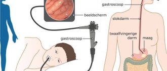
After examination by a surgeon, the patient is sent for a series of examinations: ultrasound of the hernia, gastroscopy, radiography, herniography, CT. Based on the examination results, a decision is made on treatment tactics.
Types of ventral hernias
Defects are classified by size:
- small ones, which almost do not change the appearance of the anterior wall of the peritoneum, are noticeable only during exercise;
- medium - formed in a certain part of the abdomen;
- extensive – affect the entire abdominal wall;
- giant - can be located in several areas of the abdominal wall.