Author:
Chekaldina Elena Vladimirovna otorhinolaryngologist, Ph.D.
Quick Transition Treatment of Acute Rheumatic Fever
In some cases, consultation with a cardiologist or neurologist is required.
Acute rheumatic fever (ARF) is a non-purulent complication that occurs 2-4 weeks after streptococcal tonsillopharyngitis.
Streptococcal tonsillopharyngitis (hereinafter referred to as GABHS-pharyngitis) is an acute infectious disease affecting the lymphoid apparatus and the pharyngeal mucosa.
Of particular danger are complications of GABHS pharyngitis, which are divided into:
- early (purulent), developing on the 4-6th day from the onset of the disease - otitis media, sinusitis, peritonsillar abscess, cervical lymphadenitis, pneumonia, etc.;
- late (non-purulent) - acute rheumatic fever, post-streptococcal glomerulonephritis, toxic shock.
Epidemiology of ARF
Acute rheumatic fever and rheumatic heart disease are called diseases of poverty and economic disadvantage. Complications caused by GABHS are the leading cause of death from cardiovascular disease in people under 50 years of age living in developing countries. ARF can occur at any age.
Worldwide, there are approximately 470,000 new cases of ARF and 275,000 deaths associated with rheumatic heart disease each year.
Most often, ARF occurs in children aged 5 to 15 years. ARF develops in 0.5-3% of cases if GABHS pharyngitis is not treated.
Acute rheumatic fever (rheumatism)
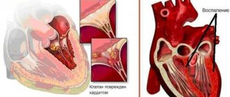
Rheumatic endocarditis clinic
extremely poor in specific symptoms. Endocarditis is always combined with myocarditis, the manifestations of which dominate and determine the severity of the patient’s condition. It is very difficult to recognize the manifestation of endocarditis at first, so the term “rheumatic carditis” is used (understanding by this damage to the myocardium and endocardium) until the final diagnosis of endocarditis. The following symptoms may indicate endocarditis: more pronounced sweating, a more pronounced and prolonged increase in body temperature, thromboembolic syndrome, a special velvety timbre of the first tone (L. F. Dmitrenko, 1921), increased systolic murmur in the apex of the heart and the appearance of diastolic murmur in the area of the apex of the heart or aorta, which indicates the formation of a heart defect. A reliable sign of previous endocarditis is a formed heart defect. “Heart disease is a monument to extinct endocarditis” (S. Zimnitsky).
Rheumatic pericarditis
is rare.
Recurrent rheumatic carditis
is characterized mainly by the same symptoms as primary myocarditis and endocarditis, but usually these symptoms manifest themselves against the background of a formed heart defect and the appearance of new murmurs that were not there before is possible, which indicates the formation of new defects. More often, rheumatic carditis has a protracted course, atrial fibrillation and circulatory failure are not uncommon.
There are 3 degrees of severity of rheumatic carditis. Severe rheumatic carditis (severe) is characterized by diffuse inflammation of one, two or three membranes of the heart (pancarditis), the symptoms of rheumatic carditis are pronounced, the boundaries of the heart are significantly expanded, and there is circulatory failure. Moderately severe rheumatic carditis (of moderate severity) is morphologically multifocal. The clinical picture is quite pronounced, the boundaries of the heart are expanded, there is no circulatory failure. Weakly expressed (mild) rheumatic carditis is predominantly focal, the clinical picture is not bright, the borders of the heart are normal, there is no decompensation.
Diagnostic criteria for carditis
- Pain or discomfort in the heart area.
- Dyspnea.
- Heartbeats.
- Tachycardia.
- Weakening of the first tone at the apex of the heart.
- Murmur at the apex of the heart:
systolic (weak, moderate or strong);
- diastolic.
- prolongation of the PQ interval;
- extrasystole, rhythm of the atrioventricular connection;
- other rhythm disturbances.
If the patient has 7 out of 11 criteria, the diagnosis of carditis is considered reliable.
To early diagnostic signs
primary rheumatic carditis include:
- Predominant development of the disease in childhood and adolescence.
- The close connection of its development with a previous nasopharyngeal infection.
- The presence of a gap (2-3 weeks) between the end of the last episode of nasopharyngeal infection and the onset of the disease, less often - a prolonged recovery after a nasopharyngeal infection.
- Frequent increase in body temperature at the onset of the disease.
- Arthritis or arthralgia.
- Auxcultative and functional signs of carditis.
- Shifts in acute phase inflammatory and immunological tests.
- Positive dynamics of clinical and paraclinical indicators under the influence of antirheumatic treatment.
The outcome of rheumatic carditis is determined by the incidence of heart defects.
Currently, the percentage of cases of the formation of heart defects after primary rheumatic carditis is 20-25%. It has been proven that the incidence of heart defects depends on the severity of rheumatic carditis.
Laboratory data
- Complete blood count: increased ESR, leukocytosis, shift of the leukocyte formula to the left.
- Biochemical blood test: increased levels of a2 and y-globulins, seromucoid, haptoglobin, fibrin, aspartic transaminase.
- Urinalysis: normal or slight proteinuria, microhematuria.
- Immunological blood tests: the number of T-lymphocytes is reduced, the function of T-suppressors is reduced, the level of immunoglobulins and titers of antistreptococcal antibodies is increased, CEC and SRP appear.
Instrumental studies
ECG:
slowing of AV conduction, decreased amplitude of the T wave and ST interval in the precordial leads, arrhythmias.
Echocardiography:
with valvulitis of the mitral valve, a thickening and “shaggy” echo signal from the valve leaflets and chords, limited mobility of the posterior valve leaflet, a decrease in the systolic excursion of the closed mitral leaflets, and sometimes a slight prolapse of the leaflets at the end of systole are detected. With Doppler echocardiography, rheumatic endocarditis of the mitral valve is manifested by the following signs: marginal club-shaped thickening of the anterior mitral leaflet; hypokinesia of the posterior mitral leaflet; mitral regurgitation; dome-shaped bend of the anterior mitral leaflet.
With valvulitis of the aortic valve, echocardiography reveals small-amplitude trembling of the mitral leaflets, thickening of the echo signal from the aortic valve leaflets.
With Doppler echocardiography, rheumatic endocarditis of the aortic valve is characterized by: limited marginal thickening of the aortic valve; transient prolapse of the valves; aortic regurgitation.
FKG:
With myocarditis, there is a decrease in the amplitude of the first tone, its deformation, pathological third and fourth sounds, a systolic murmur occupying 1/2-2/3 of systole, decreasing and adjacent to the first sound. In the presence of endocarditis, high-frequency systolic murmur is recorded, which intensifies during dynamic observation, protodiastolic or presystolic murmur at the apex during the formation of mitral stenosis, protodiastolic murmur at the aorta during the formation of aortic valve insufficiency, and diamond-shaped systolic murmur at the aorta during the formation of narrowing of the aortic mouth.
X-ray examination of the heart:
increase in heart size, decreased contractility.
Rheumatic arthritis
More typical for primary rheumatism, it is based on acute synovitis. The main symptoms of rheumatic arthritis: severe pain in large and medium-sized joints (symmetrically), often in the knees and ankles, swelling, hyperemia of the skin in the joint area, severe limitation of movements, volatile pain, rapid relief of non-steroidal anti-inflammatory drugs, absence of residual joint effects. Currently, transient oligoarthritis is more often observed, and monoarthritis is less common.
Joint damage is often combined with carditis, but can be isolated (usually in children).
Rheumatic lung disease
Gives a picture of pulmonary vasculitis and pneumonitis (crepitus, fine rales in the lungs, multiple foci of compaction against the background of an enhanced pulmonary pattern).
Rheumatic pleurisy
It has the usual symptoms. Its distinctive feature is the rapid positive effect of antirheumatic therapy.
Rheumatic kidney disease
Gives a picture of nephritis with isolated urinary syndrome.
Rheumatic peritonitis
It manifests itself as abdominal syndrome (more often in children), characterized by abdominal pain of varying localization and intensity, nausea, vomiting, and sometimes tension in the abdominal muscles. Antirheumatic treatment quickly relieves pain.
Neurorheumatism
It is characterized by cerebral rheumovasculitis, encephalopathy (memory loss, headache, emotional lability, transient disorders of the cranial nerves), hypothalamic syndrome (vegetative-vascular dystonia, prolonged low-grade body temperature, drowsiness, thirst, vagoinsular or sympathoadrenal crises), chorea.
Chorea
occurs in 12-17% of patients with rheumatism, mainly in girls from 6 to 15 years old.
The onset of chorea is usually gradual, the child becomes whiny, lethargic, irritable, then a characteristic clinical pentad of signs develops:
- Hyperkinesis - erratic, violent movements of various muscle groups (muscles of the face, neck, limbs, torso), which is accompanied by grimacing, pretentious movements, impaired handwriting, slurred speech; it is difficult for the child to eat and drink (he drops the mug, cannot bring the spoon to his mouth without spilling the soup). Hyperkinesis is often bilateral, intensifies with excitement, and disappears during sleep. The child cannot perform the finger-nose coordination test. Hyperkinesis in the hand area is easily detected if the doctor holds the child’s hand in his hand.
- Muscular dystonia with a pronounced predominance of hypotension up to muscle flabbiness (with weakening of hyperkinesis). Severe muscle hypotonia can even lead to the elimination of hyperkinesis and the development of a “paralytic” or “mild” form of chorea. A characteristic symptom is “flabby shoulders” - when lifting the patient by the armpits, the head sinks deeply into the shoulders.
- Impaired statics and coordination during movements (staggering when walking, instability in the Romberg position).
- Severe vascular dystonia.
- Psychopathological manifestations.
Currently, an atypical course of chorea is often encountered: mild symptoms with a predominance of vegetative-vascular dystonia and asthenia. Against the background of antirheumatic treatment, chorea stops after 1-2 months. In the presence of chorea, heart defects form very rarely.
Rheumatism of the skin and subcutaneous tissue
It manifests itself as annular erythema (pale pink, ring-shaped rashes in the torso, legs), subcutaneous rheumatic nodules (round, dense, painless nodules in the area of the extensor surface of the knee, elbow, metatarsophalangeal, metacarpophalangeal joints). Nodules occur rarely and are most often combined with carditis.
Currently, a point of view has emerged that a continuously relapsing course of rheumatism does not exist. A new relapse of rheumatism is possible only when the previous relapse is completely completed, and when there is a new encounter with streptococcal infection or its new exacerbation.
Features of the course of rheumatism depending on age
In childhood, acute and subacute onset of rheumatism is often observed, while chorea, annular erythema and rheumatic nodules are observed along with polyarthritis and carditis.
At high school age, mostly girls become ill; usually the disease develops gradually, and rheumatic carditis often takes a protracted course. In half of the patients, heart disease often develops and there is a tendency to relapse of the disease. In adolescents, the incidence of mitral valve insufficiency decreases and the incidence of combined mitral heart defects increases. 25-30% of adolescents experience cerebral pathology in the form of chorea and cerebral disorders.
Rheumatism in young people (18-21 years old) has the following features:
- the onset is predominantly acute, characterized by classic polyarthritis with high body temperature, but small joints of the hands and feet, sternoclavicular and sacroiliac joints are often affected;
- subjective and objective signs of rheumatic carditis are expressed;
- In most patients, rheumatism ends in recovery, but in 20% of patients, heart disease develops (usually mitral regurgitation), and in 27%, mitral valve prolapse.
Clinical features of the course
rheumatism in adults:
- the main clinical syndrome is rheumatic carditis, it is observed in 90% of patients with primary and 100% of patients with recurrent rheumatism;
- the formation of heart disease after one rheumatic attack is observed in 40-45% of patients;
- polyarthritis with primary rheumatism is observed in 70-75% of patients, and the sacroiliac joints are often involved;
- latent forms of the disease are becoming more frequent;
- In elderly and senile people, primary rheumatism practically does not occur, but relapses of rheumatism that began at a young age are possible.
Activity levels
Clinical manifestations depend on the activity of the rheumatic process. At maximum activity
general and local manifestations of the disease are bright with the presence of fever, the predominance of the exudative component of inflammation in the affected organs (acute polyarthritis, diffuse myocarditis, pancarditis, serositis, pneumonitis, etc.).
Moderate activity
is manifested by a rheumatic attack with or without moderate fever; there is no pronounced exudative component of inflammation.
Moderate or mild signs of rheumatic carditis, polyarthralgia or chorea are observed. With minimal activity
of the rheumatic process, clinical symptoms are mild, sometimes almost undetectable. Often there are completely no signs of the exudative component of inflammation in organs and tissues.
Diagnosis of ARF
To diagnose ARF, the major and minor Jones Criteria developed by the American Heart Association are used.
Five big criteria:
1. Carditis and valvulitis - develops in 50-70% of cases.
Damage to the layers and valves of the heart. Occurs within 3 weeks after GABHS pharyngitis. As a rule, it begins with endocarditis followed by the development of pancarditis; the mitral and aortic valves are most often affected. Disease progression can last for years after ARF and lead to heart failure.
2. Arthritis (migratory polyarthritis) - develops in 35-66% of cases.
Joint inflammation is the earliest manifestation of ARF; it occurs within 21 days after GABHS pharyngitis, lasts 4 weeks and goes away without a trace. The knee, ankle, elbow, and wrist joints are most often affected. Arthritis is migratory in nature - several joints are successively affected.
3. Damage to the central nervous system (rheumatic chorea, Sydenham chorea, “St. Vitus’s dance”) - develops in 10-30% of cases.
Sharp, irregular, involuntary movements of the limbs, muscle weakness, emotional disorders. Occurs 1-8 months after acute GABHS pharyngitis. Facial muscles are more often affected, and speech disturbances may occur. Emotional changes include outbursts of inappropriate behavior, including crying and restlessness. In 17-35% of patients it may develop into obsessive-compulsive disorder.
4. Rheumatic erythema - develops in less than 6% of cases.
A pink or pale red, non-itchy, ring-shaped rash. Localized on the body or limbs, but not on the face. The rash may appear, disappear, or appear again.
5. Subcutaneous nodules - develop in less than 10% of cases.
Dense, painless formations from a few millimeters to 2 cm. They persist for no more than a month. Localization of nodules is often above the bone, on the extensor surfaces, symmetrically. The skin over the nodule is not inflamed and mobile.
Minor criteria:
- Arthralgia is pain in the joints.
- Fever (above 38.5 °C).
- Increased erythrocyte sedimentation rate (ESR) - above 60 mm/h, C-reactive protein (CRP) - above 30 mg/l.
- Prolongation of the PR interval on the ECG.
The diagnosis of ARF is established on the basis of:
- the fact of having suffered GABHS-pharyngitis - confirmed by a positive rapid test, bacteriological examination at the time of acute infection or an increase in the titer of antistreptolysin-O (ASLO) already during the occurrence of complications;
- Jones criteria: 2 major criteria, 1 major and 2 minor criteria, or 3 minor criteria if the patient has previously suffered ARF.
All patients with suspected ARF must undergo an ECG and EchoCG (ultrasound of the heart) to identify morphological changes in the heart valves and signs of pathological regurgitation (backflow of blood). Laboratory tests according to indications (since they are nonspecific) - ESR and CRP. If signs of chorea are present, a neurological examination is necessary.
Publications in the media
Acute rheumatic fever (ARF) is a systemic inflammatory disease of connective tissue involving the heart and joints in the pathological process, initiated by group A β-hemolytic streptococcus, occurring in genetically predisposed people. The term rheumatism, widely used in practice, is currently used to designate a pathological condition that combines acute rheumatic fever and rheumatic heart disease.
Statistical data . Incidence: 2.1 per 100,000 population in 2001. The incidence of rheumatic heart disease in Russia is 0.17%. The predominant age is 8–15 years. Etiology. -Hemolytic streptococcus of group A, “rheumatogenic” serotypes M3, M5, M18, M24. The M protein on the streptococcal membrane has antigenic determinants similar to components of the heart muscle, brain and synovial membranes.
Genetic aspects . Ag D8/17 B-lymphocytes are detected in 75% of patients with ARF. Pathogenesis • Antistreptococcal antibodies are responsible for selective damage to the heart valves and myocardium with the development of immune aseptic inflammation, cross-reacting with heart tissue (molecular mimicry) • M-protein has the properties of a “superantigen” that causes activation of T- and B-lymphocytes without preliminary processing of Ag- presenting cells and interactions with class II molecules of the major histocompatibility complex.
Clinical picture • Onset of the disease. In more than half of the cases, 2–4 weeks after a streptococcal nasopharyngeal infection, fever, asymmetric joint pain, pain in the heart, shortness of breath, and palpitations occur. In other patients, the onset occurs as a monosyndrome (carditis, arthritis or chorea) • A repeated attack occurs with symptoms of carditis.
• Arthralgia and rheumatic polyarthritis (80% of patients) - typical reactive synovitis with fluid effusion into the joint cavity, swelling and redness of the periarticular tissues, sometimes with severe pain, tenderness and limitation of active and passive movements. Characteristic features:
•• damage to large joints (knee [most often], ankle, elbow, shoulder and much less often - wrist) •• symmetry of the lesion •• migrating, volatile nature of arthritis •• complete reversibility of the articular syndrome, no changes on radiographs, restoration of joint function • • in children, signs of arthritis, as a rule, completely disappear, and in adults a persistent course can be observed, leading to the development of Jaccoud's syndrome (painless deformation of the hands with ulnar deviation without inflammation in the joints and without dysfunction of the joint) •• with rheumatism, more often with repeated attacks, polyarthralgia rather than arthritis often occurs.
• Fever (90%). • Subcutaneous nodules (10% of patients), grain to pea-sized, localized in the periarticular tissues may appear during acute rheumatic fever. These nodules do not bother patients, they are painless, and the skin over them is not changed. Involution of the nodules occurs over a period of several days to several weeks. Observed only in children.
• Ring-shaped erythema - pale pinkish-red spots up to 5-7 cm in diameter with clear, not always smooth edges. Characterized by localization on the skin of the chest, abdomen, back and limbs, spontaneous disappearance and (rarely) recurrence. Occurs in less than 5% of patients. Erythema nodosum is not typical for rheumatism. • Chorea occurs in 10–15% of patients, more often in girls, 1–2 months after a streptococcal infection. It is chaotic involuntary twitching of the limbs and facial muscles. Characteristic is the complete disappearance of symptoms during sleep. • Rheumatic carditis (rheumatic carditis) can be primary (first attack) and recurrent (repeated attacks), with or without the formation of valve disease. Clinical symptoms of rheumatic carditis: •• Cardialgia . Characterized by prolonged stabbing, aching pain in the heart area, usually without irradiation. With pericarditis, pain is associated with breathing and intensifies in a horizontal position. •• In most cases an enlarged heart , which is not always detected physically. A chest x-ray is recommended. •• Arrhythmias as a manifestation of myocarditis ••• Sinus tachycardia (100 or more per minute), recorded at rest ••• Atrial fibrillation occurs, as a rule, when signs of mitral valve stenosis appear ••• Ventricular (rare) or supraventricular extrasystoles •• • Atrioventricular block with prolongation of the P-Q interval over 0.2 s or the appearance of Wenckebach (Samoilov-Wenckebach) periods. •• Decrease in sonority of the first tone at the apex of the heart, appearance of the third tone and murmurs. A gentle blowing systolic murmur with a tendency to increase in intensity and conducted into the axillary region is a symptom of mitral valvulitis. Protodiastolic murmur along the left sternal border is a sign of aortic valvulitis. •• Signs of congestive heart failure are rarely observed with primary rheumatic carditis; much more often they accompany recurrent rheumatic carditis. • Acute rheumatic fever lasts 6–12 weeks. There is no chronic or continuously relapsing course.
Laboratory data • Increased ESR, increased titers of antistreptolysin O, AT to DNase in a titer of more than 1:250 • Bacteriological examination of a throat smear reveals group A -hemolytic streptococcus. Instrumental data • ECG • Chest X-ray • EchoCG. Differential diagnosis • Reactive arthritis • Lyme disease • Infective endocarditis • Viral (Coxsackie B) myocarditis • Mitral valve prolapse • Functional heart murmur. Kissel-Jones-Nesterov diagnostic criteria (revised 1992) • Major criteria: •• carditis •• polyarthritis •• chorea •• annular erythema •• subcutaneous nodules • Minor criteria: •• clinical symptoms (fever, joint pain) • • laboratory changes (increased ESR, appearance of CRP, prolongation of the P–Q interval •• Additional signs: positive cultures from the tonsils for group A β-hemolytic streptococcus, increased titers of antistreptolysin-O and/or other antistreptococcal antibodies. The presence of two large or one large and two minor criteria in combination with the mandatory presence of additional signs allows us to consider the diagnosis of acute rheumatic fever reliable.Despite the presence of time-tested criteria, the diagnosis of ARF continues to be a problem, since individual criteria (fever, ESR, etc.) are not specific, and subcutaneous nodules and Annular erythema is rarely observed.
TREATMENT Management tactics . For acute rheumatic fever, hospitalization, bed rest, and a diet low in salt and rich in vitamins and protein are indicated. The basis of drug treatment is antibacterial and anti-inflammatory therapy. Drug treatment • Antibacterial therapy •• Benzylpenicillin 1.5–4 million units/day for adolescents and 400–600,000 units/day for children for 10–14 days, followed by a transition to benzathine benzylpenicillin or benzathine benzylpenicillin + benzylpenicillin procaine •• When if you are allergic to penicillins, you should choose a macrolide: roxithromycin for children 5–8 mg/kg/day, adults 150 mg 2 times/day, clarithromycin for children 7.5 mg/kg/day, adults 250 mg 2 times/day. It should be remembered that streptococcal resistance to erythromycin is currently increasing. • NSAIDs for 3.5–4 months (during treatment it is necessary to periodically conduct blood tests, urine tests, and liver function tests) •• Diclofenac 50 mg 3 times a day. • The use of GC is most justified for pancarditis. One of the treatment regimens is prednisolone 20–30 mg/day until clinical effect, then gradually reducing the dose over 20–30 days.
Prevention • Primary prevention : rational treatment of streptococcal diseases of the oropharynx for 10 days •• Aminopenicillins ••• amoxicillin 750 mg/day for children, 1500 mg/day for adults •• Cephalosporins ••• cephalexin for children with body weight <40 kg - 25– 50 mg/kg/day, adults 250–500 mg 2–4 times/day (daily dose 1–2 g) ••• cefaclor children 20 mg/kg 3 times/day, adults 750 mg 3 times/day ••• cefuroxime for children 125–250 mg 2 times a day, adults 0.25–0.5 g 2 times a day •• Macrolides ••• roxithromycin for children 5–8 mg/kg/day, adults 150 mg 2 times a day days •• Penicillins with β-lactamase inhibitors ••• amoxicillin + clavulanic acid (adults 375 mg 3 times / day) for 10 days. • Secondary prevention is indicated for patients who have had acute rheumatic fever to prevent relapses. The best results are achieved by year-round bicillin prophylaxis (benzathine benzylpenicillin 600,000–1,200,000 units (children), 2,400,000 units (adults) once every 3 weeks. Benzathine benzylpenicillin + benzylpenicillin procaine 1,500,000 units once every 10–12 days. D duration of secondary prophylaxis •• at least 5 years after the last reliable rheumatic attack for patients who have had acute rheumatic fever without carditis •• more than 5 years - for patients who have had rheumatic carditis •• with relapses for life • Prevention of infective endocarditis against the background of formed rheumatic heart disease with carrying out any surgical interventions (tooth extraction, abortion, abdominal surgery) - see Infectious endocarditis.
Synonym . Sokolsky-Buyo disease. Abbreviations. ARF - acute rheumatic fever.
ICD-10 • I00 Rheumatic fever without mention of cardiac involvement • I01 Rheumatic fever with cardiac involvement
Treatment of ARF
Eradication of GABHS infection is necessary regardless of whether there are signs of pharyngitis. Antibacterial therapy is carried out similarly to therapy for acute tonsillopharyngitis.
Symptomatic therapy:
- arthritis - non-steroidal anti-inflammatory drugs to relieve pain and prevent the involvement of new joints;
- carditis - treatment is carried out only if heart failure develops;
- chorea - usually does not require treatment, but sometimes it may be necessary to prescribe antipsychotics and anticonvulsants;
- erythema and subcutaneous nodules - no treatment.
Prevention of ARF
Primary is timely diagnosis and treatment of GABHS pharyngitis.
Secondary - prevention of new episodes of GABHS infection, including eradication of GABHS even in asymptomatic carriers.
The duration of antibiotic prophylaxis is determined based on the characteristics of the existing pathological process. If we are talking about post-streptococcal arthritis, it can be limited to 1-2 years. In case of ARF without carditis, the duration of taking antibiotics is 5 years or until the patient is 21 years old (whichever is longer), ARF with carditis without consequences is 10 years or up to 21 years old (whichever is longer), ARF with damage to the heart valves is 10 years or up to 40 years old ( whichever is longer), and sometimes for life.
Rheumatism in children
Clinical manifestations of rheumatism in children are diverse and variable. The main clinical syndromes include rheumatic carditis, polyarthritis, minor chorea, anular erythema and rheumatic nodules. All forms of rheumatism in children are characterized by clinical manifestation 1.5-4 weeks after the previous streptococcal infection.
Heart damage during rheumatism in children (rheumatic carditis) always occurs; in 70-85% of cases – primary. With rheumatism in children, endocarditis, myocarditis, pericarditis or pancarditis may occur. Rheumatic carditis is accompanied by lethargy, fatigue of the child, low-grade fever, tachycardia (less often bradycardia), shortness of breath, and heart pain.
A repeated attack of rheumatic carditis, as a rule, occurs after 10-12 months and is more severe with symptoms of intoxication, arthritis, uveitis, etc. As a result of repeated attacks of rheumatism, acquired heart defects are detected in all children: mitral insufficiency, mitral stenosis, aortic insufficiency, aortic stenosis, mitral valve prolapse, mitral-aortic disease.
In 40-60% of children with rheumatism, polyarthritis develops, both isolated and in combination with rheumatic carditis. Characteristic signs of polyarthritis in rheumatism in children are predominantly damage to medium and large joints (knees, ankles, elbows, shoulders, less often - wrists); symmetry of arthralgia, migrating nature of pain, rapid and complete reverse development of articular syndrome.
The cerebral form of rheumatism in children (chorea minor) accounts for 7-10% of cases. This syndrome mainly develops in girls and is manifested by emotional disorders (tearfulness, irritability, mood swings) and gradually increasing motor disorders. First, handwriting and gait change, then hyperkinesis appears, accompanied by impaired speech intelligibility, and sometimes the inability to independently eat and care for oneself. Signs of chorea completely regress after 2-3 months, but tend to recur.
Manifestations of rheumatism in the form of anular (ring-shaped) erythema and rheumatic nodules are typical for childhood. Ring-shaped erythema is a type of rash in the form of rings of pale pink color, localized on the skin of the abdomen and chest. There is no itching, pigmentation or flaking of the skin. Rheumatic nodules can be found in the active phase of rheumatism in children in the occipital region and in the joint area, at the sites of tendon attachment. They look like subcutaneous formations with a diameter of 1-2 mm.
Visceral lesions in rheumatism in children (rheumatic pneumonia, nephritis, peritonitis, etc.) are practically never encountered at present.
How is ARF treated at the Rassvet Clinic?
We provide timely diagnosis and adequate treatment of GABHS pharyngitis, which reduces the incidence of ARF by almost 70%. When choosing antibacterial therapy, we always give preference to penicillin antibiotics - as the most effective drugs that have been proven to reduce the incidence of ARF. We never reduce the course of antibiotic therapy when there is clinical improvement. If GABHS pharyngitis is detected, we do not prescribe local treatment (rinses, sprays) to the detriment of systemic antibacterial therapy.
We provide adequate eradication therapy for GABHS in ARF in order to prevent relapses and progression of rheumatic heart disease. We offer a full examination for diagnosed ARF - ECG, ECHO-CG, consultation with a cardiologist and neurologist with the selection of the necessary therapy.
Rheumatism
Terminology
Rheumatism is one of those diseases that everyone knows about or, at least, has heard of.
However, prevailing ideas about rheumatism vary very widely: from the childhood fear of sneakers (“You can’t always walk in them - you’ll catch a cold and get rheumatism!”) to a more adult discovery: it turns out that rheumatism affects not the legs, but the heart. A careful study of available sources sometimes only confuses the situation. In fact, how can we understand a disease in which it is not a single organ or even a functional system that suffers, but an entire type of tissue in the body? However, this is exactly what happens with rheumatism. Rheumatism is an inflammatory-immune lesion of connective tissue, which is present almost everywhere in the body.
The definition of “inflammatory-immune” should be used in this order: first inflammation, then an immune reaction to it, and then an autoimmune attack on one’s own connective tissue. This is akin to what a brave, fast, but not very accurate bodyguard can do, continuing to shoot in bursts after the invasion has long been repulsed...
As a rule, the connective tissue structures of the heart, joints, blood vessels, and subcutaneous layers are affected. Much less common (1-6 percent of the total volume of registered rheumatism) are rheumatic lesions of the central nervous system, respiratory and visual organs, and gastrointestinal tract.
Rheumatism does not show any endemicity (regional dependence of epidemiological indicators): people get sick everywhere. However, there is a clear dependence of morbidity on age - among primary patients, the category of 5-15 years predominates - as well as an inverse correlation with the standard of living of the population. Considering that the majority of children on the globe (up to 80%) live in the so-called. developing countries, one should not be surprised at the widespread prevalence of rheumatism in the “third world”.
In the last quarter of a century, morbidity and mortality in Russia have decreased by more than three times. However, if we are talking about such a serious disease, and even with a predominantly early manifestation, no epidemiological data can be considered “satisfactory”, except zero. Today, statistics are contradictory and are mainly of an evaluative nature (and this applies not only to the Russian Federation): as a rule, data from the 1960s-90s are extrapolated to the current situation. A more or less reliable estimate of the incidence among Russian children can be considered a frequency of 2-3 cases per 10,000 (and over 1.5% of all heart defects are rheumatic); for comparison, in third world countries this figure varies from 60 to 220 per 10,000.
In conclusion of a brief general overview, it should be noted that the term “rheumatism” itself is currently used mainly by Russian-speaking medicine. In the official international lexicon these are “Acute rheumatic fever”, “Rheumatic heart disease”, “Rheumatoid arthritis”, etc.; however, code M79.0 (heading “Diseases of the musculoskeletal system and connective tissue”) in ICD-10 indicates “Rheumatism, unspecified.”