General information
Elephantiasis is usually called swelling and chronic stagnation of lymph, manifested in the form of deformation and persistent diffuse increase in the size of any part of the human body, most often the limbs and scrotum. Elephantiasis is usually accompanied by painful growth - hyperplasia of the skin and subcutaneous fat.
These changes are fibrotic and irreversible, affecting the skin, subcutaneous tissue and fascia. The pathology most often affects the lower extremities, which become similar to the legs of an elephant, hence the origin of the name of the disease. The causes of elephantiasis can be different, from parasitic agents to congenital defects of the lymphatic system. These include blockage of the lymphatic capillary system and impaired lymph circulation.
Related disorders
Symptoms of the following disorders may be similar to those of elephantiasis. Comparisons may be useful for differential diagnosis.
- Hereditary lymphedema is an inherited disease of the lymphatic system characterized by abnormal swelling of certain parts of the body. The lymphatic system is a circulatory network of vessels, ducts and nodes that filter and distribute certain fluid (lymph) and blood cells throughout the body. Lymphatic fluid accumulates in the soft tissues within and under the skin due to obstruction, malformation, or underdevelopment (hypoplasia) of various lymphatic vessels. There are three forms of hereditary lymphedema: congenital hereditary lymphedema or Nonne-Milroy disease, subcutaneous lymphedema or Meige's disease, and lymphedema of Tarde. Symptoms include swelling of the affected areas and thickening and hardening of the skin in the affected areas. In most cases, hereditary lymphedema is inherited as an autosomal dominant trait.
Pathogenesis
Elephantiasis begins with slight swelling and swelling, which may subside or reappear. Eventually, after about 1-3 years, there is a two to threefold thickening of the lower leg and possibly the thigh if the leg is affected. The shape of the leg is awkward and cylindrical, turning into thickenings, for example, the deepening of the ankle joint can be smoothed out.
Stages of development of elephantiasis
The mechanism of excessive thickening of the affected parts is based on stagnant processes affecting the lymphatic system. They can arise as a result of factors such as:
- blockage of lymphatic vessels by parasites - nematodes , for example, filaria , chyluria , etc.;
- suppuration and subsequent scarring of the lymph glands, for example, the inguinal glands;
- inflammation of the lymphatic vessels with lymphangitis , phlebitis , erysipelas , varicose ulcers, chronic eczema , syphilis , lupus , with repeated frostbite and other chronic or recurrent processes leading to inflammatory edema.
The resulting stagnation of lymph leads to its penetration into tissue crevices and provides excessive nutrition to the structures of adjacent tissues, which causes hyperplasia as a result of the proliferation of connective tissue in the subcutaneous tissue. This process is chronic, lasts for many years, and therefore is practically undetectable in adolescents.
Attention! Wuchereriosis and other filariasis are caused by parasitism in the human body of very thin worms (no more than 0.3 mm in diameter, up to half a meter long) that can block the lymphatic system. They are spread and dispersed by blood-sucking insects, so it is extremely important to protect yourself with repellents and mosquito nets when on vacation in tropical countries, because they can not only cause elephantiasis, but also penetrate into the eyes, causing blindness .
What kind of disease is this
Elephantiasis (other names: elephantiasis, elephantiasis or lymphedema) is a pathology in which there is a violation of the outflow of lymph through the lymphatic vessels, which leads to its stagnation, accumulation and subsequent increasing swelling, as a result of which the affected limb increases in volume and in the extreme stage becomes disfigured . Due to stagnation of lymph, metabolic products and toxins accumulate in soft tissues, which causes protein breakdown and the formation of fibrin fibers. Subsequently, rough connective tissue forms at the site of edema, which disrupts muscle function and leads to disability of the patient. Externally, the affected limb looks like a cylinder and resembles an elephant's leg. Cracks, ulcers and warts appear on the skin.
Most often, elephantiasis of the legs develops, and, as a rule, one limb is affected.
Classification
According to the development mechanism there are:
- primary lymphedema (congenital), which is caused by congenital pathology of the lymphatic system, the development of the disease occurs in childhood and adolescence;
- secondary lymphedema (acquired), occurs more often than the primary form of the disease and is caused by previous diseases and injuries/surgeries.
There are three stages in the course of the disease:
- Stage 1 mild edema;
- Stage 2 of dense edema;
- Stage 3 elephantiasis.
Causes of elephantiasis of the feet
There are two main causes of elephantiasis:
- congenital or acquired disorders in the lymphatic system, including overproduction of lymph, defects, aplasia, insufficiency of the valve system, stenosis and occlusion of lymphatic vessels, as well as diseases such as Nonne-Milroy syndrome, Shereshevsky-Turner syndrome ;
- infectious, more often parasitic agents, which include Bancroft's filamentum - causes wuchereriosis , but there may also be a streptococcal infection;
- mechanical injuries and thermal burns (usually repeated frostbite);
- tumors that can compress the lymphatic capillaries;
- Iatrogenesis - can be caused by radiation or surgery;
- disorders of the circulatory system - chronic venous insufficiency and postthrombophlebitic syndrome ;
- damage to the central nervous system;
- endocrine disorders.
Life cycles of various representatives of filariae
Elephantiasis, caused by various parasitic roundworms (filariasis), is more common among residents of tropical zones (Indian archipelago, Arabia, the west coast of Africa, Central America), where mosquitoes spread and transmit the infection. In the former CIS countries it is very rare.
Elephantiasis of the lower extremities: causes
There are two types of elephantiasis of the feet:
- The primary form of the disease develops under the influence of lesions and dysfunctions of the lymphatic system. The prerequisites for the occurrence of pathology can be either congenital or acquired. In the first case, we are talking about genetically determined underdevelopment of lymphatic vessels, ducts and nodes, excessive lymph synthesis, and the presence of genomic pathologies such as Shereshevsky-Turner syndrome and Milroy-Mage disease. With a congenital tendency to elephantiasis, the first signs of the disease usually appear quite early: in early childhood or adolescence (in rare cases, the congenital form of the disease may appear at a later age).
- The secondary (acquired) form of the disease is more common than the primary one. It develops under the influence of parasitic infestations, injuries, streptococcal infections, and vascular pathologies.
Since the acquired form of the disease is more common, let us consider the causes of secondary elephantiasis of the lower extremities in more detail:
- Penetration of parasites into the body. The disease is provoked by infection with filaria - extremely thin (up to 0.3 mm) threadworms. Infestation occurs as a result of the bite of mosquitoes, which are carriers of these parasites. That is why filaria infection mainly occurs in countries with hot and humid climates - Africa, Central America and Southeast Asia, since these regions are habitats for mosquitoes. This localization explains the fact that the overwhelming number of cases of elephantiasis are registered in these regions. After entering the host’s body, the parasites concentrate in the lymph nodes, where they form tangles that interfere with normal lymphatic drainage. As a result, lymph accumulates, stretches the walls of lymphatic vessels and ducts, and fluid leaks into the surrounding soft tissue. As a result of the course of the disease, an inflammatory process may develop, followed by the growth of adipose and connective tissue.
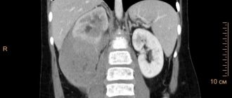
- Tumor processes. The process of development of malignant tumors is often accompanied by metastasis of altered cells in the lymph nodes. This occurs due to the ability of cancer cells to travel throughout the body with lymph flow. As a result, lymphatic drainage in one or more nodes is disrupted, lymph accumulates in nearby vessels and ducts, the pressure in them increases, some of the fluid seeps into the surrounding soft tissues and elephantiasis develops.
- Lymphadenectomy (surgical removal of a lymph node). If a lymph node has been removed (most often as a result of oncology treatment), there is a risk of developing lymphedema with the subsequent transition of edema to elephantiasis.
Expert opinion
The development of lymphedema or elephantiasis of the lower extremities due to lymphadenectomy during surgical treatment of pelvic cancer is quite common. The degree of swelling in this case can vary greatly and can be either insignificant or pronounced.
Vascular surgeon, phlebologist
Osipova Ekaterina Yakovlevna
- Streptococcal infection. Erysipelas of the skin, tonsillitis, pharyngitis, scarlet fever and other diseases caused by streptococci cause additional stress on the lymphatic system. As a result, lymphadenitis develops - enlargement and hardening of the lymph nodes, which may be accompanied by soreness, redness of the skin, and an increase in local and general body temperature. The load on the lymphatic system caused by infection causes damage to blood vessels and ducts, as a result of which some of the lymph accumulates in the surrounding soft tissues and edema forms.
- Various injuries. Fractures of the limbs, frostbite, extensive burns and other injuries can cause damage to the integrity of the lymphatic vessels, ducts and nodes. This leads to disruptions in lymphatic drainage processes and accumulation of protein-rich fluid in soft tissues.
- Vascular pathologies. The lymphatic and cardiovascular systems are closely interconnected. Therefore, if one of them does not function well enough, the second may also fail. Varicose veins, thrombosis, phlebitis, thrombophlebitis, obliterating endarteritis and other vascular pathologies of the lower extremities can provoke an increase in the load on nearby lymphatic vessels, resulting in lymphatic edema, which can lead to the development of elephantiasis.
Elephantiasis of the foot: symptoms
Symptoms of elephantiasis
In addition to hypertrophy and hyperplasia - swelling, changes in shape and increase in size of the pathologically altered part of the body, patients may experience symptoms such as:
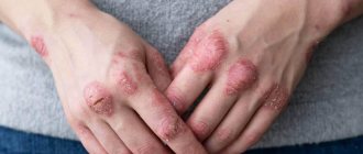
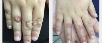
- the occurrence of papillomatous growths, ulcers and warts (as in the photo below, elephantiasis of the legs);
- the skin may become shiny, pale or bluish, and does not fold;
- lymphatic and venous stasis;
- edema;
- pain;
- stiffness of movements;
- the development of hypertrophic processes in the layers of skin and subcutaneous tissue, caused by an increase in intermuscular space as a result of the proliferation of connective and bone tissue, which ultimately causes thickening of the bone.
Patient with elephantiasis
If the disease is caused by erysipelas , then in the affected area of a sharply elevated painful ridge, a burning sensation, redness (erythema), increased temperature, pinpoint hemorrhages, erosions, trophic ulcers, blisters may occur, which may subside, forming brown crusts.
Erysipelas on the leg
Stages of the disease
At an early stage, lymphedema is quite difficult to recognize: it appears in the evening and disappears after a night's rest. The limbs feel soft to the touch, and patients may confuse them with swelling that occurs after exercise. As the disease progresses, lymphedema increases, becomes permanent, and becomes dense. The skin in the area of the second toe cannot be folded (positive Stemmer's sign). Swelling begins on the foot and spreads higher up the leg.
When the disease reaches the third stage, doctors talk about elephantism, or elephantiasis. The disease leads to the fact that one leg is seriously different in size from the other (if lymphedema is observed only on one limb), it is difficult, and sometimes impossible, for the patient to choose clothes or shoes, and it is difficult to move. The swelling becomes dense, since the lymph contains a lot of protein, and the penetration of protein molecules into the tissue leads to the formation of fibrous fibers there. Elephantiasis is accompanied by the appearance of papillomas on the skin, vesicles from which lymph may leak.
Tests and diagnostics
To confirm the diagnosis of filariasis, clinical data and epidemiological history information, results of laboratory and instrumental research methods are studied. The most important factor is the detection of microfilariae in blood tests using microscopy at low magnification. In addition, immunological techniques can be used, but they do not give strictly specific results.
If the nature of the pathology is not parasitic, then a more complete picture can be given by ultrasound of the lower extremities, rheovasography, lymphography, duplex scanning of blood vessels, radiography and MRI. In addition, general blood tests, urine tests, and a blister test may be performed during the examination.
Diagnosis of elephantiasis of the lower extremities
If the symptoms described above appear, do not delay visiting your doctor. The treatment of diseases of the lymphatic system is carried out by a specialized specialist - a lymphologist. If you do not have the opportunity to visit this doctor, make an appointment with a phlebologist or angiosurgeon: since the lymphatic and circulatory systems function interconnectedly, a vascular specialist can tell you how to treat elephantiasis in the legs.
To make an accurate diagnosis, not only a medical examination is important, but also additional research. This is especially necessary in the initial stages of elephantiasis, when its symptoms may be mistaken for manifestations of vascular diseases (in particular, thrombosis). Before starting treatment for elephantiasis, the following diagnostic procedures are recommended:
- Dopplerography of blood vessels.
- Lymphangiography.
- CT.
- MRI.
- Laboratory tests: general and biochemical blood test, serological test, blood test after taking Diethylcarbamazine. The latest study reveals the presence of filaria in the human body.
- Performing the McClure-Aldrich test.
Methods for treating elephantiasis of the feet
In men
In addition to enlargement of the lower extremities, the pathology can affect the external genitalia. In men, this is the scrotum, which as a result resembles a sac-like tumor and can reach colossal sizes - up to 54 kg of weight, hanging down to the level of the knees and even the floor. In this case, elephantiasis develops in the form of dropsy of the membranes of the testicles - hydrocele. Microfilariae are found in the fluid punctate.
Elephantiasis of the scrotum
Elephantiasis can also spread to the penis, but does not affect the cavernous bodies, urethra and testicles. As with elephantiasis of other parts of the body, the scrotum, perineum and foreskin may become covered with warty and papillomatous growths, and hyperkeratosis , ulcerations, excoriations or eczematous dermatitis If a secondary staphylococcal or streptococcal infection has been associated, the pathology may acquire a purulent-septic character.
Elephantiasis of the scrotum is extremely dangerous, because hypertrophy of the foreskin makes urination difficult, disrupts sexual function, making erection painful and causing impotence .
Diagnostics
Diagnosis of the disease consists of examining the affected limb or part of the patient’s body, collecting complaints and medical history from a phlebologist. The specialist measures the volume of the diseased limb and compares it with the circumference of the healthy one, then prescribes instrumental research methods.
Lymphangiography
The study involves injecting a special dye (methylene blue) into the space between the fingers. The dye begins to spread upward and stains the lymph vessels. Then the skin is incised and a large lymph vessel is isolated, into which a contrast agent is injected. After this, X-rays are taken.
Signs of lymphovascular damage:
- the shape of the vessels resembles beads or a spindle;
- narrowing or blockage of blood vessels;
- areas of dilated lymph vessels;
- release of contrast into tissue;
- the walls of blood vessels are thinned;
- the vascular pattern is poorly expressed - not all lymph vessels are visible.
Doppler examination of blood vessels
The examination is painless and is carried out using an ultrasound machine. Signs of elephantiasis:
- blockage and narrowing of blood vessels in places;
- the presence of varicose areas;
- detection of blood clots or accumulations of parasites in lymph vessels;
- detection of connective tissue between muscles and under the skin;
- damage to vein valves.
Laboratory research
If the parasitic nature of elephantiasis is suspected, the following laboratory tests are carried out:
- blood serology - detection of antibodies to filariae;
- general blood test - increased eosinophils (a sign of the presence of helminths in the body);
- blood clotting – increased clotting rates;
- blood microscopy - examination of a blood smear under a microscope to detect filariae;
- provocative test - taking diethylcarbamazine stimulates the release of parasites into the capillaries and their detection by microscopy of a blood smear.
Other methods
Additional diagnostic methods are also used:
- X-ray of the limb (bone thickening, calcium deposits, osteoporosis);
- thermography;
- MRI;
- lymphoscintigraphy;
- CT scan;
- McClure-Aldrich test - intradermal injection of 0.1 ml of saline solution - the resulting blister resolves almost immediately, which indicates edema.
Among women
Women can also suffer from elephantiasis and, like men, the external genital area, that is, the labia majora and minora, can be affected. Such rare cases are more typical for tropical latitudes; in our climate, elephantistic enlargement of the external genital organs occurs only in women providing paid sexual services. The affected parts can become deformed and reach the size of a fist, sometimes become covered with bubbles that can easily burst and release a clear liquid - lymph, which coagulates in the air. This usually occurs as a result of dilation of the lymphatic vessels and may be accompanied by massive discharge - lymphorrhea .
Surgery of venous and lymphatic pathology
Vein diseases
The body's venous system consists of an extensive network of veins - vessels through which blood moves from organs and tissues to the heart and lungs. When moving from the lower extremities to the heart, the blood flow overcomes the force of gravity. The function of the pump that pushes blood up through the veins is performed by the muscles that contract when walking, and the valves on the inner lining of the veins prevent the return flow of blood. Defects of the venous valves play a significant role in the development of venous diseases: varicose veins and its consequences - chronic venous insufficiency, trophic ulcers, thrombophlebitis.
Phlebology is a specialized branch of cardiovascular surgery that studies the anatomy, physiology of blood circulation, as well as diseases of the venous vessels, issues of their prevention, diagnosis and treatment. Treatment of vein diseases is carried out in departments of vascular pathology or phlebology centers. Vein diseases today are one of the most common diseases of the vascular system; these are diseases of young people of working age, mainly from 25 to 45 years. Untimely treatment of venous pathology is fraught with dire consequences, including disability and death.
Symptoms of venous disease that require immediate medical attention include fatigue, heaviness, cramps and pain in the legs, swelling, vascular dilation in the legs, the appearance of areas of discoloration and thickening of the skin and subcutaneous tissues.
To assess the degree of venous circulation disorder and select further treatment tactics, diagnostics are carried out using duplex ultrasound scanning and Doppler ultrasound - modern high-precision methods for studying the condition of blood vessels.
In the treatment of venous pathology, conservative methods (drug and compression therapy), non-surgical endovasal methods (laser coagulation, sclerosing therapy of veins) and surgical treatment methods are used.
In modern phlebology, low-traumatic methods for the treatment of vein diseases, based on the use of endoscopic, laser, and radiofrequency technologies, have become widespread, allowing to minimize complications and the rehabilitation period after the intervention. Timely treatment can achieve a good cosmetic result, which is especially important for women suffering from diseases of the veins of the lower extremities.
To prevent the development of diseases of the veins of the lower extremities, phlebologists advise following simple but very effective rules.
- Avoid prolonged standing or sitting in a cross-legged position.
- Avoid compressing the blood vessels of the legs with tight clothing and elastic bands, wearing high heels
- To improve venous outflow from the lower extremities during rest, it is necessary to give the legs an elevated position relative to the body
- To strengthen the leg muscles, you should perform self-massage, engage in physical therapy, and take a contrast shower.
- Avoid hot baths, frequent visits to baths, saunas, and excessive tanning.
- To reduce blood viscosity and the tendency to form blood clots, increase fluid intake to 2 liters per day
- For the initial manifestations of varicose veins, as well as for the purpose of its prevention during pregnancy, it is recommended to use medical compression hosiery
Varicose veins of the lower extremities (International Classification of Diseases, 10th revision (ICD-10) / IX Diseases of the circulatory system (I00-I99) / I80-I89 Diseases of the veins, lymphatic vessels and lymph nodes, not classified elsewhere / I83 Varicose veins lower extremities) is an important medical and social problem due to the tendency towards an increase in morbidity in people of working age, an increase in the number of complicated forms and, as a consequence, the formation of permanent disability. Varicose veins are characterized by high prevalence. So in Russia, according to the most rough estimates, it is diagnosed in 30 million people. In the USA and Western European countries, about 25% of the population suffers from various forms of varicose veins. After 70 years, the disease occurs in more than 70% of people. Mostly women over the age of 20 are affected (5 times more often than men). Clinical picture The main clinical symptoms of varicose veins are as follows:
- visible dilated veins;
- fatigue of the lower extremities;
- a feeling of fullness in the calf muscles, burning, soreness in the lower extremities;
- periodic and later permanent swelling of the feet and legs;
- night cramps in the calf muscles;
- general bluishness of the skin of the lower extremities;
- age spots, etc. (see pictures).
The severity of the symptom complex is greater in the evening, after physical exertion, and in hot weather. Stages of development In the development of varicose veins of the lower extremities, 4 stages are distinguished: 1st stage (compensation). There are minor cosmetic defects (spider veins), no complaints. 2nd stage. Twisted dilated veins appear, slight swelling of the ankles, mild night pain. Stage 3 (subcompensation). Reporting, night cramps in the calves, rapid fatigue of the legs, a feeling of muscle swelling, and skin pigmentation are observed. Stage 4 (decompensation). Severe swelling of the feet and ankles, a sharp increase in the width of the veins, acute pain, itching, severe cramps. Signs of thrombophlebitis and venous ulcers often appear.
Varicose veins of the lower extremities are one of the most common diseases on earth. The only radical, pathogenetically substantiated method of treatment is surgery. Conservative therapy is an addition to the main treatment.
Drawing. Physiology of venous outflow and pathophysiology of varicose veins n/c Acute venous thrombosis (ICD - I80 Phlebitis and thrombophlebitis; I82 Embolism and thrombosis of other veins) is the formation of a “blood clot”, a thrombus, in the lumen of a vein (more often observed in the deep venous system of the lower limbs, much less often - upper).
Drawing. Venous thrombosis.
Risk factors for venous thrombosis are, for example, young women taking oral contraceptives, fractures of limb bones, mass formations of the pelvis and retroperitoneal tissue, prolonged bed rest, paraplegia, the postpartum period, cancer, however, it is difficult to identify specific causes in a significant number of cases. The pathogenesis of venous thrombosis consists in the acute occurrence of an obstacle to venous outflow and redistribution of blood along collaterals against the background of inflammation of the venous wall; after which the process (period) of recanalization of thrombosed veins and restoration of pathological blood flow through them begins, which (period) ends by the sixth month. But even in recanalized veins, the blood flow does not become normal because the lumen of the vein does not reach its original diameter, and the destruction of the valves after thrombosis leads to retrograde blood flow. The consequence of venous thrombosis is Postthrombophlebitis syndrome (PTPS) or postthrombotic disease. Diagnosis of acute venous thrombosis in outpatient and inpatient settings is based on an assessment of clinical symptoms and the results of instrumental examination methods and should be carried out as soon as possible since the clinical prognosis depends on the speed of determining the fact of thrombosis, its localization, and the nature of the proximal part of the thrombus. In all cases of acute venous thrombosis, it is preferable to begin the examination with ultrasound angioscanning.
Drawing. Ultrasound angioscanning (ultrasound of n/c veins).
Treatment Goals
venous thrombosis: stop the spread of thrombosis, prevent thromboembolism of the pulmonary arteries, prevent the progression of edema and thereby prevent possible venous gangrene and limb loss, restore the patency of the veins (prevention of post-thrombophlebitis disease, prevent relapse of thrombosis. Suspicion of deep vein thrombosis of the lower extremities, especially if established diagnosis are an indication for emergency hospitalization of the patient (if possible, in a specialized angiosurgical hospital, in extreme cases in a general surgery department). The main objective of surgical treatment is to prevent pulmonary embolism. For this purpose, when an embolic (floating) thrombus is identified, depending on the specific clinical situation, direct or catheter thrombectomy, percutaneous implantation of vena cava filters of various designs, ligation or plication of the great veins/inferior vena cava.In all other cases, treatment problems are solved with the help of conservative therapy. Also, conservative therapy must be carried out after any of the listed surgical interventions.
Drawing. Scheme: a) completed operation of thrombectomy from the femoral veins supplemented by plication of the SMV according to the original method of the Bakulev Center; b) front view; c) side view; d) in cross section. Anticoagulant therapy is indicated for all patients with clinical and laboratory signs of active thrombus formation, which usually corresponds to the first three weeks of the disease. This is the most effective means of stopping the progression of thrombosis with a proven therapeutic effect. Anticoagulant therapy should be carried out with mandatory consideration of contraindications to these drugs. Anti-inflammatory drugs (NSAIDs) are used due to the fact that there is inflammation of the venous wall and perivasal tissues, as well as pain syndrome, which makes it difficult for the patient to activate. Local treatment includes local hypothermia in the projection of vascular bundles, as well as the use of ointments (gels are preferable), the main active principles of which are heparin and NSAIDs. You should not use warming alcohol and ointment compresses, which can support the phenomena of phlebitis and contribute to the progression of thrombosis.
Non-drug treatment methods: adherence to a certain motor regimen and elastic compression. The more carefully the patient follows the motor regimen and compression therapy regimen in the acute stage of the disease and during the rehabilitation period, and the longer the time it is carried out, the better the results of treatment of venous thrombosis, and the less pronounced the phenomena of chronic venous insufficiency in the long-term postthrombotic period.
Postthrombophlebitic syndrome (PTFS) or postthrombotic disease (ICD - I87 Other venous lesions / I87.0 Postthrombotic syndrome) is a chronic obstruction of venous outflow from the lower extremities, developing after deep vein thrombosis. Clinically, PTFS can manifest itself several years after acute thrombosis. Patients experience a bursting feeling in the affected limb and painful night cramps, ring-shaped pigmentation and swelling are formed, which over time acquires fibrous density. Diagnosis of PTFS is based on anamnestic data and ultrasound results of the veins of the lower extremities. Increasing decompensation of the venous circulation is an indication for surgical treatment.
With thrombosis, a blood clot forms in the lumen of the vein. After the acute process subsides, the thrombotic masses are partially lysed and partially replaced by connective tissue. If lysis predominates, recanalization occurs (restoration of the vein lumen). When replaced by connective tissue elements, occlusion occurs (disappearance of the lumen of the vessel). Restoration of the vein lumen is always accompanied by destruction of the valve apparatus at the location of the thrombus. Therefore, regardless of the predominance of one or another process, the outcome of phlebothrombosis is a persistent disturbance of blood flow in the deep vein system. Increased pressure in the deep veins leads to dilation (ectasia) and incompetence of the perforating veins. Blood from the deep vein system is discharged into the superficial vessels. The saphenous veins dilate and also become incompetent. As a result, all veins of the lower extremities are involved in the process. Blood deposition in the lower extremities causes microcirculatory disorders. Impaired skin nutrition leads to the formation of trophic ulcers.
Classification There are two variants of the course (edematous and edematous-varicose forms) and three stages of postthrombophlebitic disease.
- transient swelling, “heavy leg syndrome”;
- persistent swelling, trophic disorders (skin pigmentation disorders, eczema, lipodermatosclerosis);
- trophic ulcers.
Symptoms The first signs of PTFS may appear several months or even years after acute thrombosis. In the early stages, patients complain of pain, a feeling of fullness, heaviness in the affected leg when walking or standing. When lying down and placing the limb in an elevated position, the symptoms quickly decrease. Modern research in the field of clinical phlebology shows that in 25% of cases, PTPS is accompanied by varicose veins of the superficial veins of the affected limb. Edema of varying severity is observed in all patients. A few months after the development of persistent edema, inductive changes in soft tissues appear. One of the characteristic signs of PTFS is ring-shaped pigmentation, which begins above the ankles and covers the lower third of the lower leg. Subsequently, dermatitis, dry or weeping eczema often develop in this area, and in the later stages of the disease, poorly healing trophic ulcers appear. The course of PTFS can be different. In some patients, the disease manifests itself with mild or moderately severe symptoms for a long time, while in others it rapidly progresses, leading to the development of trophic disorders and permanent disability. Diagnostics. If post-thrombophlebitis disease is suspected, the doctor finds out whether the patient suffered from thrombophlebitis. When collecting anamnesis, it is necessary to pay attention to episodes of severe prolonged swelling and a feeling of fullness of the affected limb. To confirm the diagnosis, an ultrasound scan of the veins of the lower extremities is performed. To determine the shape, location of the lesion and the degree of hemodynamic disturbances, direct or computer radiopaque venography is used.
Treatment of PTFS. Conservative therapy During the adaptation period (the first year after thrombophlebitis), patients are prescribed conservative therapy. The indication for surgical intervention is early progressive decompensation of blood circulation in the affected limb. At the end of the adaptation period, treatment tactics depend on the form and stage of PTFS. In the stage of compensation and subcompensation of circulatory disorders, constant wearing of elastic compression devices and physiotherapy are recommended. Even in the absence of signs of circulatory disorders, patients are contraindicated in hard work, work in hot shops and in the cold, and work associated with prolonged standing on their feet.
In case of circulatory decompensation, the patient is prescribed antiplatelet agents, fibrinolytics, and drugs that reduce inflammation of the vein wall. For trophic disorders, pyridoxine, multivitamins, and desensitizing agents are indicated.
Surgery. Surgery cannot completely cure a patient with PTFS . The operation only helps to delay the development of pathological changes in the venous system. Therefore, surgical treatment is performed only if conservative therapy is ineffective.
The following types of operations for PTFS are distinguished:
- reconstructive interventions (vein resection and plastic surgery, bypass surgery);
- corrective operations (phlebectomy and miniphlebectomy - removal of dilated saphenous veins, ligation of communicating veins).
Forecast. To date, no type of treatment, including surgical interventions, can stop the further development of the disease if its course is unfavorable. Within 10 years from the moment of diagnosis of postthrombophlebitic disease, disability occurs in 38% of patients. I89 Other non-infectious diseases of the lymphatic vessels and lymph nodes/ I89.0 Lymphoedema, not elsewhere classified
Lymphatic pathology Lymphedema (elephantiasis) (ICD-10: I89.0; I97.2; Q82.0) is a polyetiological disease associated with impaired lymphatic drainage, which causes disruption of protein metabolism in tissues, progressive and irreversible formation of fibrous tissue, leading to significant enlargement and deformation of a limb or any part of the body. The reasons that lead to these changes may be associated with a congenital anomaly of the lymphatic vessels or be a consequence of their damage of various types.
Drawing. Patient M., 23 years old, with stage III congenital lymphedema of the left leg. A) before treatment and B) 4 months after resection, skin-plastic surgery.
Diseases of the venous system of the extremity, in particular postthrombotic disease, are not the cause of the development of lymphedema, but if the patency of the deep veins is impaired, secondary persistent disorders of lymph circulation in the extremity of the “pseudoelephantiasis” type are often observed.
Lymphedema is a common socially significant disease. According to WHO, about 10% of the population suffers from this pathology. More than 10 million people worldwide suffer from lymphedema caused by chronic infection. Patients with lymphedema account for 2.5-7% of all patients with peripheral vascular disease. The main group of patients are young and middle-aged women. The number of newly identified patients increases every year, and there is no downward trend in the incidence rate. The social significance of the topic under discussion is great and is due to the fact that the majority of patients are people of working age (96%), for whom rehabilitation is extremely important.
How does lymphedema develop? Lymphedema can be congenital or acquired. In the first case, it can appear in early childhood or during puberty, when hormonal levels change. But congenital pathologies are less common. Lymphedema often occurs as a complication after skin diseases, oncology and gynecological problems.
Drawing. Patient B., 62 years old, with stage III postmastectomy lymphedema of the left upper limb. Lymphedema tends to constantly progress. In areas of edema, skin changes, trophic disorders (ulcers, dermatitis, eczema), recurrent erysipelas, and fungal skin lesions (mycosis) begin to occur. The last stage of the disease is called “elephantiasis” due to the strong increase in size of the limb. It is important to stop the development of the disease, to eliminate existing problems at the initial stages of the process, when irreversible changes have not occurred in the skin and subcutaneous tissue.
Lymphedema is a chronic, progressive disease; it cannot be cured forever , but lymphedema can be significantly reduced or completely eliminated with the help of comprehensive treatment.
Clinical manifestations of this disease are: swelling of the upper or lower extremities, impaired nutrition of the skin, the appearance of trophic changes in the skin and subcutaneous fat. The most common complications of lymphedema are erysipelas, hyperkeratosis, papillomatosis, and lymphorrhea.
Diagnosis of lymphedema is based on the patient’s complaints, anamnesis of the disease and life, and features of the clinical picture. To confirm the diagnosis, special instrumental research methods are used:
- lymphoscintigraphy (radioisotope study of lymphatic vessels);
- magnetic resonance and computed tomography (allows visualization of tissues and organs layer by layer if an oncological etiology of lymphedema is suspected);
- lymphography.
- imfovenous anastomoses
- Vascularized lymph node transplantation
The choice of a rational method of treatment for lymphedema of the extremities is determined by the clinical course, stage of the disease and extent of the lesion. Treatment should be comprehensive, individual, with constant preventive measures. Currently, conservative and surgical methods are used, each of which has its own indications. The main goal in the treatment of lymphedema is to restore the outflow of lymph from tissues and reduce lymphedema. Conservative therapy as the main method of treatment is indicated for patients with stage I lymphedema. For more severe lymph circulation disorders, the method is auxiliary and is carried out when preparing patients for surgery and as a preventive course after surgical treatment.
Drawing. Patient S., 47 years old, with stage IIb-III lymphedema of the lower extremities before (photo on the left) and after (photo on the right) a course of inpatient complex anti-edematous therapy (20 bed days according to compulsory medical insurance standards). Reduction in the circumference of the left shin by 25 cm and hip circumference by 7 cm.
Among the surgical methods used:
Drawing. Scheme and intraoperative photo of lymphovenous anastomosis. The operation is performed under a microscope using microsurgical techniques and instruments.
Lymphorrhea (lympha - moisture, rhoe - flow), or lymphorrhagia, is a fairly rare pathological condition. May be traumatic or non-traumatic in nature. The traumatic form of lymphorrhea is more often diagnosed in young and middle-aged people. The non-traumatic variant of the pathology is detected mainly in patients of the middle and older age groups. Minor losses of lymph do not pose a danger to the body. With massive single lymphorrhagia or chronic leakage, exhaustion develops and death is possible.
Causes of lymphorrhea. Lymphorrhagia is a polyetiological condition that occurs during injuries, operations, and some diseases. The main causes of pathology are considered:
- open and closed injuries to lymphatic vessels of significant diameter, most often the thoracic duct;
- accidental injury to vessels or manipulation of such vessels during operations in certain anatomical areas;
- rupture of altered vessels with lymphangiectasias, vascular tumors, blockage by cancer emboli, especially against the background of lymphostasis and lymphangitis.
Classification Along with the outflow of chylous fluid, the following variants of lymphorrhea are distinguished: lymphocele - lymph accumulates in the tissues; chylothorax - fluid is detected in the pleural cavity; chylopericardium – lymph flows into the pericardial cavity; chyloperitoneum - fluid is found in the abdominal cavity. The release of lymph with urine due to its leakage into the urinary tract is called chyluria; leakage through the intestinal wall leads to the development of exudative enteropathy.
Diagnostics Depending on the cause of the development of lymphorrhea, thoracic surgeons, oncologists, cardiologists and other specialists can diagnose this pathology. The diagnosis of external lymphorrhagia is made on the basis of anamnesis, complaints and the results of a physical examination. In case of internal loss of lymph, additional research is necessary. Diagnosing external lymphorrhea is usually not difficult. Lymphorrhagia in the body cavity must be differentiated from the accumulation of other fluids. Chylothorax is distinguished from hydrothorax, pleurisy, hemothorax. Chyloperitoneum is differentiated from various variants of ascites, chylopericardium is differentiated from hemopericardium and exudative pericarditis.
Treatment of lymphorrhea The patient is hospitalized in a hospital according to the profile of the underlying disease. Treatment tactics are determined by the localization of lymphorrhagia.
NMITs SSH im. A.N. Bakuleva of the Ministry of Health of the Russian Federation has unique experience in performing all kinds of operations on the main veins and lymphatic system. We operate on patients with varicose veins of the lower extremities, perform reconstructive interventions for PTF, venous aneurysms and compression venous stenoses. To date, at the National Medical Research Center of Sports Sciences named after. A.N. Bakulev Ministry of Health of the Russian Federation performs about 100 operations on the lymphatic system per year (the largest number of operations among clinics in the Russian Federation). The Center performs almost all types of surgical interventions existing in the world for congenital and acquired pathologies of the great veins and lymphatic system, the results of which correspond to the world indicators of the world's leading clinics specializing in such operations.
Diet for elephantiasis
Diet for lymphostasis of the lower extremities
- Efficacy: no data
- Timing: as prescribed by the doctor
- Cost of products: 1500-1600 rubles. in Week
Swelling and impaired lymphatic drainage require compliance with a special drinking regime and diet. The main recommendations include:
- eating easily digestible foods;
- enriching the menu with dishes containing rice, peas, lentils, as well as vegetable soups and, in general, boiled food;
- maximum limitation of salt and white sugar consumption; for an afternoon snack, ripe fruits and salads made from them are best suited;
- ban on fried and fatty foods;
- eliminating coffee, black tea, and tobacco completely;
- Your daily diet should include a glass of light cow's or coconut milk, water with honey, orange, pomegranate or grape juice.
Treatment
How to treat elephantiasis? Therapy for elephantiasis is not an easy task, and the process itself takes a long time, sometimes a lifetime. The main thing in the treatment of elephantiasis is the patient’s mood for success. Conservative treatment methods are used only at the early stage of the pathology; if the disease progresses to the second/third stage, surgical treatment in combination with drug therapy is recommended.
Therapy for elephantiasis has the following goals:
- reduce lymph production;
- improve tissue nutrition;
- remove metabolic products and toxins from the body (affected area);
- normalize the condition of lymph vessels and lymph circulation;
- prevent the proliferation of connective tissue.
Conservative therapy
Used at an early stage of the process or as an addition to surgical treatment:
- Diet
The patient should avoid eating fatty, smoked and salty foods, canned food, fast food and marinades, strong tea and coffee, and also completely eliminate alcohol and “quit” smoking. The food should consist of fruits and vegetables, preferably fresh, and some seasonings (ginger, turmeric, garlic, coriander).
- Lymphatic drainage massage
It can be done manually (only by a specialist) or using hardware. Lymphatic drainage massage improves lymph outflow, promotes activation of lymph nodes, and strengthens lymphatic vessels. The technique involves stroking and shaking the affected area/limb. The massage begins from a place remote from the center of the pathological process (for example, from the fingers). Smooth movements of the massage therapist along the lymph vessels “drive” the lymph to the body.
- Bandaging or elastic bandaging
Used after a massage session or in the morning without getting out of bed (in case of damage to the legs). You can tightly bandage the limb with an elastic bandage or wear special compression stockings (stockings, tights).
- Physiotherapy
Prescribed for cases of limb disease. The specialist develops a set of exercises that the patient must perform daily for 15–25 minutes. Therapeutic exercises force the muscles to contract, and they, in turn, affect the blood and lymphatic vessels, normalizing blood and lymph flow.
- Physiotherapy
Physiotherapeutic procedures that are effective are: electrophoresis with lidase and magnetotherapy, phonophoresis with enzymes (longidase, streptokinase) and laser therapy. Physiotherapy normalizes blood circulation, returns interstitial fluid to tissues, improves lymph circulation, reduces swelling and promotes the resorption of connective tissue.
- Medications
For elephantiasis caused by filaria, anthelmintic drugs (diethylcarbamazine, albendazole, ditrazine) are prescribed in combination with antihistamines (suprastin, claritin) to relieve allergic reactions that have developed to toxins and waste products of parasites.
In case of erysipelas or the addition of a secondary infection, the use or parenteral administration of antibiotics (penicillins, cephalosporins) is indicated. Antihistamines, vitamins and immunomodulators are also prescribed to stimulate the immune system, angioprotectors to strengthen the vascular wall, normalize blood and lymph flow, improve the elasticity of the walls of blood vessels (troxerutin, rutoside, trental). In addition, it is necessary to take NSAIDs (butadione, indomethacin) and glucocorticoids (dexamethasone, hydrocortisone), which stop inflammation, reduce swelling, and eliminate pain.
Surgical treatment
The goal of surgery for elephantiasis is to create new pathways for lymph flow and reduce the volume (swelling) of the affected area. Indications:
- progressive stagnation of lymph;
- proliferation of fibrous tissue;
- formation of lymphatic sacs;
- relapse of erysipelas;
- severe pain syndrome.
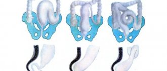
Various surgical techniques are used:
- restoration of patency of venous and lymphatic vessels;
- creation of lymphovenous anastomoses (crossed lymphatic vessels are sutured to the accompanying branches of the saphenous veins);
- excision of overgrown skin, subcutaneous fat and fascia in order to reduce the circumference of the limb.
Consequences and complications
thrombophlebitis or erysipelas (usually with streptococcal infection) can develop against the background of elephantiasis All these changes can lead to:
- lymphostasis or in other words - lymphedema ;
- possible addition of purulent-septic infections;
- malignancy of papillomas ;
- lymphorrhea , ulcers , eczema and muscle atrophy, since the upper integument and muscle tissue also suffer due to lack of trophism.
Elephantiasis can significantly limit freedom of movement, since it is much more difficult to walk on “heavy legs”; this may also be partly due to the degeneration of muscle tissue. As a result, all this can lead to complete immobilization and disability.
Causes
The causes of elephantiasis vary depending on the form of the pathology:
Primary (congenital) elephantiasis:
- dysplasia or underdevelopment of lymph vessels;
- Milroy-Mage disease (gene pathology associated with sex chromosomes);
- Shereshevsky syndrome (chromosomal abnormality);
- excessive production of interstitial fluid.
Secondary (acquired) elephantiasis:
- obstruction of the lymph nodes or its disruption (compression by the tumor or its metastases, chemotherapy, removal of lymph nodes);
- streptococcal infection caused by hemolytic streptococcus (cellulitis, erysipelas) – bacteria multiply in the lymph capillaries, where they secrete toxins that cause allergic reactions and narrowing of lymphatic vessels;
- damage to lymph vessels caused by extensive trauma, frostbite or burns;
- venous diseases that lead to malnutrition of the soft tissues of the limb and later to disruption of the patency of the lymphatic vessels (varicose veins, post-thrombophlebitis syndrome, phlebitis, thrombophlebitis);
- infection with filamentous helminths - filaria, in particular Bancroft's threadworm (Wuchereria bancrofti) - filariasis or wuchereriosis - parasites are transmitted through the bite of a mosquito/sandfly, the disease is common in the tropics and subtropics (helminths live and multiply in lymph vessels, where they intertwine into balls, which leads to blockage blood vessels; in addition, the development of a toxic-allergic reaction of the body to parasites “spurs” the proliferation of connective tissue fibers).
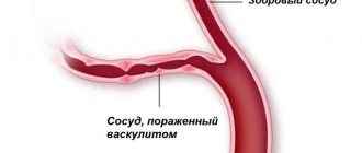
- autoimmune diseases - lead to damage to the blood and lymphatic vessels (systemic lupus erythematosus);
- chronic eczema;
- radiation;
- syphilis.
Symptoms of pathology
At first, a person does not notice that he has elephantiasis in his legs or does not pay special attention to the signs of pathology. At first there are no symptoms; there may be slight swelling of the foot or ankle, which goes away on its own. Painful sensations do not bother me. Over time, the pathology progresses and makes itself felt with the following symptoms:
- bursting sensation in the lower extremities;
- swelling in the hand or foot;
- swollen lymph nodes;
- hardening of the lower extremities;
- change in leg shape;
- skin disorder;
- the formation of fistulas through which lymph flows out;
- skin pigmentation is disturbed.
With elephantiasis, the skin is injured, which leads to dermatitis, hyperkeratosis with keratinization of the skin. A patient with elephantiasis has papillomas. Severe stages of the disease are accompanied by pain and heaviness in the lower extremities. A person gets tired quickly and feels constant weakness.
Drug therapy
At the first stage of elephantiasis the following are used:
- antihistamines to relieve allergic reactions (Loratadine);
- anthelmintic drugs that prevent parasites from multiplying (piperazine);
- angioprotectors to normalize tissue nutrition (Rutoside);
- pyridoxine to improve metabolism.
At the second stage of the disease, the following is prescribed:
- angioprotectors for relaxing vascular muscles (Troxerutin);
- enzymes to normalize the state of fiber (Lidase);
- non-steroidal anti-inflammatory drugs (Reopirin);
- desensitizing drugs to relieve inflammation (Claritin);
- biostimulants that soften connective tissue;
- vitamins.
At the third stage, to maintain the body, the following is prescribed:
- angioprotectors to reduce edema (Troxerutin);
- antibiotics to destroy infection in diseased tissues (Azithromycin);
- venotonics to improve fluid circulation in blood vessels (Detralex).