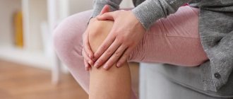
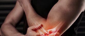
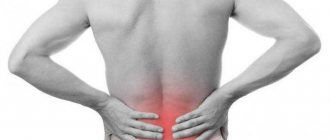
Pain in the shoulder joints is a common reason for patients to seek treatment. They arise for several reasons, and if left untreated, they lead to limited abduction in the shoulder joints, after which the person cannot raise his arm above his head. The most common cause of this pain is arthrosis of the shoulder joint. Modern treatment methods make it possible to slow down or even stop the development of degenerative joint diseases and maintain vital activity in patients.
Causes of Shoulder Arthritis
The disease is inflammatory in nature and requires effective treatment and subsequent rehabilitation. The shoulder is one of the most mobile joints in the human body. He experiences increased stress and is most often injured. As a result, inflammation can develop, which causes dystrophy and degeneration of articular cartilage.
The following are the main prerequisites for its occurrence:
- heredity;
- autoimmune disorders;
- increased load on the shoulder;
- hypothermia;
- injuries (bruises, sprains, fractures);
- diseases of the central nervous system;
- infections.
General information
Cartilage tissue is a smooth layer between the adjacent areas of bones. It ensures their easy sliding relative to each other, ensuring free and painless operation of the joint. Excessive stress, inflammation or injury can trigger a degenerative process that gradually spreads over the entire surface.
As a result, the smoothness of the articular surfaces is disrupted, and movements begin to cause pain. At the same time, bone growths begin to appear along the edges of the joint, replacing the affected cartilage. As the degenerative process progresses, it involves not only bones, but also surrounding tissues. The limb becomes deformed, the muscles spasm, and the ligaments become weak and lose elasticity. Without treatment, a person loses the ability to move his arm.
Make an appointment
Symptoms, stages and forms of arthritis of the shoulder joint
The treatment strategy depends on the etiology and nature of the pathology. The main signs of joint inflammation are:
- pain;
- limited mobility in the shoulder;
- specific crunch in the joint;
- swelling;
- local temperature increase;
- deterioration of the patient's general condition, weakness.
To treat arthritis of the shoulder joint, it is important to differentiate its form:
- post-traumatic (occurs after dislocations, sprains, fractures);
- rheumatoid (appears against the background of reduced immunity, after infectious diseases);
- osteoarthritis (associated with age-related degeneration of joint structures, develops after 50 years, often affects athletes involved in throwing equipment, wrestling, as well as people lifting weights due to their profession).
Treatment for arthritis of the shoulder joint is developed taking into account the degree of development of the pathology:
- The first stage is characterized by the appearance of pain, which increases with exertion and subsides with rest. The pain depends on the weather and intensifies at night. The hand gets tired quickly;
- on the second the pain becomes constant. There is a feeling of stiffness and clicking in the shoulder;
- on the third, irreversible changes affect the structures of the joint, it becomes deformed.
Diagnostics
Diagnosis of arthrosis of the shoulder joint requires an integrated approach. To accurately make a diagnosis and determine the extent of damage, the doctor uses the following methods:
- interview and history taking: the patient’s complaints are recorded, the circumstances of the occurrence of certain symptoms are determined; It is mandatory to clarify information about previous diseases and injuries, and the presence of joint damage in parents;
- examination: the doctor evaluates the joint visually, determines the range of movements, the area of greatest pain, etc.;
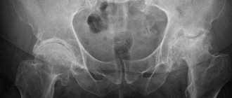
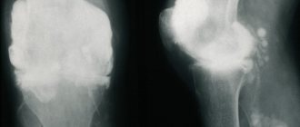
- X-ray and CT: the main diagnostic method that allows you to see the characteristic symptoms of osteoarthritis (narrowing of the joint space, cartilage degeneration, bone growths and deformities);
- Ultrasound: makes it possible to assess the condition of cartilage, bones, ligaments, joint capsule and muscles;
- MRI: allows you to obtain virtual sections of all structures of the affected area;
- laboratory diagnostics: a blood test reveals an active inflammatory process, often accompanying arthrosis;
- arthroscopy: examining the inside of a joint using a camera inserted through a small puncture.
If the disease is secondary in nature, examinations and consultations with narrow specialists on the underlying pathology are mandatory.
Types of treatment for glenohumeral periarthritis
The effectiveness of therapy largely depends on how correctly the cause of the disease has been identified. For example, if the inflammatory process started as a result of osteochondrosis of the cervical spine, then in addition to standard anti-inflammatory therapy, agents are used that improve the nutrition of nerve fibers and relieve muscle spasms.
The main task in the treatment of glenohumeral periarthritis is to relieve inflammation of the periarticular tissues and improve mobility in the joint.
They usually start with non-steroidal anti-inflammatory drugs orally and locally, in the form of ointments. Most often prescribed:
- diclofenac;
- piroxicam;
- ketoprofen;
- meloxicam;
- Celebrex.
For a mild form of the disease, this may be sufficient.
Blockades with diprospan have a good effect. They have a powerful anti-inflammatory effect and are applied to painful points near the joint.
The effectiveness of injections in the treatment of glenohumeral periarthritis can be increased by using other therapeutic measures in parallel with them - in particular, kinesiotaping, special exercises aimed at improving joint mobility, and physiotherapy.
Limiting the load on the affected joint is also an important step towards recovery.
Prevention methods
The main preventive method is reasonable physical activity. If professional duties require stress, you need to remember about rest and relaxation. It is also important to monitor the condition of the spine, because incorrect position of the neck and back for a long time can also cause the disease. Excessive sports loads can lead to sprains, torn ligaments and muscles, and other types of injuries - so it is better to conduct training under the guidance of experienced trainers.
Remember: with glenohumeral periarthritis of the shoulder joint, the pain can be so severe that it will prevent you from doing your usual activities. It is better to prevent a disease than to treat it.
If the disease does affect you, and you are bothered by pain when raising your arms or putting your hand behind your back, do not put off visiting a doctor. The sooner therapy is started, the greater the chance that you will be able to get rid of unpleasant symptoms quickly and completely.
Registration for treatment of glenohumeral periarthritis in our clinic in Moscow is made by calling the numbers listed on the website. Waiting for you!
Surgically
In cases of severe arthrosis, which is accompanied by rapid destruction of cartilage tissue, surgical intervention (arthroplasty) is used. The most common procedures are hip or knee replacements. Previously, most of the operations were performed on elderly people, whose age was 60 years or older. However, with the development of technology, artificially produced implants have become much better, and therefore young patients also undergo similar operations. However, another procedure in the form of revision surgery has been prepared for them in the future. Therefore, it is important to understand this before undergoing surgery.
What it is?
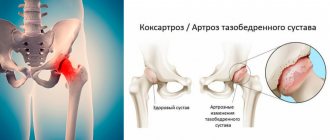
Shoulder arthrosis is a long-term, constantly progressive metabolic-dystrophic disease, leading to the gradual destruction of articular cartilage, protective growth of bone tissue with deformation of the joint and loss of its function.
A wide range of arm movements is ensured by the synchronous interaction of the joints of the shoulder complex:
- humeroscapular or simply humeral;
- acromioclavicular - between the collarbone and the acromial process of the scapula;
- sternoclavicular - between the sternum and collarbone.
The shoulder joint is very mobile, which is ensured by the convex head of the humerus and the relatively flat glenoid fossa of the scapula. The joint is strengthened by the tendons of the muscles of the upper limb, and the coracoacromial ligament is located above it. Not too reliable reinforcement allows the joint to move in different directions, but at the same time increases the risk of injury.
The code for shoulder arthrosis according to the International Classification of Diseases, 10th revision (ICD-10) is M19 (other types of arthrosis). Treatment of shoulder arthrosis should begin as early as possible. But advanced stages of the disease can also be successfully treated.
General clinical recommendations
For patients suffering from shoulder arthritis, it is recommended to:
- eat properly regularly, giving preference to low-fat animal products and vegetable and dairy foods;
- do not load the shoulder with increased physical activity, especially heavy lifting;
- regularly engage in exercise therapy: perform at home a complex of therapeutic exercises prescribed by the attending physician;
- do not overcool;
- get rid of bad habits - smoking and alcohol abuse - they disrupt blood circulation and metabolic processes;
- promptly treat all acute and chronic diseases, hormonal disorders;
- carry out maintenance treatment regularly.
We combine proven techniques of the East and innovative methods of Western medicine.
Read more about our unique method of treating arthritis
What other recommendations are given for shoulder arthrosis?
The patient is recommended to limit the consumption of spicy and salty foods, and increase collagen-containing foods: fresh herbs, poultry, seafood, salmon fish. Diet helps to eliminate pain more quickly, but is by no means an independent treatment method. An integrated approach helps reduce symptoms, eliminate inflammation and put the disease into remission.
However, traditional treatment does not affect the root causes of degenerative intra-articular changes. One of them is the lack of synovial fluid, which acts as an intra-articular lubricant. Against the background of its deficiency, excessive friction of the joint tissues and their rapid wear begin. The problem can be eliminated by intra-articular injections.
Treatment at the Energy of Health clinic
Orthopedists at the Energy of Health clinic offer patients comprehensive treatment methods for shoulder arthrosis:
- modern drug therapy regimens that combine high efficiency and minimal side effects;
- time-tested and new physiotherapeutic techniques;
- PRP therapy;
- physical therapy and massage to relieve restrictions on joint movements.
If necessary, therapeutic punctures of the joint are performed with the administration of painkillers or artificial synovial fluid to facilitate movement.
We monitor the patient throughout the treatment to keep the disease under control.
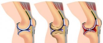
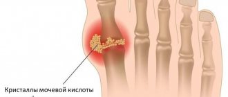
Symptoms of arthrosis
This disease usually first affects the knee joints, then spreads to the hip and shoulder joints. Arthrosis of the knee joint, the symptoms and treatment of which will be described below, is diagnosed, like other lesions, based on the results of radiography. In the picture you can see a reduction in the joint space and the presence of bone tissue damage. However, the results of radiographic examination do not always correspond to the clinical picture. In case of obvious pain, changes may not be expressed in the image, or vice versa - in the absence of pain, the image demonstrates obvious destruction of the joint tissue. Therefore, if necessary, other studies are prescribed: ultrasound, arthroscopy, MRI.
There are four main symptoms of arthrosis:
- Pain. In most cases, pain is expressed during movement, usually its intensity is quite weak. For this reason, patients do not pay attention to this at first and the disease progresses. Over time, the pain becomes more pronounced; a person experiences it even with light exertion. Ultimately, in the absence of adequate treatment, pain also manifests itself at rest.
- Crunch. When the joint wears out, a dry, characteristic crunch is heard when the surfaces rub. With further development of the disease it intensifies. It should be noted that if the sound is loud and the crunching is not accompanied by pain, we are not talking about arthrosis.
- Reduced range of motion. Muscle spasms, reduction of the joint space, and the formation of osteophytes lead to stiffness of movement.
- Modification of joints. The disease causes bone deformation and swelling caused by the accumulation of large amounts of fluid. Due to constant irritation of joint tissues, synovitis develops. It is worth noting that there is no point in pumping out the liquid without removing the cause of its excessive secretion.
With arthritis, pain does not depend on human movements and in most cases occurs at night. They should not be confused with arthrosis. Arthritic pain syndrome is more severe.
Glucosamine
It acts as an amino sugar, which gives a “start” to the biochemical synthesis of glycosylated proteins. It is also a structural component of cartilage tissue. Thanks to glucosamine, the body receives “natural” materials for the development, repair and support of tendons, ligaments and other tissues. With age, the body produces less of this substance, which leads to deterioration of the musculoskeletal system.
Rheumatologists say that taking a glucosamine supplement can reduce pain and the destruction of cartilage tissue. More than 18 studies that served as the basis for the 2005 review clinically supported the substance's effectiveness and beneficial effects on osteoarthritis.
The norm for patients is 700 mg/day. If necessary, the dosage can be increased to 1500 mg.
Mechanism of action of intra-articular injections "Noltrex"
To restore the viscosity of synovial fluid, a special artificial endoprosthesis Noltrex with the addition of silver ions has been developed. The gel is injected into the joint cavity in a medical office at intervals several times. The composition is completely biocompatible with tissues and therefore does not cause allergic or other reactions.
By introducing the drug into the joint cavity, the following results can be achieved:
- protect cartilage - the gel covers the synovial membrane and articular surfaces with an even layer, softens mechanical stress and stops further destruction;
- restore the viscosity of the synovial fluid - a drug with viscosity characteristics close to natural normalizes the properties of the joint environment;
- dilute the rubbing surfaces - the product restores the normal viscosity of the synovial fluid, as a result of which the joint spaces widen and friction stops.
Noltrex relieves pain in the shoulder joint for a year and a half
Causes of the disease
A person can raise his arms up using the upper limb girdle, which consists of the shoulder joints, shoulder blades and collarbones. Together they form mobile, active joints. Thus, at the junction of the semicircular part of the scapula (acromion) and the proximal end of the clavicle, the acromioclavicular joint is formed. It includes cartilage, ligaments, surrounding soft tissue. It is noteworthy that there is very little synovial (intra-articular) fluid in the AC joint capsule.
Specific loads on the upper body cause instability of the ligamentous apparatus of the joint, which in turn leads to a violation of the integrity of the cartilage tissue. At the next stage, pathological growth of bone cells occurs and growths (osteophytes) are formed. The combination of pathogenetic factors leads to an inflammatory, destructive process – AC joint arthrosis.
Experts have identified a category of patients among whom arthrosis of the acromioclavicular joint is most often diagnosed:
- weightlifters working with heavy weights;
- bodybuilders who load the belt of the upper limbs in order to sculpt the muscles of the back, arms, and chest;
- track and field athletes competing on specific apparatus – uneven bars, rings, horizontal bar;
- boxers;
- miners;
- welders;
- movers;
- blacksmiths.
The main factors provoking the onset of degenerative changes are:
- heavy physical labor associated with lifting and carrying heavy objects;
- prolonged forced raising of arms;
- congenital anomalies in the development of bone and cartilage tissue associated with weakness of ligaments, muscles, and improper formation of chondrocytes;
- injuries to the collarbones and shoulder blades;
- surgical interventions in the articulation area
- placing your arm bent at the shoulder under your head while sleeping.
Long-term loads on the acromioclavicular joint lead to the replacement of cartilage tissue with bone. The formation of osteophytes causes a decrease in motor amplitude. The pathological process of arthrosis takes a long time without specific symptoms. The appearance of pain indicates an advanced form of the disease.
If the root cause of acromioclavicular arthrosis is not established, they speak of the primary form of the disease. In this case, the pathological process spreads throughout all osteochondral joints. Primary ACS is most often transmitted genetically.
The secondary type of ACS is spoken of when the main pathogenetic factor of the disease is known:
- pathologies of the endocrine system;
- violation of metabolic processes;
- multiple microtraumas;
- progressive osteoporosis;
- excess weight;
- lesions of the articulation elements, acquired or congenital