Many of our patients (as well as patients, for example, of chiropractors or neurologists) do not even realize how interconnected the problems they are being treated with are. For example, the jaw joint hurts, the head hurts, shooting in the ear, clicking in the jaw, pulling at the base of the skull... it would seem that these are different symptoms, but the reason may be the same for everyone: TMJ dysfunction
.
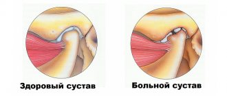
What is arthrosis of the TMJ
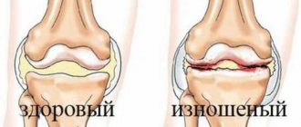
Arthrosis of the TMJ is a disease that destroys the components that form the joint (Greek arthron joint, suffix oz - destruction). First, the articular cartilage is destroyed, then the following occurs in the articular elements:
- proliferation (tissue growth);
- calcification (calcium redistribution) and ossification of cartilage;
- hyperplastic (proliferation) and destructive (destruction) processes in the epiphyseal parts of bones (these are the rounded ends of the bones - the head and fossa);
- reactive-inflammatory (from the word “response”) changes in the synovial membrane;
- fibrosis (overgrowth of connective tissue) with hardening of the joint capsule, which affects nearby muscles, tendons and ligaments.
With the destruction of cartilage, its shock-absorbing functions are reduced, and impacts are transmitted directly to the bone. Patients involuntarily increase the destruction by reacting emotionally to events - they clench their teeth, not daring to say too much, with a “stony” face and tense muscles, compressed blood vessels and stress hormones, they face the blows of fate. The amount of nutrients decreases, the TMJ would be happy to recover - but there is no building material. Instead, the epiphyseal sections of the bone are flattened under pressure, and bone growths appear on them.
Then the joint enlarges, compressing the nerve endings located nearby. The pain radiates to the ear, back of the head, and teeth. When the jaw moves, a specific clicking sound appears (occlusion-articulation syndrome).
ICD codes M.19. 0 (1, 2, 8 – last digit changes)
Causes of arthrosis of the temporomandibular joint
Arthrosis can be triggered by a one-time injury (compression, blow, bruise), as a result of which cracks and erosions appear on the articular surfaces. The disease is caused by a fracture of the condyle and condylar process if the fusion is incorrect.
Other reasons:
- prolonged stress;
- consequence of acute traumatic arthritis;
- birth trauma (arthrosis develops due to improper application of forceps);
- underdevelopment of the jaw (microgenia);
- sudden removal of molars (accident, fight);
- errors during dental prosthetics;
- impaired coordination of muscle contractions during dislocation and subsequent sharp (jerky, zigzag, circular) movements of the jaw;
- complete absence of teeth;
- deep bite;
- introduction of drugs into the joint cavity (for example, hydrocortisone, glucose solutions, novocaine).
Structure of the TMJ
Etiological factors of arthrosis (without which the disease does not develop):
- infections;
- metabolic disease;
- injuries;
- atherosclerosis of the terminal branches of blood vessels;
- prolonged spastic contraction of the lateral pterygoid muscle (responsible for moving the jaw forward and to the side).
Even children are diagnosed with TMJ arthrosis. In newborns, the disease develops as a result of birth trauma. Dysfunction in the joint due to various malocclusions is observed in 40% of children from 4 to 14 years old, but in only 1% x-rays reveal coracoid (myogenic) arthrosis.
During menopause, the likelihood of developing arthrosis due to endocrine disorders increases. With age, it is possible to develop senile, i.e. invaluable arthrosis, when cartilage tissue cannot recover, dries out and collapses.
At risk are people whose professional activities involve inadequate load on the joint (violinists), or those suffering from spasms of the masticatory muscles (bruxism).
Pain in the jaw joint: causes
- Defects in the dentition - lack of teeth, overestimation of fillings;
Pathological abrasion of teeth;- Broken bite
;
- Overload of the masticatory muscles (for example, if you chew on only one side for a long time);
- Inaccurate prosthetics, errors in orthodontic treatment;
- Other anomalies of the oral cavity and jaw structure;
- Bruxism, involuntary grinding of teeth.
- Birth trauma, trauma to the skull and jaw;
- Osteochondrosis and scoliosis of the spine;
- Severe constant stress
;
- An infection in the joint cavity that provokes inflammation of the TMJ.
Symptoms of TMJ arthrosis
Information about arthrosis of the temporomandibular joint on the Internet is 50% far-fetched descriptions of arthrosis of large joints, 30% is outdated data and obvious nonsense. And only 20% is true. Alas, texts are written by people without medical education, copying not from special educational literature or monographs, but from each other. Therefore, trust only trusted sources, and treat your health where there are no such ignorant things on the clinic websites.
First signs
A person may assume that he has arthrosis of the jaw when, after visiting doctors and following their recommendations, pain in the back of the head, ear, when chewing, hearing loss on one side, clicking, etc. does not go away.
Due to the structural features of the joint, the body manages to turn on the compensatory mechanism, so there is no long-term aching pain; due to the medications taken, it successfully disappears for a while.
Obvious symptoms
There are only 2 obvious symptoms (but it is also impossible to say 100% that this is arthrosis):
- displacement of the jaw to the side;
- pain when chewing.
You need to see a doctor immediately.
How dangerous is the disease?
TMJ arthrosis is silent and unnoticeable; people live with the disease for years without even knowing about the problem. But in vain.
Degrees of TMJ arthrosis
In the Russian Federation, the Kosinskaya classification of arthrosis has been adopted, which takes into account both symptoms and radiographic data. However, the TMJ is an exception to the rule: the joint “hangs”, held by muscles and ligaments, and does not experience weight loads comparable to other joints.
When at stage 1 according to Kosinskaya the joint space narrows, the pressure on the jaw simultaneously increases, which leads to problems with the teeth, but maintains the distance. The process is gradual, so this moment can be recorded on an MRI, but since there are no symptoms characteristic of the disease in the initial stage, it cannot be said unequivocally that this is stage 1 arthrosis. Only at stage 2, when symptoms appear (pain, facial asymmetry, etc.), and the patient finally consults a doctor, is a diagnosis made.
Stage 3 according to Kosinskaya: absence of joint space, sclerosis, necrosis, inability to open the mouth, chew and speak.
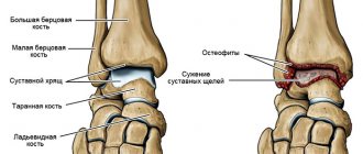
Damage to the TMJ by arthrosis
Possible complications
Arthrosis is not only a problem of the joint. Compensatory, in an effort to maintain chewing function, the body redistributes the load, which leads to tooth loss and rapid wear.
The previous diseases will be reflected in TMJ synovitis, and then the inflammatory process will affect the ear and nose (with decreased hearing, nasal congestion on one side), a headache will appear, which can radiate to the neck, back of the head and not stop.
The face will lose symmetry and become pasty (the skin appears loose, finely swollen, and grayish in color). Feeding is possible only through a tube; already at the second stage the ability to fully open the mouth is lost
Any localization and form of arthrosis has serious complications, so you should not delay treatment.
See how easily the disease can be cured in 10-12 sessions.
Exacerbations
Osteoarthritis is not arthritis; a chronic disease does not have periods of exacerbation. But this does not mean that the pain will be equally aching. The inflammatory process (cold, infection, virus) spreads to the joint with the development of synovitis. Swelling and pain appear, which can appear at any radial point (from the teeth to the back of the head). The source of inflammation expands, the oral cavity, ears, and breathing through the nose are at risk.
You need to understand that the brain is located nearby. And you shouldn’t wait for necrotic tissue to give rise to oncology.
Types of arthrosis of the temporomandibular joint
For treatment to be effective, it is important to understand that there are several types of arthrosis of the lower jaw.
Deforming arthrosis
Osteoarthritis of the TMJ usually develops after injury. The clinical course depends on the nature of growth and the location of osteophyte proliferation (towards soft tissues or the articular cavity). If bone growth is directed to soft tissues, the disease is asymptomatic for a long time. If the osteophyte grows into the cavity of the glenoid cavity, local acute pain appears, which occurs with limited jaw movement. Clicking and crunching are dull, and sometimes popping sounds appear.
The joint becomes deformed with the growth of the condyle, changes occur in the synovial membrane and are accompanied by hemorrhagic synovitis. The reason for this is irritation of the TMJ, caused by the multiple presence of dead and rejected cartilage cells (intra-articular detritus). The synovial villi on the inner lining of the joint enlarge and fat is deposited in them. Occasionally, they degenerate, forming islands of bone and cartilage tissue (metaplasia), which are separated from the articular surface and form intra-articular free bodies.
Please note: this is not salt, it is osteochondral tissue. Therefore, folk remedies for arthrosis, which can still help with gout, do not work.
Viral and infectious diseases during this period inflame the joint membrane, accelerating the destruction of cartilage and bone.
Facial asymmetry does not appear in all patients diagnosed with arthrosis deformans. This depends on the compensatory capabilities of the neuromuscular complex and on the functional grinding of the articular surfaces.
Sclerosing arthrosis
Not only vessels can be sclerotic. With arthrosis, the 2 upper layers of bone become sclerotic (bone tissue is replaced by dense connective tissue). In this case, some compaction of the head occurs, followed by expansion. Since replacement is a slow process, the body manages to compensate for the changes. Therefore, the disease goes unnoticed in the initial stages.
Neoarthrosis (post-infectious arthrosis of the TMJ)
The disease is a consequence of an acute inflammatory process in the TMJ, with repeated acute respiratory viral infections and with the presence of dysfunctional jaw syndrome (luxation, neuromuscular, occlusal-articulatory). It is asymptomatic. With exacerbation of chronic inflammation, the following is noted:
- dull, aching pain that intensifies when moving the jaw;
- crunch;
- clicking in the HFNS.
X-rays show usuria (disappearance of osteochondral tissue), defects in the articulating surfaces of bones, and sometimes the complete absence of condyles.
Myogenic arthrosis of the TMJ
In orthopedics, there is a separate type of deforming arthrosis of the TMJ, myogenic. Its difference: a beak-shaped bone growth on the anterior surface of the condyle.
X-ray shows myogenic arthrosis, the contours of the articular surface due to osteophyte resemble a bird
Myogenic arthrosis occurs due to prolonged spastic tension of the lateral (lateral) pterygoid muscle. Its middle bundles are attached to the anterior-inner surface of the condyle and its process. Prolonged muscle spasm leads to a lack of coordination of muscle contractions, the bone beams change direction, stretch, positioned along the direction of the tendon traction. If the spastic contraction of the muscle continues, the bones that form the joint will begin to break down.
Differences from other forms:
- the condyle always has a beak-like shape;
- bone growth (osteophyte) is always localized in a specific place;
- no restrictions on jaw movement;
- the disease occurs without facial asymmetry.
The initial stages of the disease are asymptomatic. The osteophyte grows gradually on the anterior surface of the condyle, does not rub against hard tissues, and forms a bed in soft tissues. In the joint area, nutrition is disrupted, there may be a slight swelling on the face, spider veins - but very often this is explained by fatigue, overload, without paying attention to the TMJ. Painful symptoms occur at the moment of dislocation, subluxation of the lower jaw. Since the movement of the jaw in such cases is atypical, the osteophyte injures the soft tissues, irritating the nerve endings - severe pain appears (it hurts to chew hard food), severe swelling, clicking, mild swelling and paleness of the skin flap (pastyness). At the moment the mouth opens, the jaw begins to shift to the side.
Metabolic arthrosis
This is a rare type of disease that occurs when salt metabolism in the body is disrupted. The reason is needle-shaped crystals of uric acid settling in the TMJ. In patients, large joints are first affected; they suffer for a long time from metabolic polyarthritis, the visual manifestation of which is “gouty bumps” on the joints.
Symptoms:
- significant deformation of the head of the lower jaw, detected by palpation;
- asynchronous movement of the condyles when opening and closing the mouth;
- hinge movements on the side of the diseased temporomandibular joint;
- crunch;
- local dull pain;
- when opening the mouth, the jaw moves to the side;
- Lateral position of the head leads to facial asymmetry.
On radiographs with metabolic arthrosis, the condyle is covered with whitish needle-shaped curls of various shapes that are not permeable to x-rays.
Crunching in joints - when to worry
Intra-articular injections of hyaluronic acid
Senile arthrosis of the TMJ
Senile, or invaliable, arthrosis occurs with age. “Aging” of cartilage tissue occurs in 3 stages:
- cartilage tissue becomes soft and loose;
- loses some of the water, dries out, becomes denser;
- The smooth surface disappears, the cartilage becomes fragile and becomes covered with cracks.
After 60 years, bone exposure begins. Patients feel uncomfortable chewing and clicks are noted in the TMJ. The x-ray shows subtle changes.
Main symptoms of TMJ dysfunction
Diagnosis of TMJ dysfunction is complex, since the disorder manifests itself in the form of multiple and varied symptoms [2]. There are often cases when they spontaneously (spontaneously) arise and disappear [1].
One of the most striking symptoms of the disorder is pain. Moreover, they can be so intense and acute that the patient loses the ability to chew and swallow food normally, speak, and open his mouth. Pain may be the only symptom of the disorder, or may be accompanied by characteristic clicks in the joint and limitations in the mobility of the lower jaw. There are often cases when dysfunction manifests itself only as sound phenomena, while other symptoms are absent [2].
Pain caused by TMJ dysfunction is localized not only in the joint itself, but in the area of the masticatory muscles [4]. In addition, pain and discomfort can manifest in the forehead and eyes, ears, temples, head as a whole, and spread (radiate) to the neck, shoulders and back [4].
Other most common symptoms include [4]:
- noise in ears;
- feeling of pressure on the eyes and sensitivity to light;
- dizziness, nausea, lack of concentration;
- clicks and other sound manifestations when the jaw moves;
- swelling in the facial area (mainly in the joint area).
Discomfort due to TMJ dysfunction can be so severe that it can significantly reduce the patient’s quality of life [4].
Diagnostics
In the initial stages, arthrosis of the jaw is asymptomatic (more precisely, if there is pain, discomfort - they are attributed to a cold, problems with teeth, inflammation of the facial nerve, etc.). When constant pain appears, the face loses symmetry, it is impossible to chew - the patient begins to visit doctors.
Remember: at the slightest suspicion of TMJ arthrosis, you should consult a doctor; it is impossible to make a diagnosis yourself (if you are not an orthopedist or a healer).
In the clinic to confirm the diagnosis you will need:
- take blood tests (clinical - to identify an infectious-inflammatory process, biochemistry - for arthrosis, biochemical parameters should be normal);
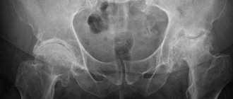
- take an x-ray in 2 projections (the image clearly shows the deformation of the osteophyte, the narrowing of the joint space, but the articular cartilage is not displayed in the image, and it is impossible to assess the degree of destruction of the TMJ in the early stages);
- undergo an MRI or computed tomography (MRI uses magnetic waves, and computer tomography uses X-rays, so in the early stages, MRI is an advantage).
Occasionally, an ultrasound of the joint is prescribed. In addition, a personal examination is required, because often it is necessary to treat not only arthrosis, but also to remove defects in the dentition, and to treat the accompanying inflammation of nearby tissues.
Contraindications
Magnetic resonance imaging of the maxillofacial joint is contraindicated:
- Patients with pacemakers, neurostimulators, cochlear hearing prosthetic systems, defibrillators implanted in the patient’s body (magnetic fields used for MRI can disable the devices);
- Patients who have metal foreign bodies in their bodies (steel or iron shavings in the eyes, vascular clips on the main arteries, fragments and fragments of ammunition);
- Children under 5 years old;
- Pregnant women up to 12 weeks of pregnancy;
- Persons with excess weight of more than 130 kg and body girth of more than 150 cm (there is no physical ability to place the patient in the tomograph).
Treatment of TMJ arthrosis
Treatment of TMJ arthrosis is complex, regardless of the stage of development of the disease. The disease cannot be overcome with one method or remedy.
Medicines for the treatment of TMJ arthrosis
In the early stages, arthrosis of the TMJ is asymptomatic, but dysfunctional syndromes may appear. Therefore, treatment should be aimed at normalizing the functioning of the lower jaw. To do this, use myogymnastic exercises (only after consultation with a doctor) and physiotherapy.
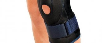
Then the position of the articular heads is normalized, the integrity of the dentition and bite are restored. For pain, clicking, crunching, and asynchronous contraction of the masticatory muscles, permanent splints, braces, and bandages are used.
At the same time, the doctor prescribes medications to restore cartilage tissue, relieve inflammation and improve metabolism around the joint.
For arthrosis of the jaw joint, consultation with a psychotherapist is indicated, because Chronic muscle spasms are always associated with problems in relationship with the world.
Medication
To restore cartilage tissue, chondroprotectors are prescribed:
- glucosamine, stimulates the production of key elements in cartilage tissue, restores the articular surface, protects from destruction;
- chondroitin sulfate, increases the ability of cartilage molecules to retain water (especially important for senile arthrosis of the temporomandibular joint), neutralizes the influence of enzymes that destroy cartilage, and stimulates the formation of collagen.
But if the cartilage is completely destroyed, chondroprotectors are not effective.
To relieve muscle spasms, the doctor prescribes mydocalm and sirdalud.
Remember: you cannot use medications on your own without a doctor. Muscle spasm is a protective reaction; without it, the TMJ will begin to deteriorate at an accelerated pace.
Drugs of this group, muscle relaxants, are used only with the simultaneous use of chondroprotectors and orthopedic treatment (splints).
Corticosteroids quickly relieve pain during synovitis, intra-articular injection only relieves inflammation, BUT the next dose is less effective (3-4 injections is the maximum), and the hormone destroys and does not heal articular cartilage. Therefore, for arthrosis of the temporomandibular joint without inflammation, drugs are not used with proper treatment.
The hyaluronic acid preparation “Ostenil mini”, a 1% solution of sodium hyaluronate (10 mg of active substance in 1 ml syringe), is also called “liquid prosthesis”. It restores the joint more effectively than chondroprotectors. 1-2 injections per year (3-4 years) are enough. There are only 2 drawbacks:
- there should be no inflammation in the joint, drugs with HA are instantly destroyed in such an environment, and the treatment will not be effective;
- this is an expensive drug (however, it is better to use it than to go under a scalpel).
For post-infectious arthrosis of the TMJ, Movalis (selective anti-inflammatory) is prescribed to suppress inflammation, as well as:
- Brufen;
- indomethacin;
- methindol;
- butadione;
- rheopirin;
- sodium salicylic acid;
- antibiotics (in the presence of low-grade fever).
Please note: long-term use of non-steroidal anti-inflammatory drugs has a negative effect on articular cartilage.
Electrophoresis with medical bile, bischofite, dimexide (compresses are also made from them), as well as with salicylic sodium (10%), lidase is indicated. Mud therapy helps a lot.
For post-infectious arthrosis, treatment is physiotherapeutic (electrotherapy with potassium iodide solution (5%) and novocaine solution (2%)). Recommended ointments are apisatron, vipratox, and an analgesic mixture.
For myogenic arthrosis, they practice novocaine blockade with vitamins B1, B12, massage using anesthetic ointments, UHF.
Metabolic arthrosis of the jaw joint is myogymnastics and the use of a splint. At the same time, salt-removing therapy (delogil, collection of salt-removing herbs) is prescribed.
Chondroprotectors: what are they, how to choose, how effective are they?
Joint pain at rest
If the condyle in the temporomandibular joint is excessively enlarged, surgical and orthopedic treatment is performed.
In addition, at any stage and for almost any type of arthrosis, vasodilators xanthinol nicotinate and pentoxifylline are prescribed, which relieve spasm of small vessels and improve blood circulation in the joint. At the same time, slight redness of the face and a feeling of heat are the norm.
Therapeutic ointments and creams do not cure advanced arthrosis of the jaw joint, but their use relieves pain, relieves swelling, and improves tissue nutrition. Finalgon and Nicoflex increase blood circulation, relieve pain and partially relax the spasmed muscle. Creams based on bee venom additionally improve the elasticity of ligaments, but due to the large number of allergic reactions, they must be used with caution. Ointments containing non-steroidal anti-inflammatory drugs (Voltaren-gel, Fastum, ibuprofen, indomethacin, etc.) are less effective than medications, but do not have as many contraindications.
Orthopedic treatment
Orthopedic devices help redistribute the load in the joint and straighten the jaw. Using splints and a sling bandage:
- functional rest is created in the joint;
- traumatic factors are eliminated;
- the activity of the chewing muscles and joints is restored.
When treating arthrosis, the dentition must be restored. Wearing mouth guards, braces, and teeth grinding are practiced.
How to treat TMJ arthrosis with exercise therapy
Physical therapy for arthrosis of the jaw joint is useful only after permission and under the supervision of a doctor.
The joint is destroyed from the inside, and the destruction of it and nearby tissues, as well as compensatory muscle spasm, intensifies with movement. Stupid exercises can cause harm, because... unknown until images are received:
- how arthrosis develops;
- what type is it;
- where are the osteophytes directed?
The joint is already receiving load - we talk, eat, opening and closing our mouths. And moving the jaw from side to side will add subluxation and swelling.
At home you can do:
- soft massage with a sponge using rotational movements around the joint to stimulate lymph outflow and blood circulation;
- gently tap a bag of raw peas or beans around the joint;
- stroke the cheek from the nose to the bridge of the nose, applying slight pressure with the palm of your hand.
Nutrition, diet
The development of arthrosis of the temporomandibular joint is not associated with dietary habits (you just don’t need to crack hard nuts so as not to break your teeth). However, it is important to pay attention to the amount of water entering the body. The individual need for clean water is calculated using the formula: 1500 ml + 20 ml per kg (over 20 kg). For example, with a weight of 60 kg, the amount of liquid is 1500 ml + 40 * 20 ml = 2300 ml
When edema occurs, diuretic herbs and herbs (birch, linden, clover inflorescences, mistletoe branches, etc.) are used.
Traditionally, for problems with joints, it is recommended to eat more vegetables and fruits (vitamins and minerals), as well as jellied meats, jelly (some patients have a special craving for soft cartilage, pig ears, etc.).
Eat a varied dietC
When pain occurs (stage 2), it is painful to eat. Food should be soft and pureed. These are juices, pureed soups, ready-made baby food in jars. Sometimes you have to feed through a tube - do not bring yourself to this state, at the first unpleasant sensations, consult a doctor.
If an operation has been performed on the joint, the food for the first time should be dietary. Food should be pureed, spicy, spicy and salty foods should be excluded.
Folk remedies
Among the folk remedies for arthrosis of the jaw, a compress with bischofite or medical bile helps a lot. But due to the fact that it is necessary to align the joint, this is a temporary measure to alleviate the condition. You still have to go to the doctor.
A compress is made only when there is no inflammation, swelling on the face or viral infectious diseases. First, place a warm (not hot!) heating pad on the sore side of the face for 3-5 minutes to warm the joint and slightly relax the spasming muscles. Then put gauze on it (attention: no colorful synthetic rags), soaked in a bischofite solution, cover first with parchment paper (cling film), then with a flannel cloth (terry towel). The compress should be kept for 1-1.5 hours, for people with sensitive skin no more than 20 minutes. If there are no negative reactions, the procedure time is increased. Course of home treatment: 10-15 compresses every other day.
A compress with medical bile cannot be used if you have pustular rashes, acne, rosacea, or rosacea. 6 layers of gauze are soaked in bile and a “sandwich” is made in the same way as with bischofite. However, they keep it for 30 minutes maximum. Course of treatment: daily for 2-3 weeks.
In case of cardiovascular insufficiency or hypertension, such procedures without medical supervision are prohibited.
When diagnosed with “metabolic arthrosis of the jaw joint,” herbal preparations that remove salts are taken. For example, collection (all herbs 100 g, grind in a meat grinder into powder):
- mint;
- buckthorn;
- dandelion;
- immortelle;
- juniper fruits;
- celandine;
- buckthorn;
- chicory (herb);
- yarrow;
- sage (leaves);
- burdock.
1.5 tbsp. collection, brew 1.5 tbsp of boiling water and infuse. Drink 0.5 tbsp. 3 times a day before meals.
This is the only type of arthritis where herbal treatment is effective. However, you can drink herbs to strengthen the immune system and for prevention during epidemics of viral infections.
Surgical operations
Surgical intervention is indicated:
- during ossification;
- with further destruction if conservative treatment does not produce results.
The joint or part of it is removed, replaced with an artificial implant or your own graft (usually part of the fibula).
Fundamentals of occlusion and biomechanics of the jaws: a new look at old concepts
A deep understanding of the basics of occlusion and biomechanics of the jaws is one of the most important and necessary components for providing comprehensive patient rehabilitation in dental practice. Knowledge of the principles of differential diagnosis of pain, planning of future iatrogenic intervention, as well as algorithms for the treatment of prosthetic disorders provides the doctor with all the necessary tools for further normalization of the patient’s dental status.
An orthopedic doctor simply cannot do without understanding how significant the concept of occlusion is not only in pathology, but also in a state of stable and adequate function. The formation of appropriate occlusal schemes is based on the redistribution of acting forces, because, in fact, it is precisely because of the excess of such indicators that diseases, pathologies and dysfunctions of the elements of the dentofacial apparatus arise.
Occlusal disorders can manifest themselves in the form of various structural damage to the dental status, such as pathological abrasion, fractures, and premature wear of restorative structures. In addition to the latter, functional pathologies are characterized by tooth mobility, loss of volume of soft and hard tissues, muscle pain, as well as pain and noise in the joints (the so-called clattering), limitation and impairment of movements of the lower jaw, remodeling changes in bone tissue in the structure of the temporomandibular joint . In such cases, patients form so-called parafunctional habits, the presence of which he himself does not know. Clinically, signs of such are manifested by excessive wear of one’s own teeth and various types of restorative structures present in the oral cavity.
There are different opinions regarding the relationship between the state of occlusion and disorders of the temporomandibular composition. According to the most extensive literature reviews, such associations are rather weakly expressed, as evidenced by the fact that when correcting occlusal relationships, it is not always possible to prevent the development and progression of joint pathologies. Based on the available data, the following conclusions can be drawn: only the absence of traumatic occlusal injuries, which are manifested by the action of excessively high parafunctional forces exceeding the adaptive capabilities of the body, ensures complete prevention of the occurrence of pathologies and dysfunctions, or the presence of such in the acceptable adaptation range. This conclusion is evidence-based, regardless of how ideal or non-ideal the occlusal schemes of each individual patient are. On the other hand, with prolonged exposure to excessive occlusal forces, the development of corresponding dysfunctions and diseases occurs regardless of the characteristics of a particular occlusal scheme. The corresponding pathological types of occlusion only further aggravate the course of related prosthetic diseases.
From the above it follows that if the doctor is fully familiar with the specifics of occlusal movements in a particular patient, and also understands their impact on the condition of soft and hard tissues, muscles and joints, then he can ensure the formation of such occlusal patterns that would be the most stable and least traumatic for each specific patient. In other words, understanding the basics of occlusion helps doctors not only plan future interventions, but also predict the functional rehabilitation of prosthetically compromised patients. The main connecting link between the pathology of the temporomandibular joint, the state of occlusion and the functional disorder of the dentofacial apparatus is the repeated action of excessive occlusal load, which goes beyond the adaptive range of the body. Based on this, the author considers it wrong to separate the dynamics of the application of force on human tissue from disorders and diseases developing in the same tissues - after all, in fact, these processes are of an indirect cause-and-effect nature.
The question is different: what is the true connection between the existing parafunction, the state of occlusion and functional deviations of the dentofacial apparatus. In order to understand how the jaw functions and where occlusion begins, you need to repeat in detail the anatomy of the masticatory muscles, the temporomandibular joint, and, of course, the teeth, taking into account the functional parameters of each of the above-mentioned components. After analyzing the anatomy, you should focus on how the relationship between the upper and lower jaws is generally formed, taking into account the occurrence of static and dynamic contacts between the surfaces of antagonist teeth. After this, the data obtained during the analysis can be implemented into a plan for future iatrogenic intervention aimed at eliminating structural disorders of the dentition and aesthetic problems, while ensuring not only the functional comfort of the dentofacial apparatus, but also the stability of the achieved results of complex rehabilitation.
In the course of analyzing the features of anatomy and intermaxillary relationships, doctors should look for key parameters of each of these components, on the basis of which they will subsequently make a decision regarding one or another possible treatment plan.
In this article, the author will refer to the concept of dental treatment planning, which takes into account changes in the facial profile during iatrogenic interventions, developed by Frank Spear. With significant destruction of the tooth structure, the main occlusal landmarks are simply lost, and the pathology goes beyond the boundaries of possible dental-alveolar compensation. Consequently, the clinician’s task is also to restore the supporting occlusal points of the intermaxillary relationship, and then, based on their stability, carry out further prosthetic rehabilitation. When implementing an approach to treatment taking into account changes in the facial profile, it is possible to ensure successful prosthetic reconstruction of the bite, based precisely on the position of the supporting occlusal landmarks.
Pankey rules and the concept of optimal occlusion
Dr. LD Pankey, being a pioneer and developer of comprehensive approaches to dentition restoration, proposed a specific concept that helps critically evaluate occlusion both during systemic dental rehabilitation and during everyday dental care:
- When the condyle of the jaw is completely in the glenoid fossa, all the posterior teeth exhibit equal and uniform contact, while the anterior teeth only lightly touch the opposing teeth.
- When the jaws are clenched, neither the teeth nor the lower jaw move.
- When the mandible moves in any direction, none of the back teeth contact faster or more strongly than the teeth in the anterior region.
Having analyzed these features, we can take a fresh look at the specifics of the anatomy of the dentofacial apparatus.
Anatomy of the temporomandibular joint
In Photo 1 you can see that the condyle of the mandible is in very close contact with the biconcave disc of the joint. These elements of the joint are located inside the capsule, which is protected behind by retrodiscal ligaments, and below, by means of capsular ligaments, is attached to the neck of the condylar process. Anteriorly, the superior portion of the lateral pterygoid muscle attaches to both the disc and the neck of the condylar process, while the inferior portion of the muscle attaches only to the neck of the condyle. Behind the joint is the external auditory canal. Anterior and superior to the condyle is the eminence, and directly above it is the glenoid fossa. The articular surfaces are covered by fibrocartilage, which is a smooth structure, and is supported by synovial fluid. The latter lubricates the surfaces of the joint, providing them with nutrients and oxygen, as well as ensuring the removal of possible debris. In the structure of the capsule, the number of blood tissues is very limited, or they may be completely absent.
Photo 1. Classic diagram of the anatomy of the temporomandibular joint.
When analyzing a joint, it is necessary to note the most important relationships of its individual components. First, there is the close condyle/disc/fossa connection. Essentially, they are in as much contact as possible, allowing the joint to withstand the necessary loads. On the other hand, this form of connection of the elements ensures the anatomical and functional integrity of this organ during dynamic movements of the lower jaw. In certain articular pathologies, this relationship is disrupted, which leads to permanent functional changes. It is obvious that the variation in the size, volume and shape of the temporomandibular joint is quite significant, and these differ greatly from person to person. Historically, we have assumed that the dimensions of the condylar process are relatively stable. However, recent research has established that the size of this anatomical structure can change and adapt over time and depending on the prevailing circumstances. A striking example is the increase in condyle size when using night guards. Due to the protrusion of the mandible to maintain patency of the upper airway, the condyle also remodels, increasing in size. Thus, it is obvious that this bone structure can not only adapt to functional conditions, but also change its shape, increasing in its geometric parameters. Consequently, the previously determined dimensional stability is very, very relative. As the first occlusal guideline, the doctor can use the position of the condyle during the registration of the centric relation, which is the most desirable. Firstly, the central ratio is a parameter that, with adequate execution technique, can be quite accurately and easily recorded. In addition, this position of the structures is repeatable, and it can be restored even if the position of the teeth changes or the contact between them is disrupted. A stable joint in this position has the ability to withstand significant loads, and the lateral pterygoid muscle can remain in a passive state even with strong clenching of the jaws (photo 2, 3).
Photo 2. Muscles that lift the mandible on the right side.
Photo 3. Muscles that lift the mandible on the left side.
Anatomy of the masticatory muscles
The function of raising the lower jaw is provided by three masticatory muscles. With parafunctional habits, such as bruxism, these masticatory muscles can develop significant force acting on all structures of the dentofacial apparatus. The masticatory muscle starts from the zygomatic arch and is attached to the lower edge of the lower jaw. The force vector of this muscle is directed upward and forward. The cross-sectional thickness of the masticatory muscles justifies the fact why they can develop the most significant strength indicators, located anterior to the temporomandibular joint. The temporalis muscle begins in the area of the temporal fossa and the deep part of the temporalis fascia. It is directed medial to the zygomatic bone and forms a tendon that is attached to the coronoid process of the mandible, and also passes into the region of the retromolar fossa distal to the last molar of the mandible. Since the muscle splits along its course, the action vectors of its force also diverge: the anterior component is directed upward and slightly anteriorly, while the posterior component is directed upward and posteriorly. This feature should be taken into account when diagnosing pain symptoms arising in the area of this particular muscle.
The medial pterygoid muscle consists of two heads: the main part of the muscle begins directly above the medial surface of the lateral pterygoid plate, while the superficial head begins from the maxillary tubercle and the pyramidal process of the palatine bone. The fibers of this muscle are directed downward laterally and posteriorly, and through the tendon are woven into the lower and posterior parts of the medial surface of the angle and ramus of the mandible. Insertable fibers connect this muscle with the masseter, forming common tendon slings, which allows both muscles to jointly perform the function of raising the mandible.
All of the above anatomical factors determine the direction vector of forces when activating the above-mentioned muscles. First, the directions of force of all three muscles shape the position of the condyle in the glenoid fossa: anteriorly and superiorly opposite the eminence and slightly medially, so that the medial pole of the condyle is the most load-bearing side of the joint. In other words, if we exclude the action of interdental contacts, then it is these muscles that determine the most superior position of the condyle, thereby ensuring close contact between the condyle, disc and glenoid fossa. The digastric muscle is one of the main muscles that lowers the lower jaw and opens the mouth. It begins in the area of the mastoid notch, forms a tendon along its path, and ends at the lower border of the mandible near the symphysis on the side of the digastric fossa. Since this muscle lowers the jaw, pain in this area is a rather unusual symptom. The lateral pterygoid muscle moves the lower jaw laterally and also forward. The upper head of this muscle starts from the infratemporal wing of the sphenoid bone, and the lower head starts from the lateral pterygoid plate. The muscle is woven in two bundles into the neck of the condylar process and the articular disc. Part of its function is to coordinate the position of the disc relative to the condyle to maintain the desired functional relationship, but in addition it provides support for the mandible in an eccentric position during intense clenching or bruxing when appropriate jaw movement is required to achieve maximum intercuspidation at existing tooth contacts . The three main levator muscles produce much more force than the lateral pterygoid muscle, therefore it must provide sufficient contraction to counteract the aforementioned elevator muscles. If these parameters do not correspond, painful sensations and even spasms may occur, which indicate a violation of muscle function.
Posterior teeth relationship
Pankey's first rule states that by positioning both condyles in centric relation, the posterior teeth form simultaneous and uniform contact, while the anterior teeth contact either slightly less or to a similar degree (Plates 4, 5).
Photo 4. Teeth contacts on the upper jaw.
Photo 5. Teeth contacts on the lower jaw.
Essentially, every type of occlusal design, regardless of condylar position, involves achieving multiple tooth contacts. With this ratio, the bite force is distributed more evenly over a larger number of tooth surfaces. In addition, maximal contact does not require activation of the lateral pterygoid muscles to maintain the mandible in the desired eccentric position. On the other hand, by ensuring simultaneous contact of the tooth cusps with flat bite pads, it is possible to guarantee the direction of the acting force down the long axis of the teeth, excluding the influence of deflecting lateral components (photo 6).
Photo 6. Areas of contact between the cusps.
It is the latter that provoke various structural damage to teeth, restorations, soft tissues and bone crest. And finally, with adequate contact of the posterior teeth, optimal distribution of the functional forces of the muscles that lift the mandible is ensured on both temporomandibular joints. When contact is ensured only in the area of the incisors, 60% of the total force generated by the muscles that lift the mandible is transferred to both joints, while with contact of the second molars this figure decreases to 5%. Posterior tooth contact is especially critical for painful and unstable joints.
Functional system assessment
Assessment of the functional system is a stage of diagnosing all conditions of the functioning of the dentofacial apparatus in each individual patient. The beginning of this stage is an analysis of how the patient talks to the doctor, and the idea of this approach was first proposed by Bob Barkley and later improved by Pankey. Barkley came to the conclusion that it is best when any disorders in a patient are diagnosed not only by the treating team of doctors, but also by the patient himself during diagnostic procedures. Therefore, a history review is a key initial aspect of treatment. A thorough and comprehensive patient examination algorithm was precisely described by William Lockard in his book “The Exceptional Dental Practice”.
Assessment of the functional state of the dental system includes diagnostics:
- temporomandibular joints
- masticatory muscles
- range and pattern of movements of the lower jaw
- analysis of static and dynamic tooth contacts.
The overall goal of a functional examination is to collect as much data as possible in order to determine whether the patient’s dental condition is stable or not. If the system is unstable, the doctor needs to determine where the structural damage occurred, what pathology could provoke it, and what type of dysfunction arose as a result. It is extremely important to consider all possible influencing factors before making a final diagnosis. Sometimes it happens that certain violations of functional parameters can be identified only at the end of the diagnostic process, or even between patient visits, based on his own complaints and comments that arose during a comprehensive examination. The order of diagnostic manipulations is determined by the attending physician himself, therefore the author developed the algorithm presented in the article himself, based on existing clinical experience. The first stage of diagnosis remains the assessment of joint function.
This step involves obtaining answers to the following questions:
- Do you feel pain when palpating your joints? If so, what is the nature of these sensations and how strong are they?
- Do you feel noises when moving your joints? If so, at what point in the movement and what is the nature of the sound? Is it painful?
- Is the movement of the lower jaw free and unrestricted? Are there any deviations? If so, how significant are they and in what direction are they observed?
- Can the joints withstand the forces or loads placed on them?
Positioning at the 12 o'clock position allows the doctor to examine the patient along the long axis of the head and at the same time analyze the existing deviations in movements and symmetrical relationships. In adult patients, full mouth opening exceeds 40 mm, and in some patients this parameter even goes beyond 50 mm. The lateral movement of the lower jaw is normally about 10 mm. In this case, the doctor must determine whether pain occurs during lateral movements? Do the ranges of lateral motion differ in any particular direction? The nature of the movements of the lower jaw is determined simultaneously by the state of the condylar process, disc, articular fossa and tubercle, as well as the stability of the lateral pterygoid muscles and the muscles that provide mouth opening. It is logical that movements in a damaged or unstable joint will be more limited than movements in a healthy state of the dental system. Therefore, assessment of the initial parameters of movements in the joint is a mandatory stage of a complex diagnostic algorithm. In addition, it is very important to record the position of the joint in the centric relation position. The author uses the bimanual centric relation technique developed by Dawson, as well as a leaf calibrator, a luchiu jig, and various types of frontal stoppers. The lack of contact in the area of the posterior teeth allows the muscles that elevate the mandible to position the condylar head more highly before the disc-fossa complex of the joint limits its position. The use of front stoppers is a fairly reliable method for recording centric relation. The patient is asked to stick the lower jaw forward and repeat this several times in the position of the frontal stopper - in this way it is possible to achieve activation of the muscles that understand the lower jaw. Initial attempts to determine the centric relation may be unsuccessful due to disc impairment, or fluid swelling within the joint capsule, not to mention possible spasms of the lateral pterygoid muscles. In such cases, the doctor will be able to determine only a preliminary central ratio. As the joints and muscles stabilize, more accurate centric relation parameters can be determined. Using the same three recording methods above, the doctor can also test the ability of the joints to tolerate force applied to them. In other words, the physician can determine whether the present condyle/disc/fossa condition is stable and healthy enough to withstand the forces of normal functional loading or even parafunctional conditions? In most cases, joint instability is caused by disorders such as inflammation, disc lesions, disturbances between the constituent surfaces, and pain in the area of attachment of the lateral pterygoid muscle to the condyle and disc. By repeated protrusive and retrusive movements of the patient using a sheet calibrator and a Lucia Jig, the condition of the joint and the lateral pterygoid muscle can be diagnosed. Adaptation to stress is tested using a bimanual technique, increasing the upward pressure force. After diagnosing the joint, they begin to evaluate muscle function. This part of the inspection is to determine the following:
- condition of the three muscles that lift the mandible. Is there any discomfort during palpation? What is the approximate cross-sectional thickness of the muscle? What level of tension is present when they are active? How does the patient react to palpation performed by the doctor?
- condition of the lateral pterygoid muscles. Does pain occur with palpation or the action of any other factor?
- condition of the muscles of the floor of the mouth.
- condition of the muscles in the neck and shoulders.
Palpation of each of these muscles is slightly different, but the doctor should palpate as many of them as possible. The physician should increase the pressure gently and slowly while observing the patient's response while rating their pain response on a scale of 1-2-3 (mild to moderate to severe). In addition, the clinician should analyze the relative cross-sectional thickness of the masseter and temporalis muscles, as this parameter is a reliable indicator of the patient's ability to generate loads of a certain force during jaw clenching or towing. Based on existing observations, it is known that patients with a shallower mandibular angle tend to have thicker masticatory muscles, which in turn allows them to generate more force during function.
And finally, the doctor needs to determine the nature of tooth contacts at different jaw ratios for a final understanding of the patient’s functional state. First, the first contact at centric relation must be determined, which will be considered the critical cutoff point, even if the patient has not reached the stage of restoration of centric relation. For this manipulation, the author uses a bimanual technique and a sheet calibrator. In addition, for the same purpose, you can use the “luchiu jig”, deprogramming the patient’s masticatory muscles, and then proceed to the bimanual technique with a progressive increase in the thickness of the calibrator sheets: this is how it is possible to evaluate the upper movement of the condyle deep into the articular fossa, and as a result, the area of occurrence of the most early contact in the dental area. This manipulation helps the clinician form an idea of the need to correct the corresponding vertical and horizontal components in order to achieve maximum contact between the antagonist teeth. To perform such a correction, it may be necessary to use various treatment methods, while at the same time it is necessary to evaluate whether such an intervention will be so effective as to compensate for all the changes in the dentofacial apparatus expected during its implementation, or whether performing it as a whole will be considered inappropriate.
conclusions
Understanding the anatomy of the joints, muscles and dentition, as well as their relationship, helps the doctor to objectively assess the parameters of the bite and the function of the dentofacial apparatus. This knowledge determines the adequacy of the choice of one or another treatment approach, based on the specific clinical conditions of each individual patient. Determining the forces generated during function and parafunctional states is a key step in diagnosing and solving the main clinical problems associated with occlusion and its constituent components. The following article will examine the features of the relationship of the anterior teeth and the dynamics of the movement of the lower jaw in case of malocclusion and various dysfunctions, including pathological abrasion of teeth. In addition, the concepts of treatment planning based on changes in the patient’s appearance remain promising in the structure of algorithms for restoring articulatory patterns, while helping to reduce the destructive influence of excessive forces on the state of the entire functional system.
Posted by Edwin A. McDonald III, DDS
Approach to treating the disease in our clinic
Our clinic is an example of integrative medicine: a synthesis of Eastern and Western approaches to treatment. In addition to neutralizing the causes of the disease and restoring the functionality of the HFNS, we restore the disturbed energy balance of the body and the integrity of its structure. Therefore, patients have the strength to cope with the disease and recover much faster than using only the usual medical protocol. All patients are different, so the appointment after the examination is individual.
We combine proven techniques of the East and innovative methods of Western medicine.
Read more about our unique method of treating arthrosis
General clinical recommendations and prevention
With arthrosis of the temporomandibular joint, it is necessary to reduce the load on the joint. To do this, you need to restore the integrity of the dentition and periodically wear braces. If you are involved in (and cannot quit) contact sports (boxing, martial arts), be sure to wear sports mouthguards.
To restore blood circulation in the joint, it is recommended to slowly (!) open and close your mouth (without sudden or lateral movements).
You will also have to get rid of habits that create additional stress on the joint:
- chew gum vigorously;
- support your cheek with your palm;
- chew seeds, nuts, hard cartilage.
Osteoarthritis of the jaw joint is called a disease of suppressed emotions. The illness can be a consequence of divorce, dismissal, or critical life situations. The most severe forms develop in nice and non-conflict people who keep their own emotions to themselves. You need to learn to enjoy life and stop seeing the world in gray colors.
Frequently asked questions about the disease
Who treats arthrosis of the temporomandibular joint?
The treatment is complex. If there is no gnathologist in the medical institution, treatment is carried out by a surgeon or orthopedic traumatologist. In this case, a dentist, a neurologist, an otolaryngologist and, if necessary, a rheumatologist and an infectious disease specialist must be involved.
Is it possible to cure TMJ arthrosis?
If bone growths have begun, the process can be stopped, but it will not be possible to defeat the disease when the joint is young and healthy. But if you start treatment at least at stage 2 of the disease, you will be able to get rid of the symptoms, stop the destruction and even restore cartilage tissue.
Why is arthrosis of the TMJ dangerous?
Deformation in the joint leads to facial asymmetry, secondary inflammation spreads to the nasopharynx and ear. Due to spasmed muscles, teeth wear out and fall out. The skin on the face becomes pasty and ages quickly.
What is the difference between arthrosis and TMJ arthritis?
Arthritis is an inflammatory process in the temporomandibular joint of infectious-allergic, traumatic, autoimmune, etc. origin, which in advanced cases can lead to arthrosis. For example, a purulent infection (purulent otitis, boil in the ear canal, flu, sore throat, mumps, etc.) infects the joint fluid. The inflammatory process spreads to the joint capsule (the local temperature rises, the blood vessels of the heads of the bones grow and dilate). The purulent process then dissolves the cartilaginous surface and meniscus, and then destroys the bone tissue, leading to arthrosis. Arthrosis destroys the joint asymptomatically at the first stage and without an acute inflammatory process. The cartilage loses moisture, dries out, and cracks. The bone then grows, changing the structure of the joint.
Literature
- Evdokimenko P.V. Arthrosis
- Petrosov Yu. A., Kalpakyants O. Yu., Seferyan N. Yu. Diseases of the temporomandibular joint
Themes
Arthrosis, Joints, Pain, Treatment without surgery Date of publication: 10/08/2021 Date of update: 11/01/2021
Reader rating
Rating: 4.5 / 5 (2)
Treatment of TMJ dysfunction
TMJ dysfunction requires complex and long-term treatment, rehabilitation and subsequent prevention of relapse of the disorder [4].
Treatment of patients with TMJ dysfunction is a very complex and multifaceted problem.
There cannot be a single and simple treatment regimen, since the same correct diagnosis requires the doctor to influence various unfavorable factors that caused and continue to maintain the syndrome of painful TMJ dysfunction in each patient “Dental rehabilitation of patients with combined pathology of occlusion and dysfunction of the temporomandibular joint” mandibular joint" Ph.D. Shemonaev V.I.
As a rule, treatment of TMJ includes several areas [4, 6].
- Dental therapy. First of all, sanitation of the oral cavity is carried out (treatment of caries, pulpitis). In addition, if necessary, foci of infection in the oral cavity, tonsils, and maxillary sinuses are eliminated.
- Orthopedic therapy. In order to normalize the bite, prosthetics and polishing of uneven fillings are performed.
- Drug therapy. Aimed at reducing chronic and acute pain with the help of a variety of painkillers and alleviating the patient's condition. In addition, anti-inflammatory therapy is also carried out.
- Physiotherapy. Includes electrical stimulation, ultrasound and magnetic therapy, phototherapy, compresses, massage. They help relax muscles and eliminate spasms.
- Physiotherapy. It should include exercises both for the masticatory muscles and for developing healthy posture and habits of correct movements (the so-called motor stereotype).
- Psychotherapy. Includes a variety of methods and means aimed at reducing the patient’s overall stress (including conversations with a psychotherapist, auto-training, sleep restoration).
- Other means. Used according to specific symptoms of TMJ dysfunction and other diseases or conditions of the patient. For example, in case of manifestations of bruxism, special protective mouthguards or splints are used, which help eliminate involuntary grinding of teeth and prevent abrasion of their surface.
Treatment of arthritis and arthrosis of the TMJ, in addition to general therapeutic measures, necessarily includes unloading of the joints, for which they try to immobilize them [6]. If the inflammation is purulent, the necessary surgical intervention (puncture) is performed. Surgical treatment is also carried out in complex cases of arthrosis, when the temporomandibular joint is subject to significant deformation [6].