Author
Vilchinskaya Yulia Anatolevna
Chief physician
Leading doctor
Ultrasound diagnostic doctor
until January 31
You get 10% cashback when purchasing a gift certificate More details All promotions
Ultrasound of the kidneys and adrenal glands
is an ultrasound examination that allows you to assess the condition of these organs and diagnose a wide range of diseases.
Kidneys and adrenal glands – JSC Family Doctor
Kidneys
They are a paired organ responsible for removing water and water-soluble substances (end products of metabolic processes) from the body. The kidneys are located on the inner surface of the abdominal wall on both sides of the spinal column. Women's kidneys are usually slightly lower than men's. The upper part of the kidney is normally located at the level of the 11th vertebra of the thoracic spine, the lower part is at the level of the 3rd vertebra of the lumbar spine. If for some reason we feel the kidneys, then the unpleasant sensations are localized, as a rule, in the lumbar region - on the right or left, depending on which kidney makes itself felt.
Adrenal glands
belong not to the urinary, but to the endocrine system. This is a paired endocrine gland involved in regulating metabolism. The adrenal glands are located above the upper pole of each kidney, hence their name.
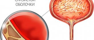
Ultrasound of the kidneys and adrenal glands is a study carried out using the echolocation method. Ultrasound is reflected differently by different tissues of the body, so using ultrasound you can obtain an image of internal organs, determine their position, size and changes in their structure. Ultrasound of the kidneys and adrenal glands is performed simultaneously, which is explained by the close proximity of these organs. At the same time, healthy adrenal glands are usually not clearly visible on ultrasound, which is explained by their acoustic properties: adrenal tissue reflects ultrasound to the same extent as the surrounding fatty tissue. But in the presence of pathological changes, the adrenal gland, as a rule, becomes clearly visible.
How is the ultrasound procedure performed?
Ultrasound waves provide data on the condition of internal organs. The results of the study help the attending physician confirm, clarify or refute the diagnosis and evaluate the effectiveness of the chosen treatment method.
An ultrasound of the kidneys is performed as follows:
- The patient lies on the couch on his side, then on his stomach;
- A special gel is applied to the lower back;
- The sensor touches the skin and slides over its surface;
- From the sensor, ultrasonic waves travel to the internal organs, and the reflected signal is displayed on the computer screen;
- The doctor takes the necessary measurements and prints out the examination results.
The procedure lasts from 15 to 20 minutes.
Carrying out the procedure
In our medical center, the ultrasound procedure is performed in a specially equipped room. First, you need to undress to the waist or expose the abdomen and lower back to provide the doctor with access to the area being examined. After this, the patient takes a lying position on a medical couch. An acoustic gel is applied to the area under study, which ensures the closest possible contact of the transducer with the skin, increases ultrasound absorption, and increases the ability of the transducer to slide. During the examination, the doctor can move the sensor over the skin to better visualize a particular organ. The doctor may ask the patient to change his body position: stand up, turn sideways, or lie on his stomach. After the procedure is completed, the remaining gel is removed with a sanitary napkin.
The sonologist enters the results of the study into a protocol,, if necessary, prints out a two-dimensional image of the organ, deciphers the study data, and draws up a conclusion. All stages of the examination take no more than half an hour.
In what cases is an ultrasound of the kidneys required?
Various disorders can occur in the urinary system - bladder, kidneys and adrenal glands:
- neoplasms: stones, polyps and cysts;
- inflammatory processes: cystitis and pyelonephritis;
- congenital features and injuries.
Symptoms may include: lower back pain, change in urine color, pain when urinating, swelling, abnormal urine tests.
Ultrasound of the urinary system and kidneys is almost always included in the preoperative examination.
Ultrasound of the kidneys and bladder: who needs it and what will it show?
Safety, accessibility, information content - these are the advantages of ultrasound examination, which is widely used in the diagnosis of diseases of various organs, including the urinary system. We talk with Anna Viktorovna Poskrebysheva, an ultrasound diagnostics doctor at Clinic Expert Orenburg, about who and for what purpose an ultrasound of the kidneys and bladder is performed.
— Anna Viktorovna, how urgent is the problem of kidney and bladder diseases?
“Based on the frequency with which patients of different genders and ages with such pathologies come to us, I can conclude that this problem is quite relevant today.
— What does an ultrasound of the kidneys and bladder show?
“This study allows us to see and evaluate the condition of the kidneys, adrenal glands and bladder. The ureters are normally not visible on ultrasound. If they are visualized, then this is a signal that there is an obstacle (stone, tumor) in them, accompanied by stagnation of urine.
First of all, we look at the presence of organs, since sometimes you can encounter, for example, the absence of one kidney. Then we evaluate their position, shape, structure, size. Next, we clarify whether there are pathologies: inflammatory processes, traumatic injuries, tumor and additional formations, calculi (stones). Ultrasound examination of the bladder also allows one to evaluate the functional activity of the kidneys by the release of urine from the ureters. That is, based on these data, it is possible to find out whether the kidney is involved in the formation of urine.
— What are the indications for this research method?
— Ultrasound of the kidneys and bladder is most often prescribed by the attending physician, based on the patient’s complaints and results of blood and urine tests. Changes in the frequency of urination, pain and pain when urinating, increased body temperature accompanied by lower back pain, changes in the color and amount of urine - all these signs may indicate a possible pathology of the kidneys and bladder. If the patient has a history of urolithiasis, as well as diseases associated with metabolic disorders (diabetes mellitus, gout, nephrocalcinosis - deposition of calcium salts in the kidney), he is also referred for an ultrasound scan of the kidneys and bladder.
Among other things, this research method is also prescribed for diseases of adjacent organs, in particular genital organs, to distinguish pathologies.
Read materials on the topic:
Why does my lower back hurt? We are convening a medical consultation “NOT the time to collect stones”: what is urolithiasis? Diabetes: how to identify and treat? “Disease of Kings” and more: what do we know about gout?
— Is special preparation required before performing an ultrasound of the kidneys and bladder?
— Special preparation is not required for examining the kidneys and adrenal glands. However, we suggest that patients with increased gas formation in the intestines (flatulence) follow a special diet 2-3 days before an ultrasound of the kidneys and bladder: it is necessary to exclude vegetables and fruits, whole grains, legumes, and whole milk from the diet. We also recommend taking sorbents and carminatives.
To better visualize the bladder, preparation is required. Ultrasound examination should be performed on a full bladder. That is, about an hour before the ultrasound, the patient should drink about a liter of water and enter the procedure with a feeling of a full bladder. If this is neglected, there is a high probability that the bladder will be indistinguishable from the intestines, and it will not be possible to conduct a high-quality diagnosis.
— How is an ultrasound of the kidneys and bladder done?
— The standard procedure involves examining these organs through the anterior abdominal wall using an ultrasound probe. We usually look at the patient in a lying position on the left side, then on the right side, and from the back - in a standing or sitting position. A special gel is applied to the study areas.
In some cases, we have to perform ultrasound examination transvaginally or transrectally. There is also a transurethral method, but this study requires special training of the doctor and special room conditions. The transvaginal and transrectal route is used for a thorough examination of the bladder in cases where the patient is overweight or flatulent. This is also advisable if there is a suspicion of urolithiasis and it is necessary to examine the exits of the ureters into the bladder.
I would like to emphasize that we first perform an ultrasound scan of the kidneys and bladder through the anterior abdominal wall for all patients, and only if necessary do we resort to the above additional methods.
Sometimes the attending physician who referred the patient asks for an assessment of the amount of residual urine. In this case, we look at the bladder in a full state and after it has been emptied.
— Anna Viktorovna, please tell us about the features of performing ultrasound of the kidneys and bladder in children
— I would like to note that there are no age restrictions for this study. In children, all organs are very clearly visible on ultrasound, so special preparation for the procedure is usually not required. The only exception is compliance with the condition of a full bladder. Young children rarely succeed in this, so examination of this organ may be difficult in them.
— Can this study be performed on pregnant women?
- Yes, you can. In any trimester of pregnancy.
— Are there any contraindications to ultrasound of the kidneys and bladder?
- Yes. Any ultrasound examination of internal organs, including the kidneys and bladder, cannot be performed in case of skin lesions in the area of study, for example, herpetic rashes, ulcerative defects, fresh postoperative scars, since the gel applied to the skin is non-sterile.
— Do I need a referral for an ultrasound scan of the kidneys and bladder?
- No, it is not necessary. Patients suffering from chronic diseases most often come directly to us and, with the diagnostic results, go for a consultation with their attending physician. In addition, patients with suspected pathologies of the kidneys and bladder can come for examination on the referral of a general practitioner, family doctor, urologist, gynecologist, etc.
Interviewed by Sevilya Ibraimova
If you need an ultrasound of the kidneys and bladder, you can sign up for the study here ATTENTION: the service is not available in all cities
The editors recommend:
We detect kidney stones. CT, ultrasound or MRI – what to choose? Urinalysis: frequently asked questions Pyelonephritis: what is this disease and how to treat it? A delicate problem: what you need to know about cystitis?
For reference:
Poskrebysheva Anna Viktorovna
Graduate of the Orenburg State Medical Academy in 2007 (currently Orenburg State Medical University)
In 2009, she completed clinical residency in the specialty “Skin and sexually transmitted diseases”
In 2011, she underwent professional retraining at the Faculty of Education and Training of Nizhny Novgorod State Medical Academy in the specialty “Ultrasound Diagnostics”
Currently, he is an ultrasound diagnostics doctor at the Expert Clinic in Orenburg. Receives at the address: Gagarin Ave., 17, room 1
How to prepare for a kidney ultrasound? Nutrition
Preparing for a kidney ultrasound involves following a diet. Three days before the examination, you must avoid foods that increase gas formation and possible inflammation. You cannot eat spicy, salty, sweet or fried foods. Cabbage, legumes and other foods that promote gas formation should also be excluded.
What you should not eat or drink before a kidney ultrasound:
- legumes;
- dried fruits;
- nuts;
- fatty meats and fish;
- sauerkraut, pickled tomatoes;
- sauces with mayonnaise;
- hot spices;
- bakery products;
- fast food;
- sausages;
- sweets;
- alcoholic drinks;
- carbonated drinks;
- mushrooms and dishes based on them;
- strong tea and coffee.
It is recommended to eat porridge - buckwheat and rice. You need to eat small portions 5-6 times a day. The last meal should be 8 hours before diagnosis. 30 minutes after it, you should take an absorbent, for example, activated carbon at the rate of 1 tablet per 10 kg of weight. Reducing gas formation in the intestines improves the visualization of the kidneys on ultrasound.
Decoding the result
Immediately after an ultrasound scan of the kidneys, a decoding of the norm in adults or children is provided to the patient. Usually this is a research protocol and several printed photographs. The protocol describes in detail the structure, shape, size and location of each kidney. The presence or presence of any inclusions or formations is also indicated. Based on the data obtained, the attending physician makes a diagnosis or prescribes additional examinations.
A significant number of kidney diseases are associated with changes in their size. Kidney sizes differ between children and adults.
Normal adult kidney size on ultrasound
First, we will describe normal kidney parameters for adults.
Kidney weight – 120-200 grams, length – 100-120 mm, width – 40-60 mm, thickness – 45-55 mm, thickness of the tissue enveloping the kidney (parenchyma) – no more than 23 mm. With age, the parenchyma becomes thinner and by old age it can reach 11 mm.
Normal kidney sizes in children on ultrasound
The size of the kidneys in children depends on age and height. If, for example, we take a child with a height of 50 to 80 cm, then only the length and width of the kidneys will be measured. As a rule, the sizes of the left and right kidneys differ. So, the width of the left kidney will normally be 2.2-2.5 cm, and the length - 4.8-6.2 cm. The width of the right kidney is 2.2-2.4 cm, length - 4.5-5, 9 cm
It must be borne in mind that in each case, the normal sizes for the child’s kidneys are determined individually, based on his physiological data.
Medication preparation
Patients who regularly take diuretics, on the day of the ultrasound, with the obligatory agreement of the doctor, should skip taking them until the examination is completed.
Medicines to normalize blood pressure and sugar levels should be taken as usual. They do not reduce the information content of the study.
In the acute phase of urolithiasis, before an ultrasound scan of the kidneys, you should take analgesics and antispasmodics prescribed by your doctor to reduce pain.
In medical, competent specialists perform ultrasound using modern expert-class equipment. We do everything to ensure that our patients receive quality services in a comfortable environment.
Call the phone number listed on the website, or leave a request in the feedback form. We will answer your questions and conduct all necessary examinations within one business day. Let's take care of your health together!
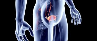
When is an ultrasound scan of the adrenal glands prescribed?
An ultrasound may be prescribed if adrenal disease is suspected. Symptoms of such diseases are:
- muscle weakness;
- weight change (loss or, conversely, excess weight gain);
- chronic hypertension (high blood pressure) or hypotension (low blood pressure);
- sexual dysfunction, decreased potency, menstrual irregularities, excess hair growth.
Ultrasound of the adrenal glands can reveal:
- cysts;
- tumors;
- hematomas;
- foci of inflammation;
- increase in the size of the adrenal glands (hyperplasia).
Where to get an ultrasound in Kazan: reviews from AM Medica patients
If you are interested in the question of where to get an ultrasound of the kidneys in Kazan, and you want to undergo diagnostics using new equipment under the supervision of a professional functional diagnostician, visit the medical one. We offer a full range of ultrasound diagnostic services at an affordable cost.
If you have never visited us before, the website provides a map of the route, the address, telephone numbers and email for contact. Make an appointment by calling the administrator and arrive at the designated time. We offer high service to our clients and guarantee impeccable quality of services provided.
For our patients, we have created a specialized service, where everyone who has undergone an ultrasound of the kidneys and urinary tract will leave reviews about their visit to the clinic on a specialized page of our website. Share your impressions of visiting the clinic, communicating with the doctor, suggestions and comments on improving our work. We will be glad if you decide to leave your feedback by sending it to the email address specified in the “Contacts” section.
Ultrasound diagnostics is a safe and painless method for studying internal organs, tissues, and structures. Thanks to this method, the doctor can quickly obtain the information he is interested in regarding the patient’s health status, in order to make further decisions on correcting health and improving well-being based on it. Ultrasound of the kidneys and bladder is a good method for studying a wide range of pathologies.
Abnormalities that can be visualized include changes in organ size, hydronephrosis, renal parenchymal pathologies, tissue scarring, and cystic formations. Ultrasound of the kidneys and bladder is performed in men and women, as well as in childhood. The use of Doppler scanning also allows one to examine the vascular system - the speed and direction of blood flow, the uninterrupted supply of blood.
Why is ultrasound the most informative research method and what diseases does it help identify?
Compared with the same palpation, of course, ultrasound is much more informative. The first method is also used, and yet the second is more reliable. In our IdealMed medical center, we use ultrasound scanners Aloka prosound alpha 6 and Mindray DC-70 - absolutely safe devices, you can conduct research with them as many times as needed.
They qualitatively and accurately visualize organs and systems and make it possible to identify pathologies almost immediately.
Using scanners, an ultrasound doctor can evaluate:
- location, shape, external size and number of organs;
- the thickness of the parenchyma and the state in which the pyelocaliceal system is located;
- are there any space-occupying formations (stones, cysts, nodes, foci of necrosis), and if so, what size are they;
- scars, recesses on the surface;
- condition of the renal arteries;
- the nature of the location and number of ureters.
And identify various diseases:
- urolithiasis;
- amyloidosis is a rare disease with a disorder of protein metabolism;
- polycystic disease - a congenital disease with the appearance of renal cysts;
- acute and chronic pyelonephritis;
- tumors;
- nephrosclerosis (“wrinkling” of the kidneys) of any etiology.
Pathologies that ultrasound can detect
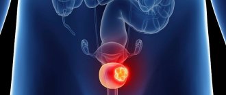
As a result of an ultrasound of the kidneys and bladder, the doctor can identify a tumor and perform a biopsy under the control of imaging equipment. In addition, thanks to the scanning device, pathologies such as the presence of cysts, stones, and obstacles to the normal outflow of urine become visible.
You can identify the size of the kidneys and urinary tract, their shape. If necessary, the study can be expanded; in addition, other visualization methods are used to provide more complete information about the detected changes. This may be MRI, CT and other types of examination.
How to keep your kidneys healthy?
To make them work like a clock, you need to follow a few simple rules.
Firstly, you need to monitor your weight and maintain it at normal levels.
Secondly, you need to set aside time for physical exercise every day.
Third, drink clean water whenever you feel thirsty. As for foods that are good for the kidneys, these are fish with fatty acids - salmon, tuna, green vegetables (spinach, broccoli), apples and paprika, olive oil.
Related articles:
- Breast ultrasound or mammography: which is better?
- What does ultrasound scan show?
What work do the kidneys do in our body?
This organ is called a blood purification “station”, since the main function it performs is the removal of waste products (what you eat), toxins and various harmful substances. If the process is delayed, over time you will begin to feel unwell and will need either treatment, diet, or both.
A few more important features:
- bone tissue formation;
- participation in phosphorus-calcium metabolism;
- regulation of blood pressure;
- participation in the formation of red blood cells (blood cells);
- regulating the amount of water and salts, maintaining water-salt balance in the human body.