Pulmonologist
Prokhina
Maria Egorovna
Experience 38 years
Pulmonologist
Make an appointment
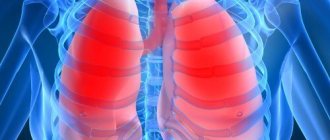
Pulmonary edema is a pathological, very serious condition, which is characterized by the release of transudate into the lung tissue. As a result, gas exchange is disrupted, which leads to serious consequences, including death.
Emergency care for pulmonary edema is the only thing that can increase the patient’s risks of survival and recovery. A person in such a situation requires immediate medical attention.
Pulmonary edema itself is most often a complication that accompanies serious problems of organs and body systems, for example, the cardiovascular system, gastrointestinal tract, etc.
Causes
In fact, there are many causes of pulmonary edema - they are different for different diseases. Let us name, for example, a few general prerequisites:
- cardiosclerosis after a heart attack, acute myocardial infarction;
- hypertension, arrhythmia;
- heart failure;
- congenital or acquired heart defects;
- chronic bronchitis, lobar pneumonia, bronchial asthma;
- complications due to ARVI, measles, influenza, scarlet fever, whooping cough and other diseases;
- prematurity in newborns;
- serious kidney problems;
- traumatic brain injury, brain surgery, etc.;
- inhalation of toxic substances.
These and many other reasons are not direct factors contributing to the development of pulmonary edema. But against the background of such conditions, it can develop, which is necessarily taken into account during hospitalization with all of the above.
BIBLIOGRAPHY
- Renkin E: Some consequences of capillary permeability to macro-molecules: tarling's hypothesis reconsidered. Am J Physiol 1986, 250:H706–H710.
- Staub N: The pathogenesis of pulmonary edema. Prog Cardiovasc Dis 1980, 23:53–80.
- Matthay MA: Pathophysiology of pulmonary edema. Clin Chest Med 1985, 6:301–314.
- Lewis FR, Elings VB, Sturm JA: Bedside measurement of lung water. J Surg Res 1979, 27:250–261.
- Sivak ED, Starr NJ, Graves JW, et al.: Extravascular lung water values in patients undergoing coronary artery bypass surgery. Crit Care Med 1982, 10:593–596.
- Sibbald WJ, Warshawski FJ, Short AK, et al.: Clinical studies of mea-suring extravascular lung water by the thermal dye technique in critically ill patients. Chest 1983, 83:725–731.
- Gallagher JD, Moore RA, Kerns D, et al.: Effects of advanced age on extravascular lung water accumulation during coronary artery bypass surgery. Crit Care Med 1985, 13:68–71.
- Bongard FS, Matthay M, Mackersie RC, Lewis FR: Morphologic and physiologic correlates of increased extravascular lung water. Surgery 1984, 96:395–403.
- Staub NC: Clinical use of lung water measurements. Report of a workshop. Chest 1986, 90:588–594.
- Schuster DP: The evaluation of pulmonary endothelial barrier function: quantifying pulmonary edema and lung injury. In: Pulmonary Edema. Edited by Matthay MA, Ingbar DH. New York: Marcel Dekker, Inc., 1998:121–161.
- Snashall PD, Keyes SJ, Morgan BM, et al.: The radiographic detection of acute pulmonary oedema. A comparison of radiographic appearances, densitometry and lung water in dogs. Br J Radiol 1981, 54:277–288.
- Gattinoni L, Presenti A, Torresin A, et al.: Adult respiratory distress syndrome profiles by computed tomography. J Thorac Imaging 1986, 1:25–30.
- Wheeler A, Carroll F, Bernard G: Radiographic issues in adult respi-atory distress syndrome. New Horiz 1993, 1:471–477.
- Halperin B, Feeley T, Mihm F, et al.: Evaluation of the portable chest roentgenogram for quantitating extravascular lung water in critically ill adults. Chest 1985, 88:649–652.
- Eisenberg PR, Hansbrough JR, Anderson D, Schuster DP: A prospecious study of lung water measurements during patient management in an intensive care unit. Am Rev Respir Dis 1987, 136:662–668.
- Hedlund LW, Vock P, Effmann EL, Lischko MM, Putman CE: Hydrostatic pulmonary edema. An analysis of lung density changes by computed tomography. Invest Radiol 1984, 19:254–262.
- Forster BB, Muller NL, Mayo JR, et al.: High-resolution computed tomography of experimental hydrostatic pulmonary edema. Chest 1992, 101:1434–1437.
- 18. Mayo JR: Magnetic resonance imaging of the chest. Where we stand. Radiol Clin North Am 1994, 32:795–809.
- Lancaster L, Bogdan AR, Kundel HL, McAffee B: Sodium MRI with coated magnetite: measurement of extravascular lung water in rats. Magn Reson Med 1991, 19:96–104.
- Cutillo AG, Morris AH, Ailion DC, et al.: Assessment of lung water distribution by nuclear magnetic resonance. A new method for quantifying and monitoring experimental lung injury. Am Rev Respir Dis 1988, 137:1371–1378.
- Morris AH, Blatter DD, Case TA, et al.: A new nuclear magnetic resonance property of the lung. J Appl Physiol 1985, 58:759–762.
- Cutillo AG, Morris AH, Ailion DC, Durney CH, Case TA: Determination of lung water content and distribution by nuclear magnetic resonance imaging. J Thorac Imag 1986, 1:39–51.
- Mayo JR, Muller NL, Forster BB, Okazawa M, Pare PD: Magnetic res-onance imaging of hydrostatic pulmonary edema in isolated dog lungs: comparison with computed tomography. Can Assoc Radiol J 1990, 41:281–286.
- Wexler HR, Nicholson RL, Prato FS, et al.: Quantitation of lung water by nuclear magnetic resonance imaging. A preliminary study. Invest Radiol 1985, 20:583–590.
- Phillips DM, Allen PS, Man SF: Assessment of temporal changes in pulmonary edema with NMR imaging. J Appl Physiol 1989, 66:1197–1208.
- Caruthers SD, Paschal CB, Pou NA, Roselli RJ, Harris TR: Regional measurements of pulmonary edema by using magnetic resonance imaging. J Appl Physiol 1998, 84:2143–2153.
- Rhodes CG: Measurement of lung water using nuclear magnetic resonance imaging [letter]. Br J Radiol 1986, 59:1135–1136.
- Cutillo A, Goodrich K, Krishnamurthy G, et al.: Lung water measure-me by nuclear magnetic resonance: correlation with morphome-try. J Appl Physiol 1995, 79:2163–2168.
- Mayo JR, MacKay AL, Whittall KP, Baile EM, Pare PD: Measurement of lung water content and pleural pressure gradient with magnetic resonance imaging. J Thorac Imag 1995, 10:73–81.
- Berthezene Y, Vexler V, Jerome H, et al.: Differentiation of capillary leak and hydrostatic pulmonary edema with a macromolecular MR imaging contrast agent. Radiology 1991, 181:773–777.
- Cutillo AG, Goodrich KC, Ganesan K, et al.: Alveolar air/tissue inter-face and nuclear magnetic resonance behavior of normal and edematous lungs. Am J Respir Crit Care Med 1995, 151:1018–1026.
- Schuster DP: Positron emission tomography: theory and its application to the study of lung disease. Am Rev Respir Dis 1989, 139:818–840.
- Schuster DP, Marklin GF, Mintun MA, Ter-Pogossian MM: PET measurement of regional lung density: 1. J Comput Assist Tomogr 1986, 10:723–729.
- Rhodes C, Hughes, JMB: Pulmonary studies using positron emission tomography. Eur Respir J 1995, 8:1001–1017.
- Schuster DP, Mintun MA, Green MA, Ter-Pogossian MM: Regional lung water and hematocrit determined by positron emission tomography. J Appl Physiol 1985, 59:860–868.
- Schuster DP, Marklin GF, Mintun MA: Regional changes in extravascular lung water detected by positron emission tomography. J Appl Physiol 1986, 60:1170–1178.
- Velazquez M, Haller J, Amundsen T, Schuster DP: Regional lung water measurements with PET: accuracy, reproducibility, and lin-earity. J Nucl Med 1991, 32:719–725.
- Spinale FG, Reines HD, Cook MC, Crawford FA: Noninvasive estimation of extravascular lung water using bioimpedance. J Surg Res 1989, 47:535–540.
- Zellner JL, Spinale FG, Crawford FA: Bioimpedance: a novel method for the determination of extravascular lung water. J Surg Res 1990, 48:454–459.
- Nierman DM, Eisen DI, Fein ED, et al.: Transthoraic bioimpedance can measure extravascular lung water in acute lung injury. J Surg Res 1996, 65:101–108.
- Adler A, Amyot R, Guardo R, Bates JHT, Berthiaume Y: Monitoring changes in lung air and liquid volumes with electrical impedance tomography. J Appl Physiol 1997, 83:1762–1767.
- Effros RM: Lung water measurements with the mean transit time approach. J Appl Physiol 1985, 59:673–683.
- Sivak ED, Wiedemann HP: Clinical measurement of extravascular lung water. Crit Care Clin 1986, 2:511–526.
- Allison RC, Carlile PV Jr, Gray BA: Thermodilution measurement of lung water. Clin Chest Med 1985, 6:439–457.
- Pfeiffer U, Backus G, Blumel G, et al.: A fiberoptics based system for integrated monitoring of cardiac output, intrathoracic blood volume, extravascular lung water, O 2 saturation, and a–v differ-ences. In: Practical Applications of Fiberoptics in Critical Care Monitoring. Edited by Lewis F, Pfeiffer U. Berlin: Springer-Verlag, 1990: 114–125.
- Schuster DP, Calandrino FS: Single versus double indicator dilution measurements of extravascular lung water. Crit Care Med 1991, 19:84–88.
- Sibbald WJ, Short AK, Warshawski FJ, Cunningham DG, Cheung H: Thermal dye measurements of extravascular lung water in critically ill patients. Intravascular Starling forces and extravascular lung water in the adult respiratory distress syndrome. Chest 1985, 87:585–592.
- Bock JC, Lewis FR: Clinical relevance of lung water measurement with the thermal-dye dilution technique. J Surg Res 1990, 48:254–265.
- Wickerts CJ, Jakobsson J, Frostell C, Hedenstierna G: Measurement of extravascular lung water by thermal-dye dilution technique: mechanisms of cardiac output dependence. Intensive Care Med 1990, 16:115–120.
- Fallon KD, Drake RE, Laine GA, Gabel JC: Effect of cardiac output on extravascular lung water estimates made with the Edwards lung water computer. Anesthesiol 1985, 62:505–508.
- Zeravik J, Borg U, Pfeiffer U: Efficacy of pressure support ventilation dependent on extravascular lung water. Chest 1990, 97:1412–1419.
- Haider M, Schad H: Effect of positive end-expiratory airway pressure (PEEP) on extravascular thermal lung water estimation in the dog. In: Practical Applications of Fiberoptics in Critical Care Monitoring. Edited by Lewis F, Pfeiffer U. Berlin: Springer-Verlag, 1990:96–104.
Varieties
There are different types of pulmonary edema:
- fulminant. It develops extremely quickly, in a few minutes - the outcome in this case is only fatal;
- spicy. Symptoms increase over four hours and the risk of death is very high. Such swelling often occurs with heart attack, suffocation, and traumatic brain injury;
- subacute. The development of symptoms alternates between active and quieter stages. Occurs in liver failure;
- protracted. It can develop within twelve hours, even several days, and not have a clear manifestation. It manifests itself in heart failure, as well as chronic lung diseases.
Obviously, each option requires different actions. If the patient can still be saved, speed of response will be a key factor.
Complications
The main thing that threatens pulmonary edema in an elderly person is the lack of oxygen in the tissues. Even if the disease has been stopped, the brain will experience serious changes, and the tissues of the heart and lungs will suffer.
Also among other serious consequences of suffering from pulmonary edema are:
- formation of congestion in the lungs;
- organ ischemia;
- emphysema.
Due to oxygen starvation, the patient’s memory will deteriorate, during the daytime he will constantly feel sleepy, general lethargy will be felt, and his mood will begin to deteriorate. You will have to carefully monitor your own condition in order to notice serious deterioration in time and consult a doctor.
Pulmonary edema in old age is a serious pathology. Even if the disease manifests itself in a protracted or subacute form, the risk of complications after therapy is high. An immediate or acute syndrome practically does not allow saving the patient. So at the first symptoms indicating the development of the disease, you need to undergo an examination and consult a doctor so that it is not too late.
Symptoms
It is possible to describe the symptoms of pulmonary edema only in general terms, since certain types of pathology occur with blurred characteristics. Signs include the following:
- severe weakness;
- shallow, very rapid breathing;
- dry cough;
- dry wheezing;
- severe shortness of breath;
- puffiness of the face and neck;
- bubbling breathing and moist wheezing;
- foam at the mouth with a pink tint;
- lethargy, confusion;
- shallow breathing;
- thready pulse.
Some signs of pulmonary edema contradict each other for the reason that everything can start with one condition and end with another. For example, rapid breathing occurs for several minutes or hours, and then it weakens. In the fastest and most dangerous forms of edema, the patient's death occurs from suffocation (asphyxia).
Are you experiencing symptoms of pulmonary edema?
Only a doctor can accurately diagnose the disease. Don't delay your consultation - call
Prognosis for pulmonary pleurisy
A small amount of exudate resolves on its own in 2-4 weeks. Pathology with the formation of purulent effusion requires more intensive and prolonged treatment. In its absence, the risk of complications increases - adhesions, pleurosclerosis, respiratory failure.
Patients who have had illnesses should undergo preventive examinations for 2-3 years after recovery. In order to prevent relapses, it is necessary to eliminate exposure to harmful factors, including toxic compounds and nicotine, avoid hypothermia, and eat a balanced diet.
Diagnostics
If the symptoms of pulmonary edema are not pronounced, additional studies are required in parallel with emergency care:
- biochemical screening. This is a blood test;
- study of blood gases;
- ECG, ultrasound of the heart;
- X-ray of the chest area;
- pulmonary artery catheterization.
In many cases, diagnosis of pulmonary edema is possible immediately - only based on the signs that appear visually in the patient and without additional examination.
Treatment
There is not and cannot be a single program for the treatment of pulmonary edema. Emergency care for a patient includes measures to reduce venous return to the heart and supply humidified oxygen. Often the patient is transferred to mechanical ventilation, and a tracheostomy may be performed.
Various drugs are also additionally administered: analgesics, diuretics, drugs that reduce pressure in the pulmonary circulation, drugs for the heart, antibacterial agents and much more. Further, treatment of pulmonary edema, if the attack is removed, comes down to treating the underlying disease that caused this pathology.
The prognosis for edema is very serious. Depending on the causes of pulmonary edema, mortality can range from 20 to 90%. The sooner the problem is identified, the higher the chances of recovery. But in many cases, the patient can only be saved by timely treatment of those diseases that contribute to the occurrence of pathology - such prevention of pulmonary edema, for example, is the only way to reduce the risks of the fulminant form of the disease, which in itself is already fatal and cannot be treated.
Health care
Emergency therapy includes the following procedures:
- Using an oxygen mask (oxygenation). If the case is urgent, then the supply of oxygen through a mask is replaced by artificial ventilation.
- The patient is given morphine as a pain reliever and sedative.
- Aminophylline is also introduced, which helps remove excess sodium from the body, dilate the bronchi and improve blood circulation in the renal glands.
- At the same time, doctors monitor the patient's blood pressure. If it exceeds the norm, then the patient is injected with sodium nitroprusside, and if the blood pressure is too low, then dobutamine is injected.
Further therapy includes taking medications. The doctor may prescribe:
- hormonal drugs;
- antibiotics;
- hepatoprotectors;
- antihistamines.
Questions and answers
What causes pulmonary edema?
There are dozens of factors that contribute to the development of this pathology. They are associated with other diseases of different body systems. Clinical recommendations for pulmonary edema are largely determined by the situation.
Can pulmonary edema lead to death?
Yes, the risk of death in the case of such a pathology is very high, and for the fulminant form it is the only possible outcome of events. This is an extremely dangerous condition that requires immediate professional help.
How to treat pulmonary edema?
This is done exclusively in medical institutions, most often in intensive care. Only doctors know everything you need to know about the symptoms and treatment of pulmonary edema in adults or children - the pathology cannot be eliminated without special knowledge and skills.
What are the features of pulmonary edema in children?
Unlike adults, this pathology occurs much less frequently in children due to other diseases. The underlying cause is usually allergies or exposure to toxins. Or the cause may be congenital anomalies of various body systems.
Stages of pathology development
The main stages of the development of the disease:
- The pressure in the capillaries will increase. During this process, the permeability of the capillary walls involved in the pulmonary circulation begins to sharply decrease, causing tissue to be excreted into the pulmonary interstitial tissue. Over time, the pulmonary alveoli will absorb a large amount of foreign fluid and lose the ability to participate in gas exchange. This is the reason why, when pulmonary edema occurs, elderly patients experience attacks of suffocation and their skin turns blue.
- Gradually, oncotic blood pressure will decrease.
- The alveolo-capillary membrane will lose its integrity.
Any suspicious symptoms indicating an exacerbation of the pathology should be accompanied by a visit to a doctor. At the first sign you need to call an ambulance.