Ascites - what is it?
Ascites is a variety of complications caused by a number of diseases.
This pathology reveals itself as the accumulation of a large amount of liquid mass inside the peritoneal cavity. Hence the second name “edema of the abdomen”. The accumulated liquid mass increases the volume of the abdomen, causing unpleasant symptoms and secondary dysfunctions of the organs of the peritoneal cavity.
In such a condition, medical intervention is simply necessary, especially if the liquid mass accumulates rapidly.
Manifestations
A small amount of liquid mass accumulated in the abdominal cavity does not reveal itself in any way. How to determine the onset of the disease, since there are no symptoms? Ultrasound comes to the rescue here.
As the disease progresses, a feeling of heaviness appears in the abdomen, and the lower part is subject to dull aching pain. Next comes difficulty breathing, after which there is indigestion in its various varieties, and urination is impaired.
A severe form of pathology can dramatically worsen your health. The state of health leaves much to be desired:
- Unpleasant sensations in the stomach become the norm;
- Shortness of breath appears;
- An umbilical hernia develops;
- Legs swell;
- Internal organs are compressed.
Medical practice is faced with the fact that some patients accumulate up to twenty liters of fluid. As a result, they have:
- Increased pressure inside the peritoneum;
- The diaphragm becomes pressed into the chest cavity;
- Significant difficulty breathing;
- Heart failure develops.
If the order of things is not restored, the following are possible:
- Impaired drainage of the lymphatic system;
- The occurrence of impaired lymphatic drainage in the legs;
- Swelling of the extremities;
- Outflow of lymph to internal organs.
The result of such changes can be disastrous. Atypical cells penetrate from the affected lymph nodes to other organs.
Pathology in a small form can be determined during a medical examination: the abdomen is enlarged, it is saggy when a person is standing, or flattened when a person is lying down. In thin patients, the navel often protrudes.
Determining the pathology at the very beginning of its development is important, since its moderate form is diagnosed in almost half of patients in the early stages of oncology.
Reasons for development
Ascites is a serious complication of stomach and colon cancer, colorectal cancer, malignant tumors of the pancreas, and oncological pathology of the mammary glands, ovaries and uterus. When a large volume of fluid accumulates in the abdominal cavity, intra-abdominal pressure increases, and the diaphragm moves into the chest cavity. This leads to disruption of the heart and lungs. There is a violation of blood circulation in the vessels.
In the presence of ascites, the patient's body loses a large amount of protein. Metabolism is disrupted, heart failure and other imbalances in the internal environment of the body develop, which worsen the course of the underlying disease.
There is always a small amount of fluid in the abdominal cavity of a healthy person. It prevents the sheets of peritoneum from sticking together. The produced intra-abdominal fluid is reabsorbed by the peritoneum.
With the development of cancer, the normal functioning of the body is disrupted. The secretory, resorptive and barrier functions of the peritoneal layers fail. In this case, either excess fluid production or disruption of its absorption processes may be observed. As a result, a large volume of exudate accumulates in the abdominal cavity. It can reach twenty liters.
The main reason for damage to the peritoneum by malignant cells is its close contact with organs that are affected by a cancerous tumor. Ascites in the presence of oncological pathology develops under the influence of the following factors:
- A large accumulation of blood and lymphatic vessels in the peritoneum through which cancer cells spread;
- Tight fit of the folds of the peritoneum to each other, which contributes to the rapid spread of malignant cells to adjacent tissues;
- Germination of a cancerous tumor through the peritoneal tissue;
- Transfer of atypical cells to peritoneal tissue during surgery.
Chemotherapy may be the cause of ascites. The accumulation of fluid in the peritoneum occurs due to cancer intoxication. If the liver is affected by a primary cancerous tumor, metastases of malignant cells from tumors of a different location, the outflow of blood through its venous system is disrupted, and portal hypertension develops - an increase in pressure inside the portal vein. The lumen of the venous vessels increases, plasma sweats from them and accumulates in the abdominal cavity.
The cause of ascites may be peritoneal carcinomatosis. In the presence of a cancerous tumor of the abdominal organs, atypical cells settle on the parietal and visceral sheets of the peritoneum. They block the resorptive function, as a result of which the lymphatic vessels do not cope well with the intended load, and lymph outflow is impaired. Free fluid gradually accumulates in the abdominal cavity. This is the mechanism of development of carcinomatous ascites.
Make an appointment
Causes
The basis for the development of the disease is pathology: during the normal functioning of the peritoneal cavity, no significant amount of liquid mass is discharged. It is only insignificant, just to avoid adhesions from sliding intestinal loops. The liquid released for this purpose constantly flows back.
When the usual operation of this mechanism is disrupted, the function of secretion of the liquid mass is destabilized, as well as the function of its outflow. The consequence of such destabilization is the accumulation of excess liquid mass inside the abdomen.
Causes of pathology in adults
In many cases, pathology develops as a symptom of a number of diseases, including:
- Portal hypertension;
- Cirrhosis;
- Hepatitis;
- Thrombosis of the liver veins.
It is possible that the cause of the pathology is blood oncology, as well as pathologies whose nature has nothing in common with the tumor.
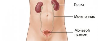
Heart failure can also cause pathology. Problems associated with lymph circulation and the activity of organs such as the thyroid gland and kidneys can also cause pathology.
Protein deficiency
Diagnostic methods
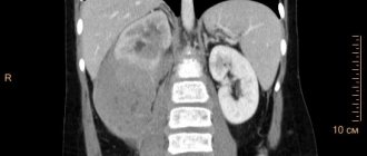
A certain amount of accumulated liquid mass (more than half a liter) can be determined during a medical examination. To confirm a preliminary diagnosis, an ultrasound is necessary.
The main problem is not to detect the liquid, but to identify the cause of its accumulation. Only then will the treatment be effective. To do this, it is necessary to conduct laboratory tests, namely:
- Blood clotting test;
- Biochemistry analysis;
- Analysis of ascitic fluid obtained during laparocentesis
Symptoms
Symptomatic manifestations of the disease depend on three main factors - the cause, the volume of accumulated liquid mass, and the rate of accumulation.
Symptoms can appear at any time: sometimes it’s weeks and months, sometimes hours.
As we have already noted, one of the visual symptoms is an enlarged abdomen. Let's add weight gain here.
The patient may develop a feeling of bursting pain, flatulence, belching, heartburn, and nausea.
An increase in the volume of the abdomen entails the appearance of severe shortness of breath, swelling of the extremities, difficulty moving and bending.
Another number of symptoms that a patient may encounter are hemorrhoids, hernias, intestinal prolapse, and the development of varicocele.
Common symptoms of the disease include:
- Feverish state;
- Toxic state;
- Losing weight and at the same time growing belly;
- The saphenous veins of the abdomen are dilated;
- Blueness on the limbs.
Types of disease
In the main classification, to differentiate pathology, the level of protein in the accumulated fluid is taken into account. According to this indicator, the disease is divided into:
- Exudative (25 g/l or more);
- Transudative (<25 g/l).
Such a division makes it possible to understand, albeit indirectly, what is the cause of the development of the disease.
Today, another indicator is used in medical practice - SAAG.
If this gradient is > 1.1, then most likely the cause of the disease is cirrhosis, heart failure, etc. All of them increase the pressure in the venous trunk.
If the gradient index is <1.1, then the cause of the disease can be considered pancreatitis or an oncological process.
Taking into account the clinical course of the disease, it is necessary to highlight the following varieties, which will be discussed below.
- Severity of the current:
- First degree. There are no symptoms, diagnosed by examination and ultrasound.
- Second degree. The abdomen is slightly enlarged.
- Third degree. The abdomen is significantly enlarged.
- An uncomplicated option. There are no signs of infection of the accumulated fluid mass and abnormalities in kidney function.
- Refractory ascites:
- Unaffected by diuretics;
- Not controlled by diuretics.
Publications in the media
Ascites is an accumulation of fluid in the abdominal cavity. It can occur in any condition accompanied by generalized edema. In adults, ascites most often occurs with liver cirrhosis, heart defects, and nephrotic syndrome. In children, ascites is more often observed with nephrotic syndrome and malignant neoplasms.
Etiology and pathogenesis • Increased hydrostatic pressure •• Liver cirrhosis •• Hepatic vein occlusion (Budd-Chiari syndrome) •• Obstruction of the inferior vena cava •• Constrictive pericarditis •• Congestive heart failure •• Heart defects (stenosis or tricuspid valve insufficiency) • Decrease colloid osmotic pressure (albumin content <20 g/l) •• End-stage liver disease with decreased protein synthetic function •• Nephrotic syndrome with protein loss •• Nutritional disorders •• Enteropathies with protein loss •• Protein starvation • Increased permeability of peritoneal capillaries • • Tuberculous peritonitis •• Bacterial peritonitis •• Malignant diseases of the peritoneum •• Metastases into the peritoneum (ovarian, colon, pancreatic cancer, etc.) •• Obstruction of the lymphatic tract (leukemia, lymphoma) • Leakage of fluid into the abdominal cavity •• Chylous ascites (secondary to rupture of the lymphatic duct due to lymphoma or trauma) •• Urinary ascites • Other causes •• Myxedema •• Meig's syndrome •• Chronic hemodialysis.
Type of fluid contained in the abdominal cavity • Transudate (for congestive heart failure, constrictive pericarditis, liver cirrhosis, nephrotic syndrome, hypoalbuminemia) •• Indicators characteristic of transudate: ••• Protein <2.5 g% • Relative density 1.005–1.015 ••• Albumin/globulin ratio: 2.5–4.0 ••• Leukocytes up to 15 per field of view ••• Rivalta test is negative • Exudate (for tumor, tuberculosis, pancreatitis, myxedema, biliary pathology, Budd-Chiari syndrome) •• Indicators characteristic of exudate: ••• Protein >2.5 g% ••• Relative density >1.015 ••• Albumin/globulin ratio: 0.5–2.0 ••• Leukocytes over 15 in the field of view • •• The Rivalta test is positive.
Clinical picture • Discomfort or pain in the abdomen • Increased abdominal volume • Increased body weight • Anorexia, nausea, heartburn • Quick satisfaction of hunger while eating • Increased body weight • Dilated veins on the anterior abdominal wall (portocaval and cavacaval anastomoses) • Dullness to percussion sound in the lateral parts of the abdomen, moving when the body position changes (with a volume of ascitic fluid of at least 2 liters) • Edema of the penis, scrotum, lower extremities • Formation of umbilical, inguinal, femoral hernia • With tense ascites - a positive symptom of fluctuation • Shortness of breath, sometimes orthopnea • Formation of pleural effusion, wheezing in the lungs may be heard • Swelling of the neck veins.
Diagnostics. Ascites is indicated by abdominal enlargement, a positive symptom of fluctuation, or moving dullness detected by physical methods. An ultrasound scan reveals fluid in the peritoneal cavity. Paracentesis can be performed followed by analysis of ascitic fluid • The diagnostic sign of exudative ascites is an increase in the total protein content in the serum of more than 2.5 g%; usually seen with tumors, infections and myxedema. The difference between the level of albumin in the serum and the protein content in ascitic fluid less than 1 g/l indicates a high probability of the malignant nature of ascites, more than 1.1 g% indicates the presence of portal hypertension • With pancreatic ascites, the amylase content in the exudate is increased • With chylous ascites, it is increased concentration of fat (in the form of chylomicrons), chylous ascites develops with cirrhosis of the liver or lymphoma • Malignant tumors are detected by cytological studies of ascitic fluid, malignant ascites is also characterized by an increase in cholesterol content above 50 mg% • The number of leukocytes in ascitic fluid, exceeding 500 / μl, suggests presence of infection. A predominance of neutrophils suggests a bacterial infection, a predominance of lymphocytes is most likely due to tuberculosis or fungal infection • A red blood cell count greater than 50,000/µl indicates hemorrhagic ascites, usually due to malignancy, tuberculosis or trauma. Hemorrhagic pancreatitis, ruptured aortic aneurysm or liver tumors can cause overt bleeding into the abdominal cavity • The presence of a bacterial infection is confirmed by bacteriological examination of the exudate • A pH of ascitic fluid <7 suggests the presence of a bacterial infection.
Laboratory tests • Ascitic fluid •• Indicators that must be determined: ••• Total number of cells ••• Number of neutrophils ••• Total protein ••• Culture for cultivation (at least 10 ml) •• Indicators that facilitate diagnosis: • •• LDH content ••• Amylase content ••• Cultivation of acid-resistant and fungal flora ••• Cytology ••• Triglyceride content •• Additional studies of ascitic fluid ••• Helminths, talc granules ••• Presence of urine, blood ••• Embryonic oncological Ag >10 ng/ml (10 μg/l) • Blood - creatinine (<1.4 mg%), electrolytes • Urine •• sodium content in one sample: ••• <10 mEq/l (diuretics are ineffective) • •• 10–70 mEq/L (diuretics prescribed) ••• >70 mEq/L (diuretics not indicated).
Special studies • Laparoscopy • Ultrasound or CT • Diagnostic paracentesis.
TREATMENT depends on the cause of ascites.
A low sodium diet bread, etc.) •• Pickles, marinades, canned food, ham, pates, sausages, cheeses, sauces, mayonnaise, ice cream •• Candies, marshmallows, milk chocolate •• All cereals, except semolina and rice • Allowed •• Salt-free bread and butter •• Beef, rabbit meat, chicken, fish (100 g/day), one egg/day •• Sour cream, milk (1 glass/day) •• Fresh vegetables and fruits or in the form of compote.
Drug therapy
• If the daily sodium excretion is 5–25 mmol, potassium-sparing diuretics are prescribed: spironolactone 100–200 mg/day • After 4 days of treatment, it is necessary to consider the indications for prescribing furosemide 80 mg/day.
• When daily sodium excretion is less than 5 mmol, potassium-sparing and loop diuretics are prescribed - furosemide 40-160 mg/day every other day in combination with potassium chloride - 50 mmol potassium per day.
• While the patient has edema, daily diuresis of up to 3 liters is safe (weight loss of no more than 1.0 kg/day is acceptable) • After the edema disappears, daily diuresis should not exceed 800–900 ml (optimal weight loss is about 0.5 kg/day).
• In case of tense ascites, it is necessary to consider the indications for therapeutic paracentesis •• Tense ascites •• Ascites with edema • Contraindications to therapeutic paracentesis •• Cirrhosis of the Child group C liver •• Blood bilirubin above 170 µmol/l •• Prothrombin index (PTI) below 40 % •• Platelet count less than 40´109/l •• Blood creatinine above 3 mg% •• Daily sodium excretion less than 10 mmol.
• Therapeutic paracentesis •• The volume of fluid removed is 5–10 liters •• Simultaneously with the removal of fluid, salt-free albumin must be administered intravenously - 6 g per 1 liter of fluid removed.
Surgery. For chronic ascites that cannot be treated, abdominal-jugular shunting (Levine shunt) is possible, but there is a high risk of infection and disseminated intravascular coagulation.
Complications and their treatment • Spontaneous bacterial peritonitis •• Develops in 8% of patients with liver cirrhosis with ascites •• 70% of patients develop abdominal pain, fever, abdominal tenderness on palpation, sharp deterioration of condition •• Protein concentration in ascitic fluid is usually less than 1 g% •• Most often caused by a gram-negative pathogen from the intestinal group •• Immediate administration of antibacterial therapy is necessary when the number of neutrophils in ascitic fluid is more than 250 per μl •• Parenteral administration of third-generation cephalosporins, oral fluoroquinolones is effective • Development of hepatorenal syndrome (see Hepatorenal syndrome) .
Prevention • Do not force diuretic therapy!
Course and prognosis • The prognosis depends on the cause of ascites • In liver cirrhosis, the prognosis is unfavorable (two-year survival rate - 40%) • The presence of hepatic cell failure significantly worsens the prognosis • Mortality in spontaneous bacterial peritonitis reaches 50%, developed hepatorenal syndrome - 95%.
ICD-10 • R18 Ascites
Pathology and cancer
Medical statistics and practice show that the most common diseases leading to the accumulation of liquid mass in the peritoneal cavity are oncological forms:
- Ovaries;
- Mammary glands;
- Uterus;
- Stomach;
- Colon.
A liquid mass accumulates with a tumor due to damage to the peritoneal lining. Atypical cells, having settled on it, cause disruption of lymphatic drainage, which, in turn, impairs fluid absorption. This is very common, for example, in gastrointestinal oncology.
If we take an organ such as the liver, the picture will be different. A tumor or metastasis localized in an organ causes compression of the venous system of the organ and disruption of normal venous outflow from the intestine. In this case, the pathology develops rapidly and is more severe. About fifteen percent of ascites in cancer processes is diagnosed in this form.
Forecast
Ascites in cancer significantly worsens the general well-being of the patient. As a rule, such a complication occurs in the later stages of oncology, in which the survival prognosis depends on the nature of the tumor itself and its distribution throughout the body.
Life expectancy with ascites depends on the following factors:
- Functioning of the kidneys and liver;
- Activities of the cardiovascular system;
- The effectiveness of therapy for the underlying disease.
The development of ascites can be prevented by an experienced doctor observing the patient. Doctors at the Yusupov Hospital have extensive experience in dealing with various types of cancer. Qualified medical personnel and the latest equipment allow for accurate diagnosis and high-quality, effective treatment in accordance with European standards.
Specifics of treatment for cancer
Conservative methods are effective in treating pathologies of only minor and moderate severity. The diuretics used here allow you to remove up to a liter of liquid mass per day.
However, the surgical option is preferable. It is used when:
- The disease cannot be treated by other means;
- A significant form of the disease, in which it is necessary to evacuate up to ten liters of liquid mass at a time;
- A form of the disease that requires combined manipulation, including evacuation of a volume of liquid mass of up to seven liters on the first day and evacuation of the remaining mass at a rate of no more than one liter per day for a week.
The operation is performed on an empty stomach and with an empty bladder. The patient is positioned in a sitting position (lying down is possible).
Before the puncture, local anesthesia is performed, then the puncture is performed under ultrasound control.
As a rule, no more than five liters of liquid mass are evacuated at a time. To maintain blood pressure, it is pumped slowly.
It is important for the patient to lie on the side free from the puncture for several hours. If during this time all the planned liquid mass is not evacuated and continues to accumulate, a special reservoir is applied.
When removing large amounts of fluid, it is important that the patient does not experience protein deficiency. To do this, the patient is injected with albumin.
In modern practice, special catheters are used to evacuate accumulated fluid, and some patients undergo the procedure of omentohepatophrenopexy. This is an important part of palliative treatment.
Treatment
Drug therapy for ascites is not carried out due to low effectiveness. Aldosterone antagonists and diuretics normalize water-salt metabolism and prevent excess secretion of peritoneal fluid. Oncologists at the Yusupov Hospital offer palliative surgery to patients with ascites in the late stages of cancer:
- Omentohepatophrenopexy;
- Deperitonization;
- Installation of a peritoneovenous shunt.
Doctors at the Oncology Clinic carry out traditional or intracavitary chemotherapy for ascites - after removing the fluid, a chemotherapy drug is injected into the abdominal cavity. Laparocentesis is performed to remove fluid. The procedure is not performed if the following contraindications are present:
- Adhesive process inside the abdominal cavity;
- Severe flatulence;
- Perforation of the intestinal walls;
- Purulent infectious processes.
Laparocentesis is prescribed in cases where taking diuretics does not lead to a positive result. The procedure is also indicated for resistant ascites.
Laparocentesis is carried out in several stages using local anesthesia:
- the patient is in a sitting position, the doctor treats the subsequent puncture site with an antiseptic and administers painkillers;
- An incision is made in the abdominal wall along the linea alba at a distance of 2-3 centimeters below the navel;
- The puncture itself is performed using a trocar using rotational movements. A special flexible tube is attached to the trocar, through which excess fluid is removed from the body. The fluid is pumped out quite slowly, and the doctor constantly monitors the patient’s condition. As the exudate is removed, the nurse tightens the patient's abdomen with a sheet to slowly reduce the pressure in the abdominal cavity;
- After the fluid is pumped out, a sterile bandage is applied to the wound.
Using laparocentesis, up to 10 liters of fluid can be removed from the patient’s body. In this case, it may be necessary to administer albumin and other drugs to prevent the development of renal failure.
If necessary, temporary catheters can be installed in the abdominal cavity, through which excess fluid will gradually be removed. It should be noted that the use of catheters can lead to a decrease in blood pressure and the formation of adhesions.
There are also contraindications to laparocentesis. Among them:
- pronounced flatulence;
- adhesive disease of the abdominal organs;
- stage of recovery after surgery on a ventral hernia.
Diuretics are prescribed to patients with developing ascites in cancer over a long course. Such drugs as Furosemide, Diacarb and Veroshpiron are effective.
When taking diuretics, medications containing potassium must also be prescribed. Otherwise, there is a high probability of developing disturbances in water and electrolyte metabolism.
Dietary nutrition primarily involves reducing the amount of salt consumed, which retains fluid in the body. It is also important to limit the amount of fluid you drink. It is recommended to include more foods containing potassium in your diet.
After removal of fluid from the abdominal cavity, patients are provided with a balanced and high-calorie diet. This allows you to meet the body's needs for proteins, carbohydrates, vitamins and minerals. Fat consumption is reduced.