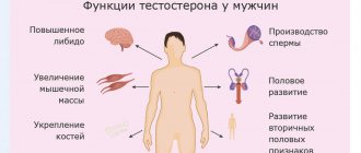
The role of estradiol in the body of men and women
Estradiol in women has the following effects on the body:
• Increased voice timbre
• Features of the figure in the form of a narrow waist and fat distribution in the hips and buttocks;
• Estradiol in women affects the condition of the skin, making it thin and smooth;
• The growth of the follicle in the ovary, as one of the most important stages of the reproductive system, is also under the influence of this hormone;
• An increase in the volume of the uterine mucosa, its preparation for egg implantation and pregnancy;
• Regulation of the menstrual cycle.
If estradiol is low, all these functions that are essential for a woman’s health will be impaired.
Estradiol is also produced in men, but not in such large quantities as in women. Its main effects in men:
• Improvement of calcium deposition in bones, so low estradiol can lead to osteoporosis - thinning of bone tissue, increased bone fragility;
• Improving cell respiration and oxygen metabolism in the body;
• Participation in the production of seminal fluid, therefore, if estradiol is low, this can lead to decreased sperm quality and infertility;
• Stimulation of metabolism;
• Increased blood clotting;
• Reducing the level of “bad” cholesterol in the blood and protecting blood vessels and the heart from atherosclerosis and other diseases.
Most of these effects also apply to women's health. Therefore, if estradiol is increased or decreased, specialist supervision and consultation with an endocrinologist or gynecologist are required. Tests for estradiol, the norm of which depends on many factors, should only be deciphered by a doctor who takes into account the characteristics of your age, health, etc., and selects treatment based on objective data obtained.
PROLACTIN
Prolactin is a hormone produced in the anterior pituitary gland; a small amount is synthesized in peripheral tissues, and during pregnancy it is also produced in the endometrium. This hormone plays an extremely important role in many processes occurring in the body, in particular, in ensuring the normal functioning of the reproductive system. Prolactin also contributes to the formation of sexual behavior. It regulates water-salt metabolism, delaying the excretion of water and sodium by the kidneys, stimulates calcium absorption, and has a modulating effect on the immune system. In general, prolactin activates anabolic processes in the body. The daily secretion of prolactin has a pulsating character. During sleep, its level increases. After waking up, the concentration of prolactin decreases sharply, reaching a minimum in the late morning hours. After noon, the hormone level increases. In the absence of stress, daily fluctuations in levels are within normal values. During the menstrual cycle, prolactin levels are higher in the luteal phase than in the follicular phase. From the 8th week of pregnancy, prolactin levels increase, reaching a peak at 20-25 weeks, then decrease immediately before childbirth and increase again during lactation. During pregnancy, prolactin supports the existence of the corpus luteum and the production of progesterone, stimulates the growth and development of the mammary glands and milk formation. An increase in prolactin levels is one of the common causes of infertility. Indications for the analysis: galactorrhea, cyclical pain in the mammary gland, mastopathy, anovulation, oligomenorrhea, amenorrhea, dysfunctional uterine bleeding, infertility, diagnosis of sexual infantilism, chronic inflammation of the internal genital organs, severe menopause, obesity, decreased libido and potency (men) , gynecomastia (men), osteoporosis. The analysis shows an increase in prolactin levels in diseases of the hypothalamus, pituitary gland, primary hypothyroidism, polycystic ovary syndrome, chronic renal failure, cirrhosis of the liver, adrenal insufficiency and congenital dysfunction of the adrenal cortex; tumors that produce estrogen and other conditions. The level of prolactin in the blood is usually reduced in case of pituitary insufficiency, true post-term pregnancy. Preparation for the study: Avoid sexual intercourse and heat exposure (sauna) 1 day before, smoking 1 hour before. Since prolactin levels are greatly influenced by stressful situations, it is advisable to exclude factors that influence the research results: physical stress (running, climbing stairs), emotional arousal. Therefore, before the procedure you should rest for 10-15 minutes and calm down.
How to get tested for estradiol
The day before the test, you should avoid smoking and sports training. In women, it is advisable to take blood samples on strictly defined days of the cycle - on days 6-7. In some cases, the period may be changed by the doctor if necessary.
They donate blood for estradiol in the morning and on an empty stomach, from 8 to 11 o’clock. You cannot eat for 8-14 hours before the test, you can only drink water. For the accuracy of the results obtained, it is also important not to overeat the day before the test.
When analyzing estradiol, the norm in women and men may vary and the result is not always accurate, so a little preparation for the study is required.
SHBG (SEX HORMONE BINDING GLOBULIN)
Most of the testosterone entering the blood binds to a specific transport protein - SHBG. A decrease in SHBG synthesis leads to disruption of the delivery of hormones to target organs and the performance of their physiological functions. In men, SHBG secretion increases with age. This can lead to a decrease in active testosterone and an increase in the effects of estrogen, which is manifested by gynecomastia (enlargement of the mammary glands in men) and redistribution of fatty tissue according to the female type. In women, the SHBG content is almost twice as high as in men, since estrogens increase the level of SHBG synthesis in the liver, and androgens, on the contrary, reduce its production. Indications for the analysis: In both sexes: clinical signs of an increase or decrease in androgen levels with normal testosterone levels, baldness, acne, oily seborrhea. In women: hirsutism, anovulation, amenorrhea, polycystic ovary syndrome, prediction of the development of preeclampsia (SHBG is reduced). In men: male menopause, chronic prostatitis, impaired potency, decreased libido. An increase in SHBG levels can be observed with thyrotoxicosis, liver cirrhosis, taking estrogens and oral contraceptives. A decrease in SHBG levels can occur with hypothyroidism, obesity, acromegaly, Cushing's syndrome, hyperprolactinemia, polycystic ovary syndrome, and after taking androgens. Preparation for the study: on an empty stomach.
Estradiol is normal in women and men
For the hormone estradiol, the norm in men ranges from 15 to 71 pg/ml. For the hormone estradiol, the norm in women varies depending on the phase of the cycle. When the cycle begins, hormone production is activated. Estradiol becomes elevated before ovulation and, when the concentration reaches its maximum, ovulation occurs. After the follicle ruptures and the egg is released, the norm for estradiol in women again corresponds to the beginning of the cycle.
Thus, in the follicular phase, estradiol can be increased to 227 pg/ml. In the preovulation phase, estradiol in women ranges from 127 to 476 pg/ml. Finally, the luteinizing phase of the cycle again returns estradiol levels to the range from 77 to 227 pg/ml. If estradiol is elevated after ovulation and remains this way, it can be assumed that pregnancy has occurred.
The estradiol norm in women during menopause decreases significantly and amounts to 19.7 – 82 pg/ml.
We must not forget about fluctuations in hormone levels throughout the day. Estradiol is reduced between 24 and 2 hours. Estradiol is usually elevated between 15 and 18 hours.
References
- Lønning, P. Estradiol measurement in translational studies of breast cancer. Steroids, 2015. - Vol. 99(Pt A). — P. 26-31.
- Butera, P. Estradiol and the control of food intake. PhysiolBehav., 2010. —Vol. 99(2). - P. 175-80.
- Vesper, H., Botelho, J., Wang, Y. Challenges and improvements in testosterone and estradiol testing. Asian J Androl., 2014. - Vol. 16(2). — P. 178-84.
- Rosner, W., Hankinson, S., Sluss, P. et al. Challenges to the measurement of estradiol: an endocrine society position statement. J Clin Endocrinol Metab., 2013. - Vol. 98(4). - P. 1376-87.
Consequences of disturbances in estradiol levels
Low estradiol in men provokes osteoporosis, heart disease, increased excitability and low reproductive ability. It can occur with chronic prostatitis, or for other reasons.
Low estradiol in women causes a prolonged absence of menstruation, dry skin and mucous membranes, discomfort during sexual intercourse, problems with conception (even infertility), and a decrease in the size of the uterus and mammary glands. The level of the hormone estradiol in women is reduced in the early stages of pregnancy.
PROGESTERONE
Progesterone is a female sex hormone produced in the corpus luteum of the ovaries and adrenal glands. Outside of pregnancy, progesterone secretion begins to increase in the preovulatory period, reaching a maximum in the middle of the luteal phase, returning to its original level at the end of the cycle. The content of progesterone in the blood of a pregnant woman increases, doubling by 7-8 weeks, and then gradually increasing until 37-38 weeks. After ovulation - the release of an egg from the follicle - a corpus luteum is formed in its place in the ovary - a gland that secretes progesterone. It exists and secretes this hormone during 12-16 weeks of pregnancy until the moment when the placenta is fully formed and takes over the function of hormone synthesis. If conception does not occur, the corpus luteum dies after 12-14 days, and menstruation begins. Progesterone is determined to assess ovulation and the viability of the corpus luteum. With a regular cycle - a week before menstruation, when measuring rectal temperature - on the 5-7th day of its rise; with an irregular cycle - several times. A sign of ovulation and the formation of a full-fledged corpus luteum is a tenfold increase in progesterone levels. Indications for the analysis : identifying the causes of menstrual irregularities, infertility, dysfunctional uterine bleeding, assessing the condition of the placenta in the second half of pregnancy, differential diagnosis of true post-term pregnancy. An increase in progesterone levels is observed with adrenal hyperplasia, corpus luteum cyst, pregnancy, and delayed maturation of the placenta. The analysis will show a decrease in progesterone levels in the absence of ovulation, insufficiency of the corpus luteum, true postmaturity, placental insufficiency, intrauterine growth restriction, or threatened abortion. Preparation for the study: the analysis is carried out on the 22-23rd day of the menstrual cycle, unless other dates are indicated by the attending physician. Blood is taken in the morning on an empty stomach, that is, when 8-12 hours pass between the last meal and blood collection. You can drink water.
Features of the study
Analysis for LH and FSH is necessary for diagnosing infertility, identifying the causes of menstrual disorders, determining ovulation, and monitoring hormonal stimulation during IVF.
For analysis, the doctor assigns the woman a specific date, approximately the 3-7th day of the onset of menstruation. Preparation for the procedure is carried out 3 days in advance. At this time, physical and psycho-emotional stress, sports activities, physiotherapy, thermal procedures, and intimacy are excluded. Venous blood is collected in the morning on an empty stomach; smoking and drinking water are prohibited.
To determine the level of LH and FSH, serum or blood plasma is analyzed using the immunochemiluminescence method, which allows even frozen samples to be examined.
If a woman has an irregular, too long or short cycle, then a single test may produce false results. To get more reliable results, do 2-3 samples at half-hour intervals. For analysis, samples of all sera are combined.
The results of studies of the hormone FSH and luteotropin may be distorted not only due to an unstable cycle.
They can be affected by hormonal or radioisotope drugs taken by the patient, pregnancy, an MRI or radiation therapy session performed the day before, drinking alcohol, smoking, severe stress, taking medications that affect the production of gonadotropic hormones, anticonvulsants, and oral contraceptives.
Calculate dates suitable for taking tests
Results sent successfully. Please check your email.
Reasons for the increase and decrease in gonadotropins
With tumors, tumor-like formations of the gonads, benign neoplasms of the pituitary gland, insufficiency of the hypothalamic-pituitary complex, prolonged hormone therapy, the production of FSH and LH can either increase or decrease. A low level, as well as the ratio of luteinizing hormone and FSH, is affected by a strict diet, fasting, malignant tumors of any location, adrenal tumor, pituitary necrosis. An increase in the concentration of gonadotropic hormones is affected by alcohol or nicotine addiction, ovarian malformations, depletion of their function, removal of the gonads, menopause, and severe renal failure. Violation of hormone production leads to endocrine infertility. If you have a menstrual cycle disorder or the inability to conceive a child, contact the AltraVita clinic. Our doctors specialize in solving these types of problems. They have successful experience in treating endocrine infertility. Here you can get tested for estrogens, luteotropin, and donate blood for FSH.
What hormones affect reproductive function?
In order for a woman of reproductive age to have regular menstruation and for the egg to mature, so that she can conceive, carry and give birth to a child, and subsequently successfully feed him with breast milk, it is necessary that the body produces certain hormones in sufficient quantities. These hormones include:
- FSH or follicle stimulating hormone;
- LH (luteinizing hormone);
- prolactin;
- estradiol;
- anti-Mullerian hormone (AMH).
17-OH-PROGESTERONE
17-OH-progesterone is a hormone responsible for human reproductive function. Produced in the adrenal glands, gonads and placenta. Indications for the analysis: diagnosis and monitoring of patients with congenital adrenal hyperplasia, hirsutism, cycle disorders and infertility in women; adrenal tumors. Most often, an increase in the level of 17-OH-progesterone is observed with congenital adrenal hyperplasia, tumors of the adrenal glands or ovaries. The level of 17-OH-progesterone is usually reduced in Addison's disease, pseudohermaphroditism in men. Preparation for the study: according to the instructions of the attending physician (in women, blood is usually taken for testing on the 3-5th day of the cycle), on an empty stomach. Our dictionary Galactorrhea is the discharge of milk, colostrum or milk-like fluid from the mammary glands. Amenorrhea is the absence of menstruation for 6 months or more. Oligomenorrhea is a menstrual disorder characterized by a short period of menstruation. Hypogonadism is a pathological condition characterized by underdevelopment of the internal and external genital organs and unclear expression of secondary sexual characteristics. Addison's disease is characterized by insufficient secretion of corticosteroid hormones by the adrenal glands. Symptoms of the disease: weakness, lethargy, hypotension, dark spots on the skin. Hirsutism is increased male pattern hair growth in women along the midline of the abdomen, face, chest and inner thighs. Adrenogenital syndrome (pseudogermaphroditism) is characterized by hyperfunction of the adrenal cortex and increased levels of androgens in the body. Anovulation is a change in the menstrual cycle characterized by the absence of an egg from the ovary. The luteal phase is the second phase of the menstrual cycle, beginning with the moment of ovulation and the formation of the corpus luteum. Duration – 12-16 days. Preeclampsia is a complication of pregnancy in which the function of vital organs, especially the vascular system and blood flow, is disrupted. Cushing's syndrome . The term is used to describe a complex of symptoms (a round, moon-shaped face that becomes reddish in color, fat deposits on the neck).
DEA-SO4 (Dehydroepiandrosterone sulfate)
DEA-SO4 is produced in the adrenal cortex. Its level is an adequate indicator of androgen-synthetic activity of the adrenal glands. The hormone has only a weak androgenic effect, but during its metabolism testosterone and dihydrotestosterone are formed in peripheral tissues. During pregnancy, DHEA-SO4 is produced by the adrenal cortex of the mother and fetus and serves as a precursor for the synthesis of placental estrogens. By puberty, the level of this hormone increases, and then gradually decreases as a person reaches reproductive age. The level of DHEA-SO4 also decreases during pregnancy. Indications for the analysis: adrenogenital syndrome, tumors of the adrenal cortex, ectopic ACTH-producing tumors, recurrent miscarriage, fetal hypotrophy, diagnosis of the state of the feto-placental complex from 12-15 weeks of pregnancy. Increased levels of DHEA-SO4 are observed in adrenogenital syndrome; tumors of the adrenal cortex; ectopic ACTH-producing tumors; Cushing's disease; feto-placental insufficiency; hirsutism in women; threat of intrauterine fetal death. The level of DHEA-SO4 is reduced in case of fetal adrenal hypoplasia (concentration in the blood of a pregnant woman); intrauterine infection; when taking gestagens.
Interpretation of meanings
The level is assessed by the attending physician in conjunction with the concentration of other hormones. Isolated values are not very informative.
All laboratory services
Make an appointment through the application or by calling +7 +7 We work every day:
- Monday—Friday: 8.00—20.00
- Saturday: 8.00–18.00
- Sunday is a day off
The nearest metro and MCC stations to the clinic:
- Highway of Enthusiasts or Perovo
- Partisan
- Enthusiast Highway
Driving directions


