The structure and functions of the head occupy one of the key positions in the study of medicine, and for good reason: the skull contains the main organs, thanks to which a person is able to perceive and understand the world around him, maintain most physiological functions and form consciousness. The brain plays the most important role here - it is the bones of the skull that are so intensely protected, trying to prevent the slightest injury, which can be fraught with serious consequences. The cavities of the skull contain the organs of hearing and vision, taste and smell, as well as blood vessels and nerves connecting the brain with the rest of the body. Articulating, the bones of the head form the upper respiratory tract and the initial section of the digestive tract (oral cavity), in which the preparatory stage is carried out - grinding and softening food.
The study of skull bones is not limited to anatomy - the structure of the head is also of interest to other scientists, including anthropologists and historians. Based on the slightest nuances of the skull, experts can determine gender, age and race, recreate the subtleties of the silhouette and predict the existing characteristics of the body. Let's look at what these or those nuances of the anatomy of the human head depend on, what role the bones of the skull play and how they perform the functions assigned to them.
The structure of the human skull: anatomy of bone, cartilage and muscle structures
It is generally accepted that the main role in the structure of the head is played by bone formations: they surround the brain tissue with a dense frame, act as protective cavities for the eye sockets, hearing organs, nasal cavity, serve as a place for muscle attachment and form openings for the passage of blood vessels and nerve fibers. Cartilaginous structures form the outer part of the nose and ears, and in infancy they also replace some parts of the bones, providing mobility and thereby preventing childhood injuries during childbirth.
The muscles of the head surround the skull with a relatively thin covering. Some facial features, features of facial expressions and the possibility of free movement of the lower jaw, due to which the chewing process is carried out, depend on their structure and degree of development. As a rule, muscle fibers are tightly attached to the bones and follow the shape of the skull throughout.
A comment
The work is devoted to the current and practically significant issue of variability in the topographic anatomy of an already anatomically complex area. Of additional interest is the issue of an interdisciplinary approach to the anterior skull base: the internal base has historically been the domain of neurosurgery, while transnasal approaches are being studied and developed jointly with otolaryngologists. Thus, the work represents the integration of knowledge from both specialties. In addition, an important age-related aspect of topographic anatomy is touched upon, which often determines the optimal access, which makes the reader think about expanding the arsenal of surgical approaches and move away from the stereotypes accepted in the specialty in favor of the most anatomically justified intervention for each individual patient. The authors rightly point out the need for pre-operative planning based on CT and MRI data not only for optimal access, but also for planning reconstruction of the skull base defect. For example, it is known that in pediatric practice many transnasal transcribriform approaches are not performed due to the limited area of the nasoseptal flap, and the choice is made in favor of the transcranial approach.
The authors rightly emphasize the significant variability in the structure of the ethmoid bone, which has been the object of study and numerous publications over the past decades in connection with the development of endoscopic rhinosurgery. It is, however, necessary to bring some terms to the generally accepted canons, according to the position document of the European Society of Rhinology - Rhinology Journal Supplement 24: European Position Paper on the Anatomical Terminology of the Internal Nose and Paranasal Sinuses - and its official Russian translation. In general, the most important “points of variability” are touched upon, which must be assessed when planning endoscopic access to the median structures of the anterior base: the depth of the olfactory fossa according to Keros, the variability in the location of the ethmoidal arteries relative to the base of the skull, the presence of Onodi cells, which must be taken into account during transethmoidal extended sphenotomy and decompression of the optic nerve canals, issues of pneumatization of the sphenoid sinus and inclined processes, which play a key role in access to the sellar and parasellar regions.
The authors’ emphasis on the age aspect is important—the degree of pneumatization of the paranasal sinuses, the presence of tooth buds, and the issue of expanding the indications for endoscopic transnasal access in a child in cases where it is surgically justified. Back in the early 2000s, we became familiar with publications about the dangers of endoscopic operations on the paranasal sinuses in children due to damage to the growth zones of the facial skeleton. This paradigm has completely changed over the past decade, when large groups of patients showed that undergoing endoscopic surgery does not affect the rate of facial growth. It is worth noting that endoscopic rhinosurgery in pediatric practice is a rapidly developing independent field - PESS (Pediatric Endoscopic Sinus Surgery), which once again proves the relevance of the research topic chosen by the authors.
G.A. Polev (Moscow)
Functions of the skull
The special structure allows the skull to cope with the functions assigned to it, among which the main ones are occupied by:
- protection of brain tissue from injury due to intense external influences;
- formation of physiognomic features of facial expression and facial expressions;
- thorough grinding and softening of food before it enters the digestive tract;
- speech function.
Human skull bones: anatomy
The following functional areas are distinguished in the human skull:
- the internal base on which the posterior, anterior and middle cranial fossae are located;
- outer base;
- temporal and infratemporal fossa;
- nasal cavity;
- eye sockets;
- solid sky;
- pterygopalatine fossa.
All these formations are formed due to various bone structures and their dense joints. In the anatomy of the human skull, there are 23 separate bones, of which 7 are unpaired and 16 are paired (8 pairs, respectively). In addition, the skull contains 3 pairs of auditory bone formations - the hammer, incus and stapes in the right and left cavities of the middle ear. The bones of the skull sometimes also include the dentition located on the upper and lower jaw. The number of teeth may vary depending on age and dental history.
Brain department
The cerebral part of the skull is the container and main protection of the brain. This area includes:
- arch formed by flat bones;
- external and internal base, consisting of mixed bones, some of which are classified as pneumatic (i.e., containing air sinuses).
The arch and base are formed due to the tight, motionless articulation of 8 bone formations - 4 paired and 4 unpaired:
- The right and left parietal bones form the lateral walls of the skull. They connect along the midsagittal line and are adjacent to the frontal bone, forming the coronal suture;
- The right and left temporal bones are located slightly below the parietal bones. On their surface there are 3 processes - zygomatic, styloid and mastoid. The zygomatic process looks like a thin bridge and connects to the zygomatic bone just above the lower jaw. The styloid protrusion serves as the attachment site for most of the muscle fibers of the neck. And the mastoid process is located directly behind the auricle;
- The frontal bone is easily palpable from the front side. It forms the surface of the forehead, the brow arches and the upper part of the eye sockets;
- The sphenoid bone represents the lower part of the eye sockets and the lateral surface of the skull. Shaped like a butterfly, this bone spans the width of the skull and supports the base of the cranial cavity;
- The ethmoid bone is located slightly below the frontal bone and forms the bony part of the nasal concha and septum;
- The occipital bone is the final part of the skull. It is located below the other bones and adjoins the first cervical vertebra at the occipital condyles at the foramen magnum through which the spinal cord passes.
Facial department
The facial skeleton is formed by paired and unpaired mixed bones. They serve as the basis of the masticatory apparatus and support for most of the facial muscles responsible for the formation of individual facial features. Each of the facial bones performs a specific function:
- Two nasal bones make up the bridge of the nose and partially provide patency of the nasal passages;
- The inferior turbinates look like thin curved plates. They separate the lower and middle nasal passages and form the lacrimal, maxillary and ethmoidal processes;
- The right and left cheekbones replace the lateral walls of the eye sockets;
- Small lacrimal ossicles are located in front of the medial part of the eye orbits. They act as the junction of the eye sockets with the nasal sinuses;
- The two maxillary bones, connecting along the midline, form the upper jaw, which holds the dentition and participates in the act of chewing;
- The palatine bones are located in the posterior region of the nasal passages, they form part of the hard palate;
- The lower jaw is one of the most powerful bones of the facial part of the skull. It is adjacent to the right and left temporal bones on both sides of the face, forming a movable joint, thanks to which the active part of chewing is carried out. In addition, the lower jaw serves as a support for the dentition and forms the visible oval of the face (cheekbones, chin, partially cheeks);
- The vomer is the main part of the nasal septum. It has a flat trapezoidal shape and occupies a central place in the nasal cavity, dividing it into two passages - right and left;
- The hyoid bone has the shape of a small horseshoe and lies under the tongue. It is one of the few bones that are not connected to others, located directly in the thickness of the muscle fibers.
Differences between the skull of a newborn and an adult, female and male
Skull of a child
Differences between the skull of a newborn and an adult, female and male There are large differences between the structure of the head of a newly born child and an adult, and quite significant:
- The newborn has a more developed brain zone, and the facial zone is much less developed. The facial part of the skull begins to actively form from the age of 13 .
- Newborn babies have fontanelles on their heads. The fontanelles are the remains of the membranous skull and are located at the junction of the sutures. That is why the pulsation of the cerebral artery is so clearly visible in this part.
- The cranial vault of a newly born baby is well developed, unlike the base. There are no sutures between the cranial bones of a newborn, so the bones are more flexible than those of an adult.
- The opening for the eyes of newborns is much larger than that of an adult.
- The baby was just born, so the jaw is poorly developed. Based on this, we can conclude that the facial zone is less high.
- There are no protrusions or bumps on the child’s face, i.e. the muscle junctions are poorly visible. The nasal cavity is very small and poorly developed. Because of this, children have breathing problems.
- The auditory openings are very large - this is one of the reasons why young children suffer from otitis media.
Skull - front view
The difference in the structure of the skull in a man and a woman, first of all, lies in size. As a rule, in men it is much larger. Here are some more differences between the female and male skulls:
- The male skull is more angular than the female.
- The attachment points of the vertebrae and masticatory muscles are strongly pronounced on it.
- A woman's skull is more even and smooth.
- In the stronger sex, the brow ridges and the bridge of the nose are well developed, while in women the parietal tubercles are better developed.
- The lower part of the face (lower jaw) is much more developed in men.
The lower part of a man's face
is uneven, rough, and the attachment points of the pterygoid muscles are clearly visible.
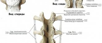
Structure of the skull: anatomy of bone joints and joints
The vast majority of the skull bones are connected using fixed sutures. Facial bone formations adjacent to each other form flat joints, invisible under the thin cover of the muscle tissue. And the temporal bone, connecting with the parietal, gives rise to the scaly suture.
There are only 3 serrated sutures in the anatomy of the skull:
- coronary, formed by the parietal and frontal bones;
- sagittal, located between the two parietal bones;
- lambdoid, located between the occipital and parietal bones.
The only movable joint of the skull is the mandibular joint. The lower jaw can perform movements in different planes: rise and fall, move to the right/left and forward/backward. Thanks to this mobility, a person can not only chew food thoroughly, but also maintain articulate speech.
Age characteristics
With age, the shape and structure of the skull changes. Thus, in newborn babies, the facial region is almost 8 times smaller than the brain, so the head may look disproportionate and large. The baby's jaws are usually underdeveloped and have no teeth, because he does not yet have the need to chew solid food.
The bones of the skull of infants do not articulate tightly, due to which the head can slightly change shape and shrink as it passes through the birth canal. This feature protects newborns from birth injuries and helps maintain normal intracranial pressure. At the interosseous sutures they have noticeable membranous areas - fontanelles. The largest, the anterior fontanelle, occupies a central position at the junction of the sagittal and coronal sutures. It usually heals by the age of two. Other fontanelles are less voluminous: the occipital, two sphenoid and mastoid membranes are not palpable by 2–3 months.
Anatomy of the skull
changes not only in infancy - formation usually takes place in 3 stages:
- Predominant growth in height, strengthening of bones and hardening of sutures - from birth to 7 years;
- The period of relative rest is from 7 to 14 years;
- The growth of the facial part of the skull is from 14 to 20–25 years, depending on puberty.
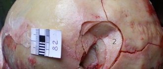
A short excursion into the anatomy of the skull allows us to clearly see that the head is an extremely complex structure, the condition of which directly affects the health of the brain, and therefore most vital functions. With the slightest injury, most of the damage is taken by the bones, but their strength is not unlimited - with a strong impact, fractures and bruises are possible, the consequences of which can be irreversible. Therefore, under any circumstances, the skull should be properly protected and protected from injury and other damage.
Consequences of skull base fractures and prognosis
Experts note that the future quality of life of patients directly depends on the quality of rehabilitation after TBI, as well as on the nature of this injury, pathologies and possible infections. In the absence of purulent inflammation, in most cases, doctors give a favorable prognosis.
If there are infectious complications, doctors talk about the possibility of developing complications such as encephalopathy, frequent headaches, uncontrolled increase in blood pressure, etc. in the future. This condition may also be accompanied by recurrent epileptic seizures.
Some traumatic brain injuries can cause excessive bleeding, coma, and even death. With such injuries, the prognosis of doctors is extremely unfavorable.
Bleeding of small volumes, intracerebral hematomas, etc. can form. It is worth noting that in this situation, the patient’s future directly depends on the timeliness and adequacy of the treatment.
What parts is the brain skull divided into?
Brain Skull
The human skull is divided into two main parts - the facial and the brain.
The bones of the medulla consist of:
- Occipital
- Sphenoid
- Lattice
- Lobnaya
- Temporal
Facial bones:
Unpaired:
- Opener
- Lower jaw
- Hyoid bone
Doubles:
- Upper jaw
- Inferior turbinate
- Palatal
- Zygomatic
- Nasal
- Tearful
The cranial bones are connected to each other and are in a stationary state. The exception is the lower jaw. It has joints and a hyoid bone, which also constantly moves.