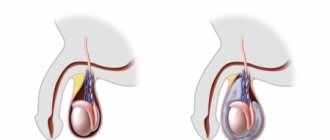
Hydrocele (hydrocele)
is a disease in which excess fluid accumulates in the membranes of the testicle.
The volume of liquid can vary - from a few milliliters to one or more liters (in exceptional cases). As a result, the scrotum enlarges, most often on one side, and in case of dropsy of both testicles, on both sides. The fluid is secreted by one of the membranes of the testicle, which is called vaginal
. The presence of fluid allows the testicle to move freely in the scrotum. However, if more fluid is released than absorbed, it begins to accumulate. This condition is called hydrocele.
Hydrocele of the testicle can be either congenital or acquired. Congenital hydrocele of the testicle is diagnosed, as a rule, during examination of the newborn, so we usually talk about hydrocele in the newborn
.
Causes of dropsy
Pathology can be congenital or acquired; accordingly, it occurs for various reasons:
- Congenital. During development in the womb, the testicles are located in the abdomen, and by the time of birth they descend into the scrotum. It forms the vaginal process - a small pocket of peritoneum. By birth or in the first days after birth, it closes, turning into a cord. If this does not happen, fluid begins to accumulate in the appendix, which forms dropsy.
Some newborns may experience a physiological hydrocele, which disappears a year after birth.
- Acquired. The cause of testicular hydrocele in men is an imbalance in the production and absorption of lymphatic fluid caused by infections of the testicle or epididymis, surgery, or injury.
Symptoms
With local dropsy, symptoms depend on the location of the swelling. With severe swelling of the eyelids, the field of vision narrows. Swelling of the feet does not allow wearing usual shoes. Dropsy of the brain leads to disturbances of consciousness, and hydrocele manifests itself as nagging pain in the testicle. Neurological edema is accompanied by dysfunction of the limbs and pain due to compression of the nerve endings.
Systemic dropsy is manifested by the following symptoms:
- dryness and swelling of the skin;
- fluctuations in weight during the day - typical for dropsy in pregnancy;
- a feeling of thirst due to blood thickening due to the effusion of fluid into the interstitial space;
- “pit” symptom: when pressing with a finger, a pit remains in the area of edema;
- the appearance of shortness of breath;
- fast fatiguability.
The absence of acute pain with dropsy masks the danger of the condition. Blood thickening increases the risk of thrombosis, which can lead to acute hypoxia and shock.
In pregnant women, whose internal edema appears only after retention of 3-4 liters of fluid, swelling of the placenta simultaneously occurs, due to which the umbilical cord vessels are compressed and oxygen starvation of the fetus develops.
Diagnostics
Diagnosis of dropsy is not difficult and is based on clinical manifestations and medical history.
If edema is detected, additional studies are carried out to identify the cause of dropsy.
Laboratory methods used:
- general urinalysis - determines the concentrating ability of the kidneys and the integrity of the glomerular apparatus;
- special tests: according to Nechiporenko, Zimnitsky, Addis-Kakovsky - they can detect kidney damage;
- general blood test - allows you to identify inflammatory and allergic processes in the body;
- biochemical tests - determine liver function and the amount of protein in the blood plasma;
- Hormone tests are done to confirm or refute the diagnosis of a tumor of the adrenal gland, pituitary gland, as well as hypofunction of the thyroid gland.
Surgical treatment of hydrocele
There are 4 types of hydrocele removal surgery:
According to Winckelmann
Plastic surgery of testicular membranes for small volumes of dropsy. The operation allows you to completely restore the functionality of the testicles. During surgery, the doctor makes an incision in the front of the scrotum and dissects the tissue to remove the testicle with its membrane to drain out the fluid. The surgeon everts the membranes after visual inspection and palpation, and stitches the tissues without squeezing the spermatic cord.
According to Bergman
The operation is indicated for large-scale hydrocele or in situations where the pathology is complicated by thickening of the testicular membrane. The formation can be on either side, so one part of the scrotum becomes enlarged. On the side with the hydrocele, the testicle may not be palpable, although the other part of the scrotum remains unchanged. Unlike the Winkelmann operation, the membranes are not everted, but excised.
Stages of severity
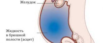
There are three stages of abdominal hydrops depending on the amount of accumulated fluid:
- Initial stage - up to one and a half liters of fluid accumulates in the abdominal cavity;
- Moderate ascites - manifested by an increase in the size of the abdomen, edema of the lower extremities. The patient is concerned about severe shortness of breath, heaviness in the abdomen, heartburn, constipation;
- Severe dropsy - from 5 to 20 liters of fluid accumulates in the abdominal cavity. The skin on the abdomen tightens and becomes smooth. Patients experience heart failure and respiratory failure. When the fluid becomes infected, ascites-peritonitis (inflammation of the peritoneal layers) develops.
Diagnostics
Doctors identify ascites during an examination of the patient. Oncologists at the Yusupov Hospital conduct a comprehensive examination of patients, which allows them to identify the cause of fluid accumulation in the abdominal cavity. One of the most reliable diagnostic methods is ultrasound. During the procedure, the doctor not only clearly sees the liquid, but also calculates its volume.
In case of ascites, oncologists necessarily perform laparocentesis. After puncturing the anterior abdominal wall, the doctor aspirates fluid from the abdominal cavity and sends it to the laboratory for testing. Computed tomography radiology scans are used to determine the presence of malignant tumors in the liver that cause portal hypertension.
Magnetic resonance imaging makes it possible to determine the amount of accumulated fluid and its location.
Rules for the postoperative period
After the patient has undergone surgery, it is recommended to avoid physical activity and sexual relations for 2-3 months. The surgical suture dissolves in one to two weeks. Swelling and the presence of a hematoma are complications of surgical intervention. Our medical center uses modern methods, so the development of complications is minimized.
A month after surgery, it is recorded that spermatogenesis has significantly worsened. Over time, it will gradually recover. If low-traumatic methods were used, this will happen in 8-12 weeks, if invasive surgery - in six months.
Forecast
Ascites in cancer significantly worsens the general well-being of the patient. As a rule, such a complication occurs in the later stages of oncology, in which the survival prognosis depends on the nature of the tumor itself and its distribution throughout the body.
Life expectancy with ascites depends on the following factors:
- Functioning of the kidneys and liver;
- Activities of the cardiovascular system;
- The effectiveness of therapy for the underlying disease.
The development of ascites can be prevented by an experienced doctor observing the patient. Doctors at the Yusupov Hospital have extensive experience in dealing with various types of cancer. Qualified medical personnel and the latest equipment allow for accurate diagnosis and high-quality, effective treatment in accordance with European standards.
Prices
Euroonko has a special offer for drainage of ascites in a day hospital - 63,300 rubles.
The program includes:
- Examination and consultation with an oncologist surgeon.
- Complete blood count, biochemical blood test, ECG.
- Ultrasound of the abdominal organs with determination of the level of free fluid
- Conducting laparocentesis with ultrasound navigation.
- Complex drug therapy aimed at restoring water and electrolyte balance.
Book a consultation 24 hours a day
+7+7+78
Bibliography:
- Pharmacotherapy of tumors. Dedicated to the memory of Mikhail Lazarevich Gershanovich // A.N. Stukov and the team of authors / Ed. A.N. Stukova, M.A. Blanca, T.Yu. Semiglazova, A.M. Belyaeva. St. Petersburg: Publishing House ANO “Oncology Issues”, 2021, 512 p.
- Willert A.B., Kolomiets L.A., Yunusova N.V., Ivanova A.A. Ascites as a subject of research in ovarian cancer. Siberian journal of oncology. 2019; 18 (1): 116–123. – doi: 10.21294/1814-4861-2019-18-1-116-123.
- Willert A. B., Kolomiets L. A., Yunusova N. V. Ascites as a tumor microenvironment in ovarian cancer: relationship between prognosis and chemoresistance. Advances in Molecular Oncology 2019;6(2):8–20.
- Yu.M. Stepanov, I.N. Kononov, T.A. Skorokhod, Ascites associated with intraperitoneal rupture of the bladder and urinary peritonitis. Clinical decline of current gastroenterology, 4 (66) • 2012.
- T.A.Baeva, D.N.Andreev, E.M.Mironova, D.T.Dicheva - Ascites: differential diagnosis and treatment. — Directory of a polyclinic doctor. No2, 2021.
- Alekseichik, S. E. Ascites. Differential diagnosis: method. recommendations / S. E. Alekseychik. – Mn.: BSMU, 2005. – 28 p.
- V.T. Ivashkin, M.V. Mayevskaya, Ch.S. Pavlov, E.A. Fedosina. Clinical recommendations of the Russian Society for the Study of the Liver and the Russian Gastroenterological Association for the treatment of complications of liver cirrhosis. Ros journal gastroenterol hepatol coloproctol 2016; 26(4).
- J. T. Tamsma, H. J. Keizer, A. E. Meinders. Pathogenesis of malignant ascites: Starling's law of capillary hemodynamics revisited. - Annals of Oncology 12: 1353-1357. 2001.
- Rony A Adam, Yehuda G Adam. — Malignant ascites: past, present, and future. - VOLUME 198, ISSUE 6, P999-1011, JUNE 01, 2004. DOI: https://doi.org/10.1016/j.jamcollsurg.2004.01.035
- Michelle Meier, Frank V. Mortensen, Hans Henrik Torp Madsen. — Malignant ascites in patients with terminal cancer is effectively treated with permanent peritoneal catheter. — Acta Radiologica Open 4(7) 1–7. DOI: 10.1177/2058460115579934.
- RC Oey, HR van Buuren, RA de Man. — The diagnostic work-up in patients with ascites: current guidelines and future prospects. OCTOBER 2021, VOL. 74, NO. 8
Non-cancerous ascites
Ascites is a consequence of various disorders that occur in the body. Treatment tactics depend on the pathological process that caused the accumulation of fluid in the abdominal cavity:
- To treat acute heart failure, cardiologists at the Yusupov Hospital prescribe metabolites, beta blockers, and ACE inhibitors to patients;
- For infectious and toxic liver lesions, therapy with hepatoprotectors is carried out;
- If ascites has developed due to low protein levels in the blood, albumin infusions are performed;
- Ascites that develops as a result of peritoneal tuberculosis is treated with anti-tuberculosis drugs.
To remove fluid from the body, patients with ascites are prescribed diuretics. The main method of eliminating ascites is to remove accumulated fluid by puncturing the abdominal wall, followed by installing drainage. For persistent ascites, peritoneal fluid is reinfused after filtration. A peritoneovenous shunt for abdominal ascites ensures the flow of fluid into the general bloodstream. To do this, surgeons form a structure with a valve, through which fluid from the abdominal cavity enters the superior vena cava system during inhalation.
Omentohepatophrenopexy for abdominal ascites is performed to reduce pressure in the venous system. The surgeon sutures the omentum to the diaphragm and liver. Then, during breathing movements, the veins are unloaded from blood. As a result, the release of fluid through the vascular wall into the abdominal cavity decreases. As a result of deperitonization (excision of areas of the peritoneum), additional outflow pathways for peritoneal fluid are created.
What can hydrocele of the testicles lead to if it is not operated on?
- disturbance of spermatogenesis with the development of male infertility due to compression of the testicle or both testicles;
- testicular atrophy due to compression of the testicles by fluid, development of circulatory disorders in the testicle;
- erectile dysfunction and decreased potency;
- premature ejaculation or rapid, accelerated ejaculation;
- an increase in the volume of the scrotum causing an aesthetic defect.
You should consult a doctor promptly. The outcome of the disease in such situations is favorable in most cases. It should be remembered that in young people, prolonged compression of the testicle with fluid can lead to testicular hypotrophy and impaired spermatogenesis.
Pathology and cancer
Medical statistics and practice show that the most common diseases leading to the accumulation of liquid mass in the peritoneal cavity are oncological forms:
- Ovaries;
- Mammary glands;
- Uterus;
- Stomach;
- Colon.
A liquid mass accumulates with a tumor due to damage to the peritoneal lining. Atypical cells, having settled on it, cause disruption of lymphatic drainage, which, in turn, impairs fluid absorption. This is very common, for example, in gastrointestinal oncology.
If we take an organ such as the liver, the picture will be different. A tumor or metastasis localized in an organ causes compression of the venous system of the organ and disruption of normal venous outflow from the intestine. In this case, the pathology develops rapidly and is more severe. About fifteen percent of ascites in cancer processes is diagnosed in this form.