- How does it manifest?
- What causes fluid to accumulate?
- Risk factors
- Classification and types
- How to treat?
- Clinical case
- What causes ascites to develop in cancer?
- Features of the treatment of ascites in cancer patients
- Conservative therapy
- Surgery
- Diagnostic methods
- Possible complications
- Forecast
- Prevention
- Prices
Euroonco doctors specialize in working with patients with ascites. We have the following treatment features for this disease:
- We provide comprehensive treatment. During laparocentesis (a puncture of the abdominal wall to remove fluid from the abdomen), we install temporary or permanent peritoneal catheters, as well as port systems. This allows the patient not to be limited in movement.
- If this is indicated, the patient is prescribed a special diet with limited water and salt load.
- If this condition occurs against the background of cancer, chemotherapy may be performed. Thanks to this, we achieve improvement in the condition of such patients with advanced ovarian and colon cancer.
- Intracavitary chemotherapy is effective. After the fluid is removed, a chemotherapy drug is injected into the abdominal cavity. In approximately half of the cases, repeated evacuation of the fluid is not required for at least 2 months.
When a patient with cancer and ascites switches to complex therapy, laparocentesis is required 2–3 times less often than usual.
How does it manifest?
The presence of a small amount of fluid in the abdominal cavity does not manifest itself clinically. In addition, the human body normally produces and absorbs approximately 1.5 liters of fluid in the abdominal cavity per day. At the initial stage of this condition, patients have no special complaints, and fluid can only be detected during an ultrasound examination.
As the disease progresses, there is more fluid in the abdominal cavity, the person feels heaviness in the abdomen, and dull aching pain in the lower part. Subsequently, difficulty breathing, indigestion (nausea, belching, stool disorders) and urination problems occur. In the most severe forms, health deteriorates significantly, discomfort appears in the abdomen, shortness of breath occurs, early satiety occurs, an umbilical hernia forms, and swelling of the lower extremities appears.
5–10 liters of fluid, and sometimes 20 liters, can accumulate in the abdominal cavity. Because of this, internal organs are strongly compressed, intra-abdominal pressure increases, and the diaphragm is pushed into the chest cavity. This entails severe difficulty breathing. Due to the fact that resistance to blood flow increases in the abdominal organs, heart failure occurs. The consequence of long-term ascites is a violation of the drainage of the lymphatic system. Because of this, there is also a violation of lymphatic drainage in the lower extremities and, as a result, their swelling. There may also be a backflow of lymph into the internal organs. As a result, cancer cells enter healthy organs from the affected lymph nodes. This can trigger the development of metastases in the liver, stomach, pancreas and other organs. [4.8]
When there is more than one liter of fluid in the abdominal cavity, this can be noticed during a routine examination: the abdomen is enlarged or deformed, in an upright position it looks saggy, in a lying position the abdomen is flattened, the sides look swollen (the so-called “frog belly”). In thin patients, the navel often protrudes. A person may also experience hydrothorax, the presence of fluid in the pleural cavity. Typically, this condition develops in patients with congestive heart failure with long-standing ascites.
Mild or moderate ascites develops in 15 to 50 percent of patients in the early stages of cancer. Severe occurs in advanced stages in 7 to 15 percent of patients. [1]
In patients with advanced advanced cancer, exudative pleurisy is most common.
Book a consultation 24 hours a day
+7+7+78
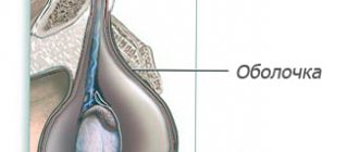
Symptoms of hydrocephalus
Congenital dropsy is diagnosed much less frequently than acquired dropsy. Usually, at the age of six months, pediatricians note that the baby’s head circumference and shape differ from the norm.
- The head circumference exceeds the child's chest circumference.
- The head takes on a pear-shaped shape, the elongation of the skull is visually noticeable, while the skin is expanded, the skin is thinned, the fontanel is enlarged,
- The child eats poorly, is extremely nervous, and cries often. During crying or bathing, the baby's chin and upper limbs may shake.
- Spasticity of the lower extremities, hyperreflexia and the presence of pathological reflexes.
- Epileptic seizures.
The symptoms of hydrocephalus in children over 2 years of age and adult patients are somewhat different:
- Severe headache, including nausea and vomiting. The pain intensifies when the patient changes a horizontal position to a vertical one.
- Fainting.
- An ophthalmological examination reveals a congestive optic disc.
- Memory and consciousness disorders, loss of already acquired skills, inability to learn new ones.
- Optical-spatial agnosia develops - it is difficult for a person to determine which object is closer/further, lower/higher.
What causes fluid to accumulate?
Pathological accumulation of fluid in the abdominal cavity occurs in certain diseases in which the regulation of water-salt metabolism and normal circulation are disrupted. The reason may be:
- Oncological diseases: secondary peritoneal carcinomatosis, lymphoma and leukemia, metastases in the hepatic portal area, primary mesothelioma.
- Diseases of the liver and its vessels: liver cancer, portal hypertension, liver cirrhosis, veno-occlusive disease, Budd-Chiari disease.
- Peritonitis (inflammation of the peritoneum) of various origins: pancreatic, fungal, parasitic, tuberculous.
- Congestive heart failure, constrictive pericarditis.
- Other diseases: ovarian tumors and cysts (Meigs syndrome), pancreatic cyst, Whipple's disease, sarcoidosis, systemic lupus erythematosus, myxedema.
Euroonco treats ascites of various origins. But since our main work is related to the treatment of malignant neoplasms, a significant part of our patients are cancer patients. [4,5]
Symptoms of dropsy
Under the pressure of the watery fluid located in the subcutaneous tissue, the skin swells, resulting in an increase in the volume of certain parts of the sick person’s body. In the area of swelling, the skin may resemble dough. When palpating edematous tissue, the pits that form with light pressure remain on the skin for a long time after the cessation of exposure. Pallor and coldness of the skin are noted, which is associated with impaired blood supply to tissues due to compression of blood vessels by edematous fluid. Dropsy is formed by a clear liquid that contains protein in very low concentrations.
Edema is a characteristic symptom of a wide variety of diseases and pathological conditions. It is an important diagnostic sign for doctors when examining patients suffering from general or local circulatory disorders, kidney diseases, and disorders of the systems regulating water-salt metabolism.
According to the location, dropsy is divided into local and general. Local edema is caused by a violation of the inflow and outflow of fluid in a separate area of tissue or in a certain organ; the cause of such dropsy in most cases is compression of the venous vessels. Blockage (compression) of the portal vein causes ascites, which is also called dropsy of the abdominal cavity, and poor circulation in the femoral vein causes swelling of the legs.
General dropsy leads to an imbalance of water balance throughout the body, which can be judged by cardiac edema. The main reasons that lead to a change in the balance of fluid flow in limited areas are: an increase in fluid pressure in small vessels (capillaries), a decrease in oncotic pressure of plasma, an increase in oncotic pressure of interstitial fluid, a decrease in pressure on tissue, high permeability of capillary vessels, a violation of reverse flow plasma.
Considering the factor that becomes leading in the development of the pathological process, dropsy is divided into mechanical, hypooncotic, membranogenic and lymphatic. Mechanical, or congestive, edema occurs due to high hydrostatic pressure in small blood vessels and disruption of the return flow of venous blood caused by blockage or mechanical compression of the blood vessels. Such pressure can be exerted by a pregnant uterus and an enlarged liver. The cause of vein blockage may be phlebothrombosis.
When the protein concentration in the blood decreases, hypooncotic edema may develop, while the protein content does not exceed 50 g/l. In this case, the low content of albumin in the blood (below 25 g/l) is of greatest importance, since they are characterized by greater osmotic activity than globulins. A maximum drop in oncotic pressure and extensive edema accompany nephrotic syndrome.
An increase in oncotic pressure of the interstitial fluid, accompanied by a change in the permeability of capillary membranes and sweating of protein-saturated filtrate into the tissue, is an important factor in the formation of edema of any origin. Edema that occurs in acute nephritis, cardiac and respiratory failure is closely related to increased membrane permeability. Cell membranes can be affected by toxins (snake venom, bacterial toxins, toxic substances, fever).
Symptoms of hydrocele of lymphatic origin occur when the reverse flow of lymph is disrupted, which causes the accumulation of protein-saturated fluid. Changes in lymph flow and associated edema accompany congenital hypoplasia of the lymph nodes, their malignant degeneration, nephrotic and starvation edema, as well as ascites.
Risk factors
Among the risk factors for the development of ascites, pathologies that can lead to liver cirrhosis are of greatest importance. First of all, these are viral hepatitis B and C, alcoholic hepatitis. Other most common risk factors:
- congestive heart failure;
- renal failure;
- obesity;
- diabetes mellitus type II;
- increased levels of “bad” cholesterol in the blood. [eleven]
Classification and types
Classically, depending on the level of protein in ascitic fluid, ascites is divided into exudative (25 g/l or more) and transudative (<25 g/l). This allows us to indirectly judge the reasons. Currently, a more accurate indicator is used - the serum albumin-ascitic albumin gradient (SAAG):
- When SAAG is more than 1.1, the cause of ascites is usually pathologies such as cirrhosis, congestive heart failure, and Budd-Chiari disease. They are associated with increased pressure in the portal vein.
- If SAAG is less than 1.1, it can be assumed that the accumulation of fluid in the abdomen is caused by pancreatitis or cancer.
According to the clinical course, the following types of ascites are distinguished:
- Depending on the severity of the current:
- 1st degree. There are no clinical manifestations; the diagnosis is established by ultrasound (determining the level of free fluid in the abdominal cavity).
- 3rd degree. Significant visual enlargement of the abdomen.
2nd degree. Slight visual enlargement of the abdomen.
- Resistant to diuretics: treatment with diuretics in combination with a diet in which sodium chloride intake is limited to less than 2 g per day is ineffective (no effect after 1 week of therapy).
Uncontrolled by diuretics: It is impossible to use diuretics because they cause significant side effects. [6,7]
Watery formations on the skin
Watery pimples are scientifically called vesicles. This is when a pimple appears on the skin with clear liquid inside. There can be one or many pimples. Pimples can merge into groups or be at a distance from each other. The location can be any: on the skin of the whole body and on the mucous membranes of the genital organs, on the scalp, only on the lips, or only in the form of an isolated lesion of a segment on the skin like a twig, strewn with such pimples. The pimple itself, with clear liquid inside, may or may not have a light red rim on the skin at the base of the watery vesicle. There may also be a slight depression in the center of the watery pimple or none at all. Doctors call this an umbilical indentation.
The size of a watery pimple can vary from small to large. After the appearance of a pimple, its further development occurs. The watery blister may burst instantly and become crusty or wet. It can also have dense walls and not open for a long time. And sometimes the transparent contents are replaced by cloudy and purulent ones.
Pimples can cause severe pain at the site of the rash, a feeling of fullness and itching. There may also be changes in body temperature, runny nose, sore throat and general malaise. Or maybe none of this will happen.
So what do these many-sided watery pimples mean? What will a pediatrician, dermatologist or therapist say with each variant of the appearance of a pimple with watery contents and with its subsequent transformations? What causes such diverse types of pimples filled with clear liquid?
This can occur with allergies, with purulent skin infections, with herpes infections in the form of herpes types 1 and 2, and with herpes called varicella zoster.
All these reasons have different clinical manifestations. And only on the basis of the totality of clinical manifestations will a dermatologist or pediatrician make a diagnosis. But to confirm the diagnosis, the doctor will suggest taking tests. This may be a study of the contents of the bladder in the form of PCR for viruses or culture for flora, a blood test for the presence of antibodies for viruses or infectious diseases.
And, of course, treatment directly depends on the diagnosis and tests.
If the diagnosis is “herpes simplex”, or “genital herpes”, or “shingles”, then the doctor has many effective antiherpetic drugs in his arsenal. These include ointments, tablets, syrups, and solutions for administration both intravenously and intramuscularly. The doctor may prescribe medications to correct the immune system. Your doctor may also recommend vaccines for herpes simplex and chickenpox. The fact is that the chickenpox and shingles viruses have the same origin and name, varicella zoster. And those who have suffered from live varicella zoster virus, have had chickenpox, remain carriers of the varicella zoster virus in the nervous ganglia for life. Only it is dormant and, under adverse influences, can be activated and cause an unpleasant disease in the form of shingles.
Well, what if the diagnosis is “chickenpox”? For chickenpox herpes, the doctor will prescribe treatment for pimples and isolate the patient for 11 days from the moment of the rash. By the way, it is better for contact people to receive the chickenpox vaccine within 72 hours after contact with chickenpox to avoid getting sick.
What if it's a skin infection? Doctors call this skin infection pyoderma. In the case of a purulent lesion, the doctor will prescribe local ointments and solutions and decide on the need for antibiotic therapy depending on the situation.
Well, what if it’s an allergy that manifests itself this way? For allergies in the form of eczema or other manifestations, local treatment with ointments and solutions with antiallergic properties is indicated. Everything necessary will be prescribed by the doctor based on the individual characteristics of the disease. The doctor will also prescribe a diet and medications that cleanse the body of allergens and toxic substances and prevent the development of allergies.
The main thing is not to let the situation get worse and consult a doctor in time (at the earliest manifestations of the disease).
How to treat?
There are several main methods for treating ascites in patients with cancer:
- conservative therapy (aldosterone antagonists, diuretics) - aimed at normalizing water-salt metabolism and reducing the formation of fluid in the abdominal cavity;
- laparocentesis - puncture of the abdominal wall under ultrasound control; used not only for removing fluid, but also for installing drainage, which will serve for long-term removal of fluid;
- palliative surgical operations - peritoneovenous shunt, omentohepatophrenopexy, deperitonization of the abdominal walls and others. [1.9]
Traditional methods of treating ascites caused by cancer do not have proven effectiveness and safety, therefore they are not used at Euroonko.
If you come to our clinic with ascites due to cancer, we recommend getting a second opinion regarding the treatment of the underlying disease from our clinical oncologists and chemotherapists.
Treatment methods
Treatment of various forms of dropsy begins with treatment of the underlying disease that caused the swelling, and symptomatic therapy is carried out for acute manifestations.
Treatment of acute hydrocele includes the use of antibiotics and analgesics, cold and thermal procedures, and wearing a suspensor. The chronic form of testicular hydrocele is treated by puncture of accumulated fluid and administration of hydrocortisone. Most often, surgical intervention is used to avoid complications.
To treat dropsy in pregnant women, optimization of nutrition, restriction of liquid and table salt intake, fasting days, and drug treatment are used.
When treating hydrothorax and pneumothorax, the emphasis is on treating the underlying disease. If a patient is diagnosed with hydrocele of a joint, a puncture is performed to remove intra-articular fluid.
Treatment of ascites depends on the severity of the underlying disease that caused it. Usually the patient is prescribed diuretics, cardiac glycosides, oxygen therapy, and a salt-free diet is recommended. In difficult cases, surgery is used to remove fluid from the peritoneum.
Elimination of the manifestations of hydrocephalus can be either surgical or conservative.
To reduce intracranial pressure use:
- General strengthening procedures;
- Pine-salt baths;
- Drug treatment for dehydration, prevention of inflammation, desensitization;
- Relief of mental disorders.
Surgical treatment aimed at creating an artificial pathway for the outflow of cerebrospinal fluid from the ventricles of the brain is more effective.
Auxiliary methods for the treatment of all types of dropsy can be scaling, hydrotherapy, compresses and wraps, and the use of traditional medicine recipes.
Clinical case
A 59-year-old woman, Sh., was diagnosed with stage IV ovarian cancer (adenocarcinoma), ascites, and chronic pain syndrome 2 b according to the ShVO. The patient noticed an increase in the abdomen up to 120 cm in circumference, difficulty breathing, and weight loss. Specific treatment at the place of residence was denied. According to the patient, she was “sent home to die.” Read more…
A 59-year-old woman, Sh., was diagnosed with stage IV ovarian cancer (adenocarcinoma), ascites, and chronic pain syndrome 2 b according to the ShVO.
The patient noticed an increase in the abdomen up to 120 cm in circumference, difficulty breathing, and weight loss. Specific treatment at the place of residence was denied. According to the patient, she was “sent home to die.” Patient Sh. was urgently hospitalized in the specialized department of Euroonko, after active symptomatic therapy aimed at normalizing blood counts and restoring water and electrolyte balance, a peritoneal port was installed. Resolution of ascites was carried out under the control of plasma protein levels. The use of peritoneal ports allows for the removal of ascitic fluid in fractional doses, which ultimately eliminates the occurrence of serious complications in the form of hemorrhagic syndrome associated with hemodilution and coagulopathy as a result of massive entry of ascitic contents into the venous bed.
After stabilization of the general condition, against the background of nutritional support, antiemetic and antisecretory therapy, patient Sh. received specific chemotherapy treatment with good effect. Upon resolution of ascites in the presence of a peritoneal port, intra-abdominal chemotherapy became possible.
Six months after the described hospitalization, the patient returned to her usual lifestyle and continues to receive systemic treatment on an outpatient basis under the supervision of a team of Euroonco specialists. The response to treatment is regarded as positive in the absence of ascites and a total reduction in the size of the lesions by more than 70%. Combined treatment in the format of systemic and local (intra-abdominal) therapy with implantation of a port system is the optimal treatment regimen for this group of patients. In the practice of Euroonco doctors, such cases occur on a regular basis. Hide
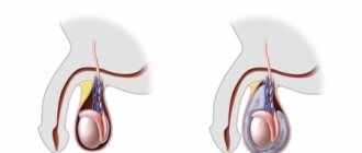
In what cases is surgery needed for hydrocele?
Surgery for hydrocele is a complex but necessary stage of treatment. It is needed:
- With a significant amount of dropsy, causing inconvenience and cosmetic discomfort
- For pain syndrome
- In case of infertility or pathological changes in the spermogram
- With a rapid increase in the volume of dropsy
By contacting me with a problem such as hydrocele, you can be sure that you will receive the most effective and safe treatment using modern technologies.
What causes ascites to develop in cancer?
The most common cancers that cause fluid accumulation are:
- ovarian cancer (in 25–30 percent of patients),
- mammary cancer,
- uterine cancer,
- stomach cancer,
- colon cancer. [2]
The accumulation of fluid in the abdominal cavity during cancer occurs due to the fact that the peritoneum (the membrane lining the inside of the abdominal wall and covering the organs located in it) is affected. Tumor cells settle on its parietal and visceral layers, resulting in disruption of lymphatic drainage. This causes deterioration in fluid absorption. Typically caused by tumors of the gastrointestinal tract and ascites in ovarian cancer.
When a tumor or metastases forms in the liver, the reason for the accumulation of fluid in the abdominal cavity is different: the venous system of the liver is compressed and the natural venous outflow from the intestine is disrupted. This condition develops quickly and usually lasts longer and is more severe. 15 percent of cancer cases occur in this particular form.
Abdominal lymphoma causes ascites through blockage and effusion (leakage) of lymph from the intra-abdominal lymph ducts. [2.6]
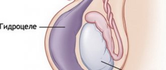
Acute and chronic testicular hydrocele
In most cases, dropsy occurs in a chronic form: fluid gradually accumulates in the membrane of the testicle. The patient may not notice changes for a long time, especially if the volume of fluid is small. He does not experience any pain; only certain inconveniences are possible (especially when the volume of fluid increases significantly) during urination, sexual intercourse, wearing underwear, etc. The asymptomatic nature of the disease often makes a man think that nothing bad is happening. Meanwhile, untimely contact with a urologist can lead to negative consequences, including dysfunction of the reproductive system, infertility, erectile dysfunction, testicular atrophy, etc.
In the acute form, fluid accumulation occurs much faster; usually such dropsy occurs due to injury, inflammation or tumor of the testicle and is accompanied by a sudden onset of pain. If left untreated, the acute form becomes chronic. But sometimes there is too much fluid, and the walls of the testicular membrane rupture. Rupture of the walls is accompanied by acute pain and requires mandatory medical intervention.
Features of the treatment of ascites in cancer patients
In hospitals that do not specialize in the treatment of cancer, the approach to patients with ascites may be ineffective due to the characteristics of this condition. For example, the main treatment may be the use of diuretics, aldosterone antagonists, and dietary changes to limit water and salt load. The effectiveness of this approach for reducing portal hypertension is relative; in cancer patients, fluid accumulation in the abdominal cavity is caused by peritoneal carcinomatosis. Therefore, conservative therapy cannot be the main treatment method in such patients.
Typically, fluid is removed from the abdomen using laparocentesis (abdominal paracentesis). This is a surgical procedure that is performed by a surgeon and an anesthesiologist-resuscitator. [3.10]
Indications for surgery for hydrocele
- pain in the perineal area;
- feeling of pressure, fullness in the genitals;
- significant increase in size of the scrotum;
- difficulty urinating;
- unaesthetic appearance of the genitals.
Refusal of surgery in men can lead to a number of complications that worsen reproductive function and potency. In addition, hydrocele can cause physiological changes in the structure of the testicles and adjacent organs.
Contraindications
- cardiovascular diseases;
- oncological pathologies;
- diabetes;
- acute inflammatory diseases of the genitourinary system;
- damage or inflammation of the skin of the testicles;
- blood clotting disorder, etc.
Most contraindications are temporary, that is, surgical intervention will be postponed until they are eliminated.
Preparing for surgery for hydrocele
Preoperative examination
Includes laboratory tests: general blood and urine tests, PCR diagnostics for STIs, coagulogram, spermogram, tests for tumor markers, etc. In addition, the patient may be prescribed instrumental types of examination - ultrasound and diaphanoscopy (examination of subcutaneous formations with a narrow beam of light).
Consultation with a urologist-andrologist
The specialist will examine the patient, determine the cause of the disease and choose the optimal method of surgical treatment. Many patients are interested in the question: is it possible to do without surgery? In most cases this is not possible. Medicinal treatments, and even more so folk remedies, are absolutely ineffective.
Consultation with a therapist
The specialist will assess the patient’s general health and check test data to exclude possible contraindications to surgical treatment. Some deviations can be corrected within 1-2 weeks before surgery to eliminate testicular hydrocele. Subsequently, the therapist monitors the patient’s recovery process.
Consultation with an anesthesiologist
The specialist will determine the type of anesthesia based on the indications and the chosen method of surgical intervention. During the consultation, you will have to answer a number of questions about height, weight, allergies, etc. In addition, the anesthesiologist assists the surgeon during the operation, monitoring all the vital functions of the patient’s body.
Hospitalization on the day of surgery
The patient has the opportunity to ask questions to the surgeon and anesthesiologist, and, if indicated, undergo preparatory procedures. In most cases, elective hospitalization is not required for testicular hydrocele. The patient arrives at the clinic on the day of the operation and leaves it a few hours after the surgery.
Conservative therapy
Conservative therapy is used in the treatment of small and moderate ascites. In other words, if there are no tiring and debilitating symptoms: pain, rapid breathing (tachypnea), etc. Up to 65% of patients have an improvement in their condition when treated with diuretics - this way you can remove up to 1 liter of fluid per day. [5]
In the later stages of cancer, reducing salt and water intake can reduce quality of life. Therefore, at Euroonko such diet correction is rarely prescribed.
Book a consultation 24 hours a day
+7+7+78
Treatment
In the treatment of dropsy, attention is paid to the underlying disease. Treatment of pathologies of the kidneys, liver, and endocrine glands is carried out.
To relieve acute conditions - cerebral and pulmonary edema - resuscitation measures are used using antifoam drugs and a complex of anti-shock therapy.
Pathogenetic treatment of dropsy is based on the mechanism of edema formation. In order to “return” the liquid medium to the vascular bed, an infusion of crystalloid (saline) solutions is performed. After which - colloidal (protein) solutions in order to increase the oncotic pressure of the blood and retain the fluid absorbed into the blood. After the infusion, diuretics are administered. The exception is pregnant patients, for whom taking any diuretic drugs is contraindicated.
Surgery
Ascites in cancer must be treated surgically when it:
- Refractory, that is, not amenable to conservative treatment.
- Large, that is, if it is necessary to remove up to 6–10 liters of fluid at a time (this difficult procedure is carried out for strict medical reasons).
- Giant. In this case, a combined operation is needed, which includes removing a large volume of fluid (up to 5–7 liters) on the first day and removing the remaining volume at a rate of no more than 1 liter per day for 7–10 days.
In the classic version, laparocentesis is performed on an empty bladder, the patient sits down, and the seriously ill person is placed on his side. [4]
It is dangerous to perform laparocentesis without observing the rules of asepsis and antisepsis. Therefore, the release of fluid is carried out only in a specialized medical institution with a license to perform surgical interventions and with a hospital. If the patient is in serious condition, it is difficult for him to move, an ambulance team is called for him.
First, local anesthesia is performed, then, under ultrasound guidance, a puncture is made with a trocar (an instrument in the form of a thin tube with a sharp end) along the midline of the abdomen or along the line connecting the navel to the iliac crest. Usually no more than 5–6 liters of liquid are removed at a time. To prevent blood pressure from dropping sharply and vascular collapse, the fluid is released slowly.
In accordance with the classical technique, the patient needs to lie for several hours on the side free from the puncture. If at this time a small amount of liquid continues to be released, then a reservoir is applied, which is removed after a day or two.
If it is necessary to remove a large amount of liquid, then a loss of protein and salts occurs, which becomes the cause of protein deficiency. To prevent such a complication, the patient is given albumin. With repeated puncture, another complication may arise - fusion of the omentum (part of the peritoneum) or intestine with the anterior wall of the abdomen. Because of this, intestinal function deteriorates significantly, and during subsequent punctures serious complications can develop. [4,6]
With the modern approach to laparocentesis, fluid drainage occurs primarily through a permanent peritoneal catheter. At the same time, the deficit in circulating blood volume is replaced by plasma expanders (from the English plasma expander - increasing plasma volume). Typically, 10–20% albumin solutions are used for this. In some cases, aminosteril, polyglucin, rheopolyglucin (dextran-40), hemacell and new starch-based preparations (refortan, stabizol, HAES-steril) can be used instead of albumin. This alternative only helps to compensate for fluid deficiency in the blood, but these drugs do not affect protein deficiency.
Some patients with ascites undergo omentohepatophrenopexy. This is a laparoscopic operation in which the omentum is sutured to areas of the surface of the liver and diaphragm. Due to the fact that contact occurs between the omentum and the liver, conditions appear for the absorption of ascitic fluid by nearby tissues. If the patient has peritoneal carcinomatosis, surgery is performed to a limited extent. Typically, in such patients, omentohepatophrenopexy becomes part of palliative treatment. [6,7]
Testicular hydrocele surgery
The operation for hydrocele of the testicle consists of opening the hydrocele cavity and excision of its walls (in order to exclude the possibility of relapse).
The operation is usually performed on an outpatient basis, that is, without hospitalization. The type of anesthesia (local or general) depends on the patient's preference. After the operation, it is recommended to wear tight swimming trunks (or a jockstrap - a special bandage that presses the scrotum to the perineum) and limiting heavy lifting.
After surgery for dropsy (hydrocele), it is sometimes possible for long-term, up to 2 months, postoperative swelling of the soft tissues of the scrotum.
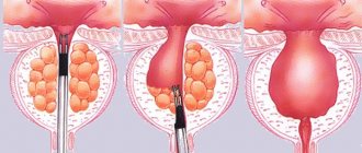
Three types of surgery for hydrocele
Winkelmann operation. A layer-by-layer dissection of the tissue of the corresponding half of the scrotum is performed to the tunica vaginalis of the testicle. The membrane is separated from the surrounding tissue and opened. A thorough examination of the testicle, epididymis and inner surface of the membrane is carried out. After this, the tunica vaginalis is turned inside out and sutured behind the epididymis. This maneuver prevents the hydrocele from forming again. Careful hemostasis is performed. The wound is sutured layer by layer. If necessary, a thin drainage is installed to ensure the outflow of ichor and wound discharge from the operated half of the scrotum. A bandage is applied. The scrotum is fixed in a position pressed to the perineum using a suspensor.
In the postoperative period after Winkelmann's operation, daily dressings and wound treatment with antiseptic solutions are performed. The drainage is removed, as a rule, on the first or second day.
Sutures are removed 8-10 days after surgery. Sexual abstinence and limitation of physical activity are recommended for 10-15 days.
Complications of the Winkelmann operation are bleeding, wound suppuration and recurrence of dropsy (hydrocele). Bleeding may be due to the abundance of blood vessels both in the testicle and in its membranes. To prevent bleeding, I use radiosurgical instruments, which allows the operation to be performed almost bloodlessly, and also, which is very important, prevents the formation of a hematoma in the postoperative period.
The main method of preventing postoperative inflammatory complications is careful adherence to asepsis (sterility). In our clinic, this is achieved by using plasma sterilization, which is the current standard for processing surgical instruments.
Relapses with a correctly performed Winkelmann operation are practically excluded, since during this operation the cavity in which fluid can accumulate is eliminated. The Winkelmann operation is ideal for small or medium-sized dropsy, in the absence of pronounced thickening and compaction of the testicular membranes.
Bergman's operation. The main stages of the operation are similar to the intervention described above. An important difference between the Bergmann operation is that the testicular membranes are not everted, as in the Winkelmann operation, but are excised and removed as much as possible. This reduces to zero the likelihood of repeated formation of dropsy (relapse).
Bergmann's operation requires more careful isolation of the testicular membranes, compared to Winkelmann's operation, so it is somewhat longer. The postoperative period in both cases proceeds similarly. Complications with a correctly performed Bergmann operation are extremely rare. Bergmann's operation is the standard for large dropsy, with thickened, dense walls.
Lord's operation is the most minimally invasive method of surgical intervention for hydrocele. The incision during the Lord's operation is usually smaller than during the Winkelmann and Bergmann operations. Careful isolation of the testicular membranes is not required, therefore, there is less trauma to the soft tissues of the scrotum. After opening the vaginal membrane and evacuating the hydrocele, the walls of the cyst are corrugated using special sutures. In this case, the membranes are not turned inside out or excised. The postoperative period after the Lord's operation is usually easier than after the Bergmann and Winkelmann operations.
Sutures are also removed after 8-10 days. Complications and relapses are rare. Lord's operation is indicated for small-volume dropsies with thin walls.
Puncture of hydrocele is currently rarely used due to the high rate of relapses, as well as the significant risk of complications (hematoma formation, suppuration, testicular damage).
Puncture of the hydrocele is carried out using a thin needle under local anesthesia. To minimize the risk of testicular damage, the puncture is best performed under ultrasound guidance. Through the puncture, the hydrocele cavity is emptied and the needle is removed. There is a technique for cavity sclerosis, however, the results of this intervention are contradictory.
Diagnostic methods
If more than 500 ml of fluid has accumulated in the abdomen, the doctor may identify symptoms during an examination. Ultrasound is used to confirm the diagnosis. Sometimes ascites is discovered accidentally during an ultrasound or computed tomography scan of the abdomen, which is performed for another reason.
Euroonco operates a comprehensive gastrointestinal screening program, which helps assess the condition of the digestive system. It, in particular, includes an ultrasound of the abdominal cavity with determination of the level of free fluid. This helps diagnose ascites at an early stage.
It is important not only to identify fluid in the abdominal cavity, but also to understand the reasons for its accumulation - this helps to assess the prognosis and prescribe effective treatment. In most cases, the examination includes the following laboratory tests:
- Biochemical blood test - complete biochemical panel. It helps assess the condition and function of the liver, kidneys, and electrolyte levels.
- Blood clotting study.
- Study of ascitic fluid obtained during laparocentesis. For analysis you need a little of it - usually 20 cm³, less than a tablespoon. It examines the level of red and white blood cells, total protein, albumin, amylase, glucose, and examines for pathogenic microorganisms. A cytological examination is performed to help identify cancer cells. [6]
Causes of hydrocephalus
Hydrocephalus in the fetus may be associated with problems with the development of the central nervous system, or with intrauterine infection. Therefore, it is so important to undergo a comprehensive examination for various infections - herpes, cytomegalovirus, before planning a pregnancy.
Acquired hydrocephalus in most cases is a consequence or complication of diseases such as:
- Meningitis - viral or bacterial etiology.
- Meningoencephalitis is an inflammatory process that affects the membrane and medulla.
- Subarachnoid hemorrhage is a common consequence of skull trauma or occurs as a consequence of rupture of an arterial aneurysm.
- Sarcoidosis is a granulomatous lesion of the meninges.
- Intraventricular hemorrhage is a consequence of difficult and traumatic obstetric care.
Possible complications
If a lot of fluid accumulates in the abdominal cavity, the functioning of the internal organs is disrupted, difficulties arise during breathing, as the mobility of the diaphragm is limited, and effusion forms in the pleural cavity.
With increased portal vein pressure, bacteria from the intestine can spontaneously enter the ascitic fluid and cause spontaneous bacterial peritonitis .
In rare cases, a very serious complication develops - hepatorenal syndrome . This term refers to impaired renal function with severe liver damage, up to severe renal failure. The exact mechanism of development of this condition is unknown, but it is believed to occur due to impaired renal blood flow, excessive use of diuretics and intravenous contrast agents. [4]
Causes
Dyshidrosis was first described as a clinical deviation from the norm by Thomas Fox in 1873.
It is worth noting that its causes and pathogenesis remain unclear to this day. Modern researchers are inclined to define dyshidrosis as a result of toxidermia (drug or food), an allergic reaction in patients with mycosis of the feet, eczema, pyoderma. The most susceptible to dyshidrosis are people prone to sweating, unstable to stress, who have suffered nervous shocks, fears, due to activation of the sweat glands, with exacerbations in the hot season.
The disease is accompanied by difficulty in permeability of the sweat ducts, exudative inflammation of the epidermis and the formation of subcutaneous blisters.
Forecast
Ascites in cancer significantly worsens the prognosis. From the moment of diagnosis, only half of the patients remain alive within 1–4 months. The average life expectancy is from 20 to 58 weeks. Timely treatment in a clinic that specializes in working with such patients helps improve survival. If the accumulation of fluid in the abdominal cavity is caused by cirrhosis of the liver, when there is no cancer, the prognosis is better, and if there is chronic heart failure, with appropriate treatment, you can live for years. [6]
Classification of hydrocephalus
Hydrocephalus can be congenital or acquired. It is generally accepted that the disease is diagnosed in children, but there is an increasing increase in cases of dropsy in adult patients.
According to the nature of the course, hydrocephalus can be:
- Acute - this form of the disease is characterized by a rapid course, the signs become obvious literally within three days.
- Subacute – symptoms increase and develop within one month.
- Chronic – the pathology develops for more than one month.
Prices
Euroonko has a special offer for drainage of ascites in a day hospital - 63,300 rubles.
The program includes:
- Examination and consultation with an oncologist surgeon.
- Complete blood count, biochemical blood test, ECG.
- Ultrasound of the abdominal organs with determination of the level of free fluid
- Conducting laparocentesis with ultrasound navigation.
- Complex drug therapy aimed at restoring water and electrolyte balance.
Book a consultation 24 hours a day
+7+7+78
Bibliography:
- Pharmacotherapy of tumors. Dedicated to the memory of Mikhail Lazarevich Gershanovich // A.N. Stukov and the team of authors / Ed. A.N. Stukova, M.A. Blanca, T.Yu. Semiglazova, A.M. Belyaeva. St. Petersburg: Publishing House ANO “Oncology Issues”, 2021, 512 p.
- Willert A.B., Kolomiets L.A., Yunusova N.V., Ivanova A.A. Ascites as a subject of research in ovarian cancer. Siberian journal of oncology. 2019; 18 (1): 116–123. – doi: 10.21294/1814-4861-2019-18-1-116-123.
- Willert A. B., Kolomiets L. A., Yunusova N. V. Ascites as a tumor microenvironment in ovarian cancer: relationship between prognosis and chemoresistance. Advances in Molecular Oncology 2019;6(2):8–20.
- Yu.M. Stepanov, I.N. Kononov, T.A. Skorokhod, Ascites associated with intraperitoneal rupture of the bladder and urinary peritonitis. Clinical decline of current gastroenterology, 4 (66) • 2012.
- T.A.Baeva, D.N.Andreev, E.M.Mironova, D.T.Dicheva - Ascites: differential diagnosis and treatment. — Directory of a polyclinic doctor. No2, 2021.
- Alekseichik, S. E. Ascites. Differential diagnosis: method. recommendations / S. E. Alekseychik. – Mn.: BSMU, 2005. – 28 p.
- V.T. Ivashkin, M.V. Mayevskaya, Ch.S. Pavlov, E.A. Fedosina. Clinical recommendations of the Russian Society for the Study of the Liver and the Russian Gastroenterological Association for the treatment of complications of liver cirrhosis. Ros journal gastroenterol hepatol coloproctol 2016; 26(4).
- J. T. Tamsma, H. J. Keizer, A. E. Meinders. Pathogenesis of malignant ascites: Starling's law of capillary hemodynamics revisited. - Annals of Oncology 12: 1353-1357. 2001.
- Rony A Adam, Yehuda G Adam. — Malignant ascites: past, present, and future. - VOLUME 198, ISSUE 6, P999-1011, JUNE 01, 2004. DOI: https://doi.org/10.1016/j.jamcollsurg.2004.01.035
- Michelle Meier, Frank V. Mortensen, Hans Henrik Torp Madsen. — Malignant ascites in patients with terminal cancer is effectively treated with permanent peritoneal catheter. — Acta Radiologica Open 4(7) 1–7. DOI: 10.1177/2058460115579934.
- RC Oey, HR van Buuren, RA de Man. — The diagnostic work-up in patients with ascites: current guidelines and future prospects. OCTOBER 2021, VOL. 74, NO. 8
Treatment of dropsy
The choice of treatment method for dropsy depends on its type, as well as the causes that caused it, and primarily consists of treating the underlying disease.
When pregnant women have dropsy, they are advised to stay hydrated and adhere to a rational, balanced diet. The diet must contain foods rich in protein (fish, cottage cheese, lean meat), fresh vegetables and fruits. Significantly limit the use of table salt. If necessary, drug therapy is prescribed (antispasmodics, sedatives and antihistamines). Prescribing diuretics for dropsy in pregnancy is undesirable, since their use can further intensify existing disturbances in water and electrolyte balance. If there is significant swelling, there are indications for hospitalization of the woman to the department of pathology of pregnant women.
For hydrocele of the gallbladder, the main treatment method is its surgical removal (cholecystectomy).
There are currently no effective methods of treating the non-immune form of hydrops fetalis. Immediately after birth, the child needs intensive care (transfusion of red blood cells, tracheal intubation and transfer to artificial ventilation, laparocentesis, pericardial or thoracic puncture). According to indications, anticonvulsant, metabolic, immunocorrective, antibacterial or antihemorrhagic therapy is carried out.
In case of hydrocele of the gallbladder, removal of the organ is indicated
Treatment of the immune form of hydrops fetalis begins before birth. Under ultrasound control, the doctor punctures the fetal umbilical vein through the anterior abdominal wall of the pregnant woman and installs a catheter into it. Through it, a transfusion of up to 50 ml of Rh-negative blood of the same group as the fetal blood is performed. In cases where the fetal blood type cannot be determined, a transfusion of Rh-negative blood of group I (0) is performed.
With hydrops fetalis, the condition of the newborn is usually assessed as very serious. Due to the significant enlargement of the brain part of the skull, which is associated with hydrocephalus, the head becomes disproportionately large.
Treatment of testicular hydrocele begins with conservative methods (wearing a jockstrap, rest, injection of sclerosing agents into the hydrocele cavity). If therapy is ineffective, surgical intervention is indicated. There are different methods of surgery for testicular hydrocele (according to Ross, Bergman and Winkelman).