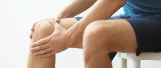
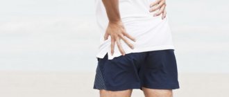
Arthrosis of the knee joint is a degenerative-dystrophic pathology that leads to deformation and destruction of articular cartilage. Gradually the limb loses mobility. According to statistics, almost every third person on the planet suffers from arthrosis, and this number is not decreasing. Elderly people, especially those who are overweight, are at risk. After 65 years, arthrosis is diagnosed in 70-85% of cases of treatment for knee pain.
A rheumatologist helps maintain a patient’s quality of life with joint pathology.
Causes of arthrosis
- Destruction of the joint due to natural wear and tear (aging of the body).
- Hormonal disorders (menopause, endocrine diseases).
- Congenital defects of the musculoskeletal system.
- Injuries, surgery on the knee joint.
- Professional sports.
- Monotonous physical work with increased load on the knee joints.
- Excess body weight.
- Genetic predisposition.
- Autoimmune diseases.
What should I do?
It is better to select a complex of drugs together with your doctor. It will probably include chondroprotectors with chondroitin and glucosamine. They are a building material for cartilage tissue, and also relieve inflammation, stabilize the condition of the joint and help reduce the amount of pain medication.
In the initial stages of arthrosis, intra-articular injections of a synovial fluid prosthesis, for example Noltrex, are indicated. While the cartilage is not sufficiently destroyed, this process can be stopped by restoring the fluid deficit. The gel is distributed inside and performs a shock-absorbing function. The joint stops hurting during movements, and most importantly, its degeneration stops.
After one or two Noltrex injections, painkillers will no longer be needed for a very long time.
2. Exercise through pain.
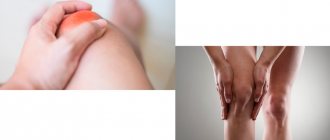
If the joint hurts, it means that the disease is in an acute stage. What it is - arthrosis or arthritis - the diagnosis will show. In any case, until the pain subsides, you should not load the joint. For such diseases, running, jumping, squats, intensive walking on uneven terrain, climbing hills and stairs, especially with heavy objects in your hands, are contraindicated.
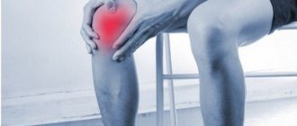
Symptoms of the disease
Deforming arthrosis of the knee joint (gonarthrosis) develops slowly and is chronic. In the early stages, the disease does not cause pain: the person feels only discomfort and stiffness in the lower limb. Gradually, motor restrictions increase. Without adequate treatment, the knee becomes noticeably deformed. Motor functions are so impaired that it is difficult for a person to walk, sit down, or stand up. Deforming arthrosis progresses to the point of disability of the patient. To preserve the joint, you must consult a doctor when the first symptoms of pathology appear.
Depending on the severity, there are three degrees of arthrosis of the knee joint:
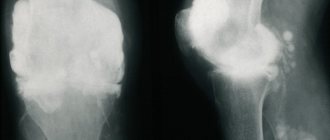
- 1st degree. Clinical manifestations of the disease are mild. Most patients do not pay attention to the symptoms and continue to lead their usual lifestyle. With grade 1 arthrosis, a person may feel discomfort in the knee after standing for a long time, intense walking, or physical activity. On an x-ray, a narrowing of the joint space is visible, osteophytes growing inside the joint are visible. If arthrosis is accidentally detected in the first stage, for example, during a medical examination, its development can be significantly slowed down and even stopped.
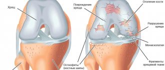
- 2nd degree. Pain with arthrosis of the knee joint becomes intense and difficult to ignore. The leg is especially bothersome early in the morning or in the evening. During the day, at rest, aching pain persists. Degenerative processes in the joint are reflected in the gait: the person begins to limp. When moving, a crunching sound is heard in the knee. Arthrosis of the 2nd degree can be complicated by a “joint mouse” - this is a condition in which a particle of bone or destroyed cartilage enters the synovial cavity. A foreign body causes severe pain that interferes with the movement of the limb. On examination, deformity of the knee is obvious. Possible inflammation and swelling. The x-ray shows a narrowed joint space and osteophytes, thickening of the bone.
- 3rd degree. A severe form of the disease that develops without treatment. Arthrosis of the 3rd degree is the cause of permanent disability. The pain in the knee is very severe, mobility is limited, the person cannot walk independently, every step is painful. The leg becomes deformed and begins to crunch heavily. On an x-ray, the doctor determines degeneration of cartilage tissue, destruction of ligaments, menisci, and proliferation of connective tissue.
At-risk groups
Many people are susceptible to osteoarthritis of the knee, because no one can predict a knee injury in advance. This most often happens among professional athletes leading an active lifestyle: the likelihood of injury is very high, but since this category of people is accustomed to bruises and injuries, at first they do not pay attention to the pain until it becomes unbearable, due to why treatment of the joint is delayed.
In addition, elderly people are at risk of developing osteoarthritis. Any organ and any tissue in the body wears out over time. This happens with the cartilage of the knee joint; it thins out and becomes fragile, susceptible to damage from any blows or falls. Therefore, it is quite easy to develop this disease in old age. The main percentage of people suffering from arthrosis are women over 50 years old: the disease is often inherited, along the maternal line.
Get a consultation on MRI diagnostics. Consultation on the service does not oblige you to anything.
People over 40 years of age who are overweight and workers whose task is to carry heavy loads are also at risk. Since each knee bears half of the body's weight and load every day, excessive stress can damage the joint.
Treatment of arthrosis of the knee joint
Therapy includes a set of procedures, medication, and recommendations for lifestyle changes. It is important not to try to treat arthrosis on your own. Often, patients in the first stages of the disease use painkillers and consult a doctor when the joint is already destroyed. The earlier treatment is started, the more effective it is.
Drug treatment
The doctor prescribes medications to relieve inflammation, swelling, reduce pain, activate metabolic processes and tissue regeneration. Medicines are selected individually.
The following medications can be taken:
- Nonsteroidal anti-inflammatory drugs (NSAIDs) in the form of tablets, ointments, injections. The drugs relieve pain and swelling well, and improve the patient’s well-being.
- Glucocorticosteroids in the form of injections directly into the knee joint. Injections are indicated for severe cases of the disease, when the limb is practically immobilized.
- Painkiller blockades. They help cope with symptoms and alleviate the course of the disease.
- Chondroprotectors. The drugs help restore cartilage tissue and slow down the destruction of the joint.
Conservative treatment
Shock wave therapy
The method is non-invasive, helps remove salt deposits and improve the trophism of connective tissue. Physiotherapy improves blood circulation and has a beneficial effect on the elasticity of ligaments. Shock wave therapy is carried out in courses of 4-10 procedures.
Plasmolifting (PRP therapy)
The patient's own platelet-rich plasma is injected into the joint. A course of plasma lifting accelerates tissue regeneration.
Phonophoresis
The method combines the effects of ultrasound and medicinal ointments. Physiotherapy products, as a rule, have a complex composition and are prepared in a pharmacy according to a prescription. Ultrasound increases the penetrating ability of the active substance.
Massage
The procedure is contraindicated in the acute stage of arthrosis. When the inflammation is relieved and the pain is reduced, you can begin a massage course. Lymphatic drainage technique helps prevent the accumulation of synovial fluid. Massage also improves blood circulation in the knee and relieves muscle spasm. The procedure is most effective after performing special exercises for arthrosis of the knee joint.
Taking a bath
You can conduct the course at home as prescribed by a doctor or as part of a spa treatment. For arthrosis, radon, turpentine, and hydrogen sulfide baths are indicated. The procedures have a beneficial effect not only on the knees, but also on the hip and ankle joints.
Hirudotherapy
Medical leeches are placed around the deformed joint. The saliva of these creatures contains active substances that promote the restoration of cartilage. Hirudotherapy is usually prescribed for grade 1 and 2 arthrosis to relieve swelling and reduce pain.
Physiotherapy
Gymnastics for arthrosis of the knee joint is an essential part of complex treatment. Special exercises help maintain muscle tone in the sore limb and prevent congestion. They begin to do gymnastics in the morning, without getting out of bed. Then during the day, perform another 3-4 sets of exercises for several minutes. It is useful to supplement therapeutic exercises for arthrosis of the knee joint with swimming.
Surgery
Surgery is indicated for grade 2 and 3 arthrosis:
- Puncture. Using a syringe, the accumulated fluid is pumped out of the joint cavity. Internal pressure decreases, swelling and inflammation decrease, and mobility improves. The procedure is carried out on an outpatient basis, at the surgeon's appointment.
- Arthroscopy. The method is used for rehabilitation of the knee joint. Arthroscopy is performed through small punctures, so the operation is quite easy to tolerate and the recovery period is short.
- Corrective osteotomy. A classic method of treating deforming arthrosis, which consists of correcting the deformed anatomical axis of the lower limb, followed by fixation of the wedge-shaped resection of the bone with a titanium plate. After osteotomy, the patient requires rehabilitation for several months.
- Endoprosthetics. Installation of an artificial joint is performed in extreme cases of deforming arthrosis of the knee joint and allows the knee to return to its previous range of motion without pain. After total endoprosthetics, the patient requires long-term (about 2-3 months) rehabilitation.
Treatment methods for grade 1 osteoarthritis
The essence of the treatment of osteoarthritis at the very beginning comes down to the return of synovial fluid to its molecular composition. This way you can improve the nutrition of cartilage and restore the surface of bones. Once mobility is restored, pain and discomfort disappear and the patient recovers.
Complex treatment:
- Eliminate the causes of the disease, if possible. It is necessary to lose weight, improve nutrition, focusing on benefits and vitamins. Those who spend a lot of time on their feet are advised to change their lifestyle and monitor the alternation of work and rest.
- Restoring hormonal balance. If there is a suspicion that the cause of the disease is the influence of hormones, the rheumatologist sends the patient to an endocrinologist. Tests show where the imbalance is hidden, and, if possible, it is corrected with medications or nutrition.
The diet for osteoarthritis consists of low-carbohydrate foods. Protein, calcium and collagen are shown. Usually they recommend adding to the menu:
- Calcium: cheese, cottage cheese
- Protein: veal, white fish, beans
- Collagen: jelly, lentils
- Vitamins: fruits and vegetables
What are the stages of gonarthrosis?
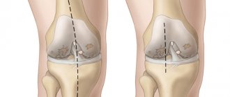
Based on changes in clinical and radiological parameters, arthrosis of the knee joint is divided into 3 stages:
Stage 1 – minor changes in the cartilage and narrowing of the medial part of the joint space of the knee joint. Marginal osteophytes begin to appear along the periphery of the glenoid cavity.
Stage 2 – the joint space is noticeably and unevenly narrowed, the cartilage is loose, thinned, the bone tissue of the epiphysis is deformed and flattened at the ends, the articular surface is covered with multiple bone growths, congruence is impaired.
Stage 3 – total changes affecting the deep layers of bone tissue. Necrosis of most of the cartilage, subchondral sclerosis, cystic lesions, and usuria are observed. There may be separation of intra-articular growths.
Diagnosis of knee osteoarthritis
In order to determine the severity of the disease, X-rays of the knee joints are performed using new generation devices. Mandatory radiological signs of knee osteoarthritis include narrowing of the joint space, subchondral osteosclerosis, osteophytes at the edges of the articular surfaces, and optional ones include clearing of bone tissue.
Arthroscopy allows doctors to assess the integrity and tone of the cruciate ligaments, the severity of thickening of the synovial membrane, reveals bone growths, and the presence of fixed or loose bodies in the joint cavity. The study is carried out at an early stage of the disease, before the appearance of characteristic radiological signs. During this procedure, a targeted biopsy is performed followed by microscopy of intra-articular structures.
Using ultrasound, the pathology of soft tissues is identified, the thickness and structure of cartilage, the condition of the synovial membrane, the presence of free fluid in the joint cavity, defects of the articular surfaces and bone growths are determined. Magnetic resonance imaging is the main method for imaging hyaline cartilage. Using the study, you can obtain information about all structures of the knee joint, clarify the integrity of the ligaments, see the pathology of the tendons, and the presence of bone marrow edema as a manifestation of osteitis. There were no changes in laboratory tests specific for osteoarthritis. In some patients, C-reactive protein levels increase slightly. Sometimes rheumatoid factor is detected in a low titer, especially in elderly and senile people. Synovial fluid is sterile, viscous, transparent or slightly turbid, the number of cells does not exceed 2000 per mm3.
What should I do?
Instead, you need to add dairy and fish dishes, buckwheat, beans, lentils, as well as products containing fiber and sulfur (flax seed, bran, onions, garlic) to the menu. They take an active part in the construction of cartilage and connective tissue.
Do your joints hurt? Eat less meat
4. Orthopedic shoes without prescription.
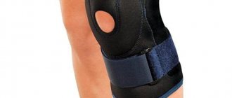
For arthrosis of the ankle, knee or hip, orthopedists often recommend wearing orthopedic insoles, or arch supports. They support the arch of the foot and reduce stress on the joints of the lower extremities. This is true provided the selection is correct. If insoles are chosen at random, they can cause harm.
Classification
Clinically, the disease is divided into three degrees.
Grade 2 gonarthrosis is characterized by an increase in the above-described signs of the disease. The pain intensifies and its duration increases. Movements are further limited. Short walking causes discomfort.
Gonarthrosis of the 3rd degree is manifested by unbearable pain, which is not relieved by taking non-narcotic analgesics, and bothers the patient at rest. Gait disturbance occurs. Upon examination, deformation of the joint and an increase in its volume are noted.
Based on localization, it is divided into left-sided and right-sided gonarthrosis.
Gonarthrosis is a chronic disease of the knee joint, in which the cartilage becomes thinner and destroyed, followed by involvement of the bone surfaces in the process.
Causes and mechanisms of development of knee osteoarthritis
Mechanical, biochemical, genetic factors and inflammation are involved in the development and progression of osteoarthritis of the knee joint. The pathological process is localized in the subchondral bone, hyaline cartilage, synovial membrane and soft tissues located around the joint. The degenerative-inflammatory process in the joint develops under the influence of the following risk factors:
- Elderly;
- Female;
- High physical activity;
- Obesity, which is an independent risk factor for the development of knee osteoarthritis, especially in women with bilateral lesions.
Osteoarthritis of the knee joint can develop with high bone mineral density, after injuries, due to the use of hormone replacement therapy, smoking, low intake of antioxidants, vitamin D and C, weakness of the lower leg muscles, and intense sports activity. The disease progresses in older people, with indolent synovitis and subchondral bone edema according to magnetic resonance imaging.