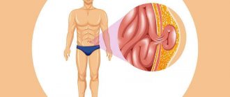
A testicular cyst is a cavity filled with fluid, which is located mainly in the area of the appendages or closer to the vas deferens, although it can be localized directly on the testicle. It looks similar to varicocele or dropsy, but has other symptoms. Urologists at the Yusupov Hospital diagnose the disease using the latest equipment from leading manufacturers in the USA and European countries.
Thanks to the operating rooms being equipped with the latest instruments and high qualifications, surgeons masterfully perform all surgical interventions. Doctors use modern methods to treat testicular cysts in men without surgery. The medical staff is attentive to the wishes of patients.
A congenital cyst appears during fetal development up to 20 weeks. It can develop due to the following reasons:
- Hormonal imbalance in a woman during pregnancy;
- Threats of abortion;
- Injuries during pregnancy.
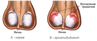
The cause of an acquired testicular cyst in men can be inflammatory diseases of the genitourinary system (orchitis, epididymitis). Testicular cysts in men can be single-chambered or multi-chambered (having septa in the cavity). Is it possible to play sports with a testicular cyst? Urologists at the Yusupov Hospital decide this issue individually in each specific case.
Statistics
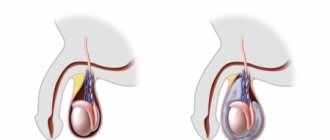
Due to the anatomical features of the veins in the testicular area, varicocele most often forms on the left side. 80-95% are allocated to these cases. Right-sided testicular varicose veins are not so common (up to 8%). On both sides, varicocele occurs in 2-12 cases out of a hundred. Male disease is “linked” to the age of the patients. A large percentage (19) of problems with testicular veins are noted by doctors in adolescents. If we consider the reproductive age of men, then the statistics of varicose veins reach 35%. These are sad numbers, because... Varicocele in sexually mature men can cause infertility. Treatment of the disease should begin at the earliest stages.
What does it look like
A photo of a varicocele clearly demonstrates a male problem - the scrotum on one side (usually the left) is slightly lower. The skin covering the testicle does not hide the contours of the veins. When self-palpating the venous vessels, a man can feel their expansion. The patient may not even be aware of other symptoms of the disease. Palpation of the scrotum and visual examination are necessary actions for all men who care about their health. This is especially true for males planning fatherhood. You need to be regularly examined by a urologist. Medical examinations and consultations with qualified specialists are offered by the Global Clinic Center, which has extensive experience in the treatment of varicocele.
History of the disease
Varicocele is a disease with deep historical roots. The first mention of the problem dates back to the 16th century and is associated with the name of Paré, a famous surgeon of the Renaissance. He drew attention to the pathology of the venous vessels near the male testicle. Many medical historians speak of the involvement of Hippocrates and Celsius in establishing the facts about this problem. The topic of varicocele is very closely related to infertility. In the 19th and 20th centuries, scientists confirmed the possibility of curing male infertility in direct proportion to the elimination of testicular varicose veins.
Varicocele. Symptoms
The danger of the first stages of the disease is that it is asymptomatic. The problem is discovered by chance, during medical examinations or when patients address other male issues (for example, in connection with infertility). Symptoms of varicocele characteristic of other degrees of the disease:
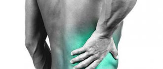
- nagging pain and discomfort in the left side of the scrotum, testicle and groin area;
- drooping left (mostly) testicle, interfering with walking;
- decrease in the size of the testicle on the left;
- manifestation of the contours of venous vessels;
- appearance of pain.
Varicocele is characterized by symptoms manifested in a decrease in the number of sperm and a decrease in their motility, in this case it is worth talking about male infertility. When doctors try to find out the cause of male infertility and analyze various symptoms in a patient, they often discover a varicocele, using the most modern diagnostic methods.
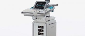
How is an ultrasound examination performed?
Testicular ultrasound is a painless procedure. Pain in a man can only occur with acute orchiepididymitis. In this case, it is better to wait a few days and do a diagnostic procedure after the inflammatory process has passed. In other cases, no unpleasant sensations occur during ultrasound scanning. The patient is usually in a supine position. The doctor applies the gel to the area of study and to the sensor of the device for smoother gliding and greater information content. The gel is water-based and hypoallergenic, so there is no need to worry about an allergic reaction of the body. The sensor is installed on the organs, and with smooth movements in various directions, the diagnostician scans the area, receiving information in the form of an image on the monitor. The ultrasound procedure lasts 10-15 minutes. The examination time may increase if a tumor is suspected, since in this case additional examination of adjacent organs is carried out. The results are delivered to the patient 10-15 minutes after the examination.
Previous NextCauses and risk factors for the disease
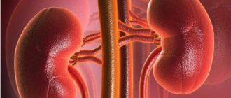
In the veins surrounding the spermatic cord, a malfunction of the valve occurs, which prevents blood from flowing back. Instead of rising upward, the blood stagnates, because... is transmitted in the opposite direction. Because of this, the venous vessels become dilated and weak. The causes and risk factors of varicocele are associated with the appearance of primary and secondary reflux. The abnormal structure of the veins associated with congenital pathology leads to the complete absence of valves for the reverse outflow of blood or their atypical operation. During puberty, blood pressure on the vessels increases, and varicose changes occur in the veins near the spermatic cord. The weak walls of blood vessels inherited from birth by boys cannot cope with the flow of blood during physiological changes. As a rule, varicocele is inherited, so this should be taken into account in case of male infertility and comprehensive treatment of testicular varicose veins should be carried out. Secondary reflux occurs when the blood outflow valves stop functioning normally due to problems in the renal or vena cava. Varicose veins most often occur on the left side, because it is on the left that the venous vessels meet the renal vein. Narrowing of the lumen in the latter structure can cause varicocele. Unfavorable factors for testicular varicose veins:
- haemorrhoids;
- phlebeurysm.
The following are considered as a trigger for the appearance of varicocele:
- Hard physical labor.
- Extreme loads in sports or weight lifting.
- Lack of normal sex life.
- Professions that require long periods of standing.
- Chronic constipation.
Main functions
The main functions of the testicles are the production of testosterone and the formation of sperm. In medicine, there are two types of functions - endocrine and spermatogenesis. Testicles simultaneously perform the function of internal secretion organs - they form hormones and carry them into the blood.
Endocrine
The endocrine function of the testicles is intrasecretory; it consists in the formation of sex hormones: testosterone, androsterone, dihydrotestosterone, dehydroapiandrosterone, which enter directly into the blood. Androgens are synthesized by Leidig cells lying in the interstitium between the convoluted seminiferous tubules.
The endocrine function of the testicles is as follows:
- development of a male body type, for example, broad shoulders, a rough voice, physical endurance, strong muscles;
- formation of courageous character, activity, endurance, desire for leadership, strong muscles;
- regulates cholesterol levels;
- responsible for the normal level of sexual activity;
- development of internal and external genital organs;
- hair development.
Reference! The production of testosterone is influenced not only by the gonads, but also by a man’s lifestyle - his diet, weight, physical activity, health and environmental factors.
The endocrine function of the testicles precedes the function of spermatogenesis, since under the influence of sex hormones the formation of the functioning of the genital organs occurs.
Spermatogenesis
Spermatogenesis is an important process that is responsible for the function of childbirth and occurs in men throughout their lives. During puberty in boys, the process of development of germ cells occurs, which lasts until old age.
There are cases where hundred-year-old men became fathers.
Spermatogenesis consists of four periods:
- Reproduction (carried out in the prenatal period, represents the division of germ cells after birth and until puberty. Upon reaching sexual development, some of the cells continue the process of forming new cells, the second part moves closer to the center of the tubule).
- Maturation (biochemical processes that are involved in preparing cells for the formation of sperm).
- Formation (sperm produce special enzymes that dissolve the shell of the egg, which allows them to penetrate inside and carry out the fertilization process).
Spermatogenesis lasts 75 days. This process is very delicate at the biochemical level, overly sensitive to the effects of adverse factors, such as:
- increased body or air temperature;
- chronic stress;
- taking hormones, antibiotics and other medications;
- excessive drinking;
- smoking;
- sedentary lifestyle;
- prolonged abstinence or sexual abuse.
Complications
The main thing that patients suffering from testicular varicose veins need to understand is the serious situation with the appearance of offspring. Infertility can become a sad complication of varicocele, so you should not delay going to the clinic for diagnosis and treatment. Doctors from the Global Clinic Center can provide qualified assistance. Statistics show that patients diagnosed with varicocele in 60 cases out of a hundred are faced with the problem of sperm dysfunction. 40% of men with testicular varicose veins become infertile.
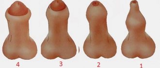
Reasons for downsizing
Small size of the tissues is a signal that pathological changes are occurring in the male body. The normal size is 4-6 cm in length and 2.5-3.5 cm in width. Size reduction in men occurs for the following reasons:
- influence of harmful factors during intrauterine development;
- pathologies of pregnancy, in particular from the fact that a woman uses different medications;
- infectious pathologies or autoimmune processes negatively affect the tissue (sometimes the human immune system is able to “eat” the tissues of its own body, including the testicles);
- injuries;
- harmful effects of ionizing radiation;
- cancer processes;
- Klinefelter or Shereshevsky-Turner syndrome (and other hereditary diseases);
- hypotrophy or atrophy of the testicles due to cryptorchidism (this often results in small genitals);
- inflammation of the testicles, varicocele and other diseases of the male genital area;
- uncontrolled use of anabolic steroids by athletes.
A true decrease in size indicates that the tissue is gradually atrophying. Accordingly, the production of sperm and testosterone decreases in them. These processes negatively affect fertility and male hormonal balance. The disease is diagnosed by palpation (palpation). The doctor makes conclusions about testicular disease only after a comprehensive examination.
The danger of severe atrophy is that it stops producing enough hormones, which negatively affects health. In some cases, this is a prerequisite for cancer.
Diagnostics
To effectively treat the disease, you need to use all diagnostic methods. To detect varicocele, experienced specialists only need to visually examine the patient and palpate the plexus of testicular venous vessels in the form of a cluster. The man is examined in a standing position, in a calm state. To clarify the results of palpation, the patient is asked to tense the abdominal muscles (Valsalva maneuver) to improve the filling of testicular vessels with blood. In this case, the study of the enlarged area will be more complete. This will help in diagnosing grade 2-3 varicocele; if the problem is not expressed, you will need the help of modern equipment. Ultrasound and doplegraphic examination of the scrotum is necessary to clarify the diagnosis. The patient must be examined in two positions - standing and sitting. To rule out infertility, a man needs to have a spermogram. One semen examination is not enough; examination in the interval from 4 to 12 weeks will make the diagnosis more accurate. This diagnosis is very informative; it can be used to determine the type of varicocele, obtain data on the size of the testicle, the location of diseased veins, and thickening of the spermatic cord.
Diagnosis of testicular cyst in men
It is not possible to diagnose a testicular cyst based on the patient’s complaints. At the appointment, the urologist palpates the scrotum and, if there is a mass formation, prescribes an ultrasound examination. During an ultrasound, the doctor who conducts the examination specifies the type of mass formation, location and size of the cyst.
Simple cysts have several characteristic ultrasound signs:
- No long-range gain effect;
- They are anechoic structures with thin-walled membranes;
- They have good echo conductivity.
With color Doppler ultrasound, there is no blood flow in them. The cyst and spermatocele do not have distinctive features on ultrasound examination. Some cysts are septated or multi-chambered. Sometimes functional diagnostic doctors find bilateral cystic lesions of the epididymis during ultrasonographic examination.
Sometimes difficulties arise when examining small cysts. In such situations, the sonologist palpates the cyst with one hand, and visualizes the cystic formation with a sensor with the other. When assessing ultrasound data, doctors sometimes encounter situations, the incorrect interpretation of which leads to an unjustified expansion of indications for surgical treatment. Thus, with anomalies in the development of the epididymis (retracted epididymis), ligamentous apparatus or sinuses of the epididymis, the fluid normally contained in the scrotal cavity can simulate a cyst.
Diaphanoscopy is a method of diagnosing a cyst using a special flashlight. The cyst has a pink color when illuminated, which is a pathognomonic diagnostic sign. Diaphanoscopy allows you to differentiate the cyst from other testicular formations and assess the amount of fluid in its cavity.
In diagnostically difficult cases, magnetic resonance imaging is performed. This research method provides a layer-by-layer analysis of testicular tissue. Used for differential diagnosis of testicular cysts and malignant neoplasms of the male reproductive gland. After receiving the examination results, urologists at the Yusupov Hospital collectively determine the patient’s treatment tactics.
Classification
The degrees of the disease are classified based on the dilation of the veins of the spermatic cord and testicle. The development of varicocele is divided into 4 degrees. They speak of zero degree if the symptoms of the disease cannot be determined visually or by manual palpation. Only instrumental examination (most often when diagnosing other problems) reveals varicocele. Grade 1 is diagnosed when the dilation of the veins cannot be palpated with the patient lying down, only standing. In patients with the 2nd degree of the disease, the doctor feels with his hands an increase in the venous vessels of the spermatic cord and testicle in men who are sitting and standing. To determine the third degree of varicocele, a visual examination of the patient is sufficient. If we consider testicular varicose veins from the point of view of the location of the pathology, then we distinguish left-, right- and bilateral varicoceles. Subclinical and clinical types of the disease are classified depending on physical examination methods.
Functions of the right and left testicles
The right and left eggs in men have other names - testes and testicles, which create the paired gonad - the sex gland, which is located in the soft scrotum. It produces testosterone and sperm. In any of the eggs there are 200 lobules, separated by partitions where the seminiferous tubules are located. Sperm maturation occurs in them.
The spermatic cords are located at the top of the right and left eggs. They contain nerves, blood vessels and the vas deferens. The appendages are located on the side of the testes. They transport seminal fluid into the vas deferens and bring sperm to maturity.
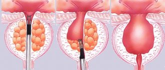
Treatment
The problem of varicose veins in the vessels of the spermatic cord and testicle cannot be eliminated. Improper operation of the blood outflow valves can be corrected through surgery. Surgical treatment of varicocele is the main method. The effectiveness of medication use is insignificant. Only the first stages of the disease can be treated conservatively. Consultation with a doctor and a joint decision on surgery for varicocele are mandatory. Surgical intervention is not always necessary for the patient. If the disease does not bother the man too much, or the patient is of advanced age, then doctors advise not to undergo surgery. A variety of surgical methods helps doctors find the only correct method. The essence of the operation for varicocele is to ligate the venous pathological vessels and normalize the outflow of blood. To save the results obtained during the operation, you should:
- limit physical and sexual stress;
- come to your doctor for examination.