Useful articles / September 7, 2020
Pregnancy is not only a happy time of waiting to meet your future baby, but also a test of the strength of the female body. One of the common problems that expectant mothers face is anemia. This physiological condition occurs quite often during pregnancy, so it is necessary to control the level of iron in the body at the stage of planning for replenishing the family. One of the important indicators that will tell you about iron deficiency is a ferritin test.
Anemia during pregnancy - ironclad arguments and dispelling myths
Anemia is one of the most common complications that occurs during pregnancy. In Russia, this diagnosis is made to every third pregnant woman. However, not everyone, when faced with this diagnosis, understands what we are talking about and what needs to be done to make the treatment as effective as possible.
Anemia is a disease in which the level of hemoglobin in the blood decreases, often with a simultaneous decrease in the number of red blood cells. The main reason for the development of anemia is the discrepancy between the intake of iron in the body and its expenditure.
During pregnancy, the costs of meeting the needs of the growing fetus cause a significant increase in the need for iron. In addition, a less common but possible cause of anemia may be insufficient intake of folic acid or vitamin B12.
Risk factors for the development of iron deficiency anemia during pregnancy include:
- history of heavy menstruation;
- diseases of the gastrointestinal tract;
- infectious and inflammatory diseases;
- anemia in the past;
- short interval between pregnancies, including conception during lactation;
- multiple pregnancy.
Since the main task of hemoglobin is to deliver oxygen - a vital element - to all tissues and cells of the woman and fetus, it is easy to imagine the harm caused by its decrease during pregnancy. However, even after childbirth the issue cannot be considered closed. It has been proven that low hemoglobin levels are associated with decreased lactation, as well as the development of anemia in the child.
Iron deficiency anemia is manifested by weakness, dizziness, pathological fatigue, distorted perception of tastes and smells, rapid heartbeat, shortness of breath, headache, and fainting. The skin becomes dry and pale, and hair and nails become brittle.
Anemia is diagnosed based on an assessment of the hemoglobin level in a general blood test. The lower limit of normal hemoglobin during pregnancy is 110 g/l. However, before hemoglobin decreases, iron stores are depleted, which is manifested by a decrease in serum ferritin levels. This condition is called latent iron deficiency and also requires correction.
Anemia and latent iron deficiency are treated with iron supplements, which are most often prescribed in the form of tablets or oral solution, but sometimes intravenous solutions are used. This need arises when the hemoglobin level is very low or in case of impaired absorption of iron from the gastrointestinal tract.
It is also important to remember the potential of a protein diet in correcting iron deficiency. Since hemoglobin is a bond of two subunits - the metal-containing heme and the globin protein - then if there is insufficient protein intake, even an adequate amount of iron in the body has nothing to bind with.
When diagnosed with anemia, it is important for the patient to remember that consuming foods high in iron will only help maintain the existing hemoglobin level, but will not be able to increase its level and saturate iron stores sufficiently.
Separately, I would like to dwell on which products contain really high iron content. A common misconception is that if you have anemia you need to eat apples, beets and pomegranates, and also drink pomegranate juice. 100 grams of apples contain 0.5 - 2.2 mg of iron; 100 grams of beets – 1.0 – 1.4 mg of iron; 100 grams of pomegranates - 0.78 mg of iron. Cucumbers, strawberries, pumpkin and other fruits and vegetables contain approximately the same amount of iron. For comparison, buckwheat contains 8 mg of iron per 100 g of product, dried fruits (dried apricots, prunes, dried apples) - from 12 to 15 mg of iron. The leader in iron content is pork liver. In addition, the content of this microelement is high in beef liver, cocoa, lentils, egg yolk, and heart.
Prevention of iron deficiency anemia in pregnant women is the study of iron reserves and hemoglobin levels at the stage of pregnancy planning, and if deviations from the norm are identified, their timely correction, consumption of foods high in iron, and intake of vitamin-mineral complexes containing preventive dosages of iron.
Obstetrician-gynecologist at antenatal clinic No. 14 Khivrich E.B.
Ferritin and pregnancy: from myths to truth
In the last few years, it has become fashionable to determine ferritin levels in all patients - from children to the elderly. Ferritin has been endowed with some kind of magical meaning/property around which diagnoses are formed, especially such a mysterious one as “hidden anemia”. Is there a place for such a diagnosis in modern medicine?
The most interesting thing is that most people suffering from “hidden anemia” find out about their disease completely by accident, without complaining about anything at all. They simply underwent a comprehensive examination for preventive purposes or in the presence of complaints unrelated to anemia, and it turned out that their iron reserves were “very bad”, which means they had hidden anemia.
It’s strange to receive letters with similar content: “I couldn’t get pregnant, the doctor said that I was infertile due to low ferritin. I took iron supplements for three+ months and became pregnant.” This is despite all other blood parameters being normal! It is unpleasant to watch how a pregnant woman is frightened by the terrible consequences of anemia, without knowing the physiology of anemia in pregnant women - in the vast majority of cases, anemia in pregnant women is not iron deficiency! One gets the impression that the craze for yet another microelement or mineral, in this case iron, arose against the background of the loss of popularity of other “vitamins” and “minerals”. If something stops generating income, you need to urgently find a replacement.
A few words about anemia
There are 8 types of anemia:
- Iron deficiency
- Pernicious
- Aplastic
- Thalassemia
- Hemolytic
- Fanconi
- sickle cell
- Gestational
The most common anemia is iron deficiency. In pregnant women, anemia is often physiological, and in most cases not due to iron deficiency.
In the past, anemia was often called anemia, meaning a condition that occurred after blood loss. Very little was known about other anemias.
Determining the type of anemia a person suffers from is key to evaluation and treatment . If a doctor diagnoses anemia based on one indicator of one analysis, or does not diagnose a specific type of anemia, then this indicates his incompetence in matters of hematology.
All types of anemia have many subtypes, when the breakdown can be both at the molecular level of structure, for example, hemoglobin, and at the level of iron absorption and or oxygen transfer by red blood cells. Blood is a dynamic system that contains a huge number of structures and substances that determine the unique structure and function of this human tissue .
The symptoms of different types of anemia are similar, so different factors are taken into account, in particular family (some anemia is hereditary), lifestyle (diet, alcohol abuse), and the presence of other diseases (autoimmune, cancer).
After collecting the history, an examination is carried out. Mild cases of anemia may be asymptomatic, so in such cases anemia can be detected accidentally by the result of a general blood test.
A few words about hemoglobin
The simplest and therefore most popular test is a general blood test. On the one hand, this is a cheap diagnostic method that does not require too much reagents or time. On the other hand, the abuse of this analysis depreciates its practical significance.
A quick look at the result of this test will show the level of hemoglobin , which is supposedly used to diagnose anemia. This is actually a relative indicator of anemia.
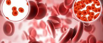
The human body contains 750 g of hemoglobin, which is mainly found in red blood cells (erythrocytes). One red blood cell contains 270 million hemoglobin molecules. Each molecule can combine and carry four oxygen molecules. Thus, one red blood cell can carry more than one billion oxygen molecules.
Several factors influence the ability of red blood cells to carry oxygen. Without focusing on biochemical reactions and substances produced directly in the body, several environmental factors affect oxygen transport. For example, being at high altitudes, where oxygen levels are lower, the human body is able to absorb and transfer more oxygen to the tissues. Also, cold temperatures increase the level of oxygen saturation of hemoglobin.
In adults, 97% of hemoglobin is type HbA, consisting of two alpha and beta chains, and about 2.5% of hemoglobin is type HbA2, 0.5% is fetal hemoglobin HbF.
It is not true that there are some cycles and periods of blood renewal (especially by which the gender of the child can be calculated). Red blood cells are destroyed and created daily. When red blood cells are destroyed, free hemoglobin appears in the blood - haptoglobin and hemopexin. There is always a certain level of free hemoglobin in the blood.
Haptoglobin is captured by monocytes and macrophages, special types of white blood cells that transport this type of hemoglobin (heme) to various tissues, which use it to produce bile pigments, iron and other substances.
Iron can circulate in the blood and be used for a variety of purposes, including recycling to make red blood cells. If there is too much free iron in the blood, it can cause kidney damage.
In determining the state of the blood, it is important to know not only the number of red blood cells (their concentration), or the level of hemoglobin, but also the size of the red blood cells and their color, as well as the volume, because with different types of anemia the structure and color of the red blood cells may be different.
A few words about iron deficiency
There are two different concepts - iron deficiency anemia and iron deficiency . In recent years, the second concept has begun to be abused due to increased testing of ferritin levels, which supposedly reflect iron stores in the body.
Iron deficiency anemia is accompanied by clinical signs, and the diagnosis is confirmed by several changes that can be found in a general blood test (low number of red blood cells, low hemoglobin, red blood cells are smaller than normal, the color of red blood cells is paler, etc.). In fact, iron deficiency anemia is microcytic hypochromic anemia (small and light red blood cells). With anemia of pregnancy, due to a significant increase in plasma (almost 40%), the concentration of erythrocytes decreases, but the size and color of erythrocytes does not change. On the contrary, the oxygen saturation of erythrocytes in pregnant women increases.
Iron deficiency anemia always implies iron deficiency, and when it is detected, doing a comprehensive examination with numerous tests, including checking iron reserves, is in most cases impractical. It is important to evaluate a person's nutrition first. If there are no serious reasons for anemia (nutrition is normal, cancer is eliminated, there is no bleeding due to uterine fibroids, for example), then a violation of iron absorption can be suspected.
Iron deficiency can be a hereditary or acquired disease associated with impaired absorption of iron due to a breakdown at the level of production of enzymes involved in this multi-stage process, as well as for other reasons. In such cases, serum ferritin, soluble transferrin receptor (sTfR), zinc protoporphyrin, reticulocyte hemoglobin, serum iron, hepcidin, total transferrin iron saturation, and a number of other biomarkers can help determine the cause.
Thus, iron deficiency will be accompanied by clinical symptoms and cannot be determined only by the level of ferritin in the blood.
A few words about ferritin
Ferritin is a protein containing iron, the level of which depends on the level of iron in the cells that use it (virtually all human cells). The more intracellular iron, the higher the ferritin level. It can also increase with inflammation, liver damage and a number of other diseases.
It is assumed that ferritin, which is determined in blood serum, is largely derived from macrophages (a type of monocyte or white blood cell) in the bone marrow. There is a lot of ferritin in the cells of the spleen and liver. It contains 24 subunits of light and heavy types. Different cells contain different types of these protein structural units.
Serum ferritin levels were first measured in 1972; in 1973, it was proposed that 1 μg/L serum ferritin was equivalent to approximately 8 mg of iron; and in 1982, Dr. Warwood and colleagues that ferritin levels reflect the level of iron reserves in the body. But is it?
In fact, serum is extremely low in ferritin. Serum ferritin plays absolutely no role in the transport and use of iron by cells. Transferrin plays this role. Serum ferritin consists of almost light chain subunits (L-form), its half-life is 30 hours, it contains virtually no iron and is almost 80% glycated (associated with sugar). In other words, serum ferritin is a plasma protein and the mechanism of its origin in the blood is still unknown. The half-life of non-glycated ferritin in cells when accidentally released into the blood is approximately 9 minutes.
A few words about diagnoses
To make a diagnosis, it is necessary to have diagnostic criteria, which is actually the basis of the diagnosis, that is, the definition of the disease. Pale skin, or dizziness, or a slightly decreased hemoglobin level are not enough to make a diagnosis of iron deficiency anemia. A combination of complaints, signs and examination results is required.
Determining ferritin levels is not enough to diagnose iron deficiency!
A few words about the reliability of the data
For a long period of time, doctors referred to the recommended hemoglobin levels proposed by the WHO in 2001 and the Centers for Disease Control and Prevention (CDC) in 1998. But few took into account that the recommendations were based on data obtained from studies in developing countries where people are malnourished, often go hungry, and therefore lack many vitamins and minerals.
All modern clinical research is still carried out in third world countries due to the low cost of biomaterials and the almost free work of medical personnel involved in research. The largest number of studies are conducted in India and China, where quality control of studies is low. Again, we take into account that most of the volunteers whose blood I study belong to low social levels. Research conducted in African countries does not meet the criteria of evidence-based medicine, as in former post-Soviet countries and South America. Therefore, any serious analysis (meta-analysis) and review of such studies from tens of thousands excludes almost 99% of publications not on the topic of iron, ferritin, and anemia. The last such analysis of publications on the topic of iron, ferritin and anemia in pregnant women was carried out in 2021 and it confirmed the fact that even the recommendations of professional societies are not based on reliable data and require revision.
Thus, in medicine still
- There is no clear definition of the value of serum ferritin and its role in determining iron stores,
- There is no knowledge about iron metabolism in detail, and especially in pregnant women,
- There are no reliable normal levels of serum ferritin, and especially for pregnant women,
- There is no clear definition of iron deficiency, especially in pregnant women.
In other words, the absorption and metabolism of iron in pregnant women has remained a blank spot in obstetrics and hematology, and all existing recommendations are based either on theoretical assumptions or on unreliable, inaccurate data.
Therefore, manipulation of serum iron levels, the concentration of which physiologically decreases during pregnancy, to make a non-existent diagnosis of “hidden anemia” or iron deficiency should not be used by doctors. Unfortunately, a total check of ferritin levels in everyone, including pregnant women, can easily be considered a commercial action that does not have a strict evidence base for the rationality and effectiveness of such testing.
Share link:
- Click to share on WhatsApp (Opens in new window)
- Click to share on Telegram (Opens in new window)
- Click here to share content on Facebook. (Opens in a new window)
- Click to share on Twitter (Opens in new window)
- Click to share on Skype (Opens in new window)
- Send this to a friend (Opens in new window)
- Click to print (Opens in new window)
By
Iron deficiency anemia during pregnancy - prevention and treatment
M.A. VINOGRADOV
, Ph.D., T.A.
FEDOROVA, Doctor of Medical Sciences, Professor, Scientific Center of Obstetrics, Gynecology and Perinatology named after.
acad. IN AND. Kulakov Ministry of Health of Russia This review is devoted to the problem of prevention and treatment of anemia during pregnancy. Iron deficiency anemia (IDA) is the most common deficiency condition and the most common form of anemia in pregnant women. Its clinical consequences are extremely important, since the adverse effects of iron deficiency affect not only the woman’s body, but can also affect pregnancy outcomes and the health of newborns. The first line of treatment for iron deficiency is iron preparations intended for oral administration, the most effective and safe form of which is currently considered an iron-polymaltose complex. In case of insufficient effectiveness and severe anemia, the preferred alternative method is intravenous iron supplementation. Timely diagnosis and adequate therapy make it possible to restore iron metabolism in a pregnant woman in the shortest possible time and prevent the development of complications.
Introduction
Iron deficiency is known to be the most common nutritional deficiency condition in the world [1] and the most common cause of anemia in pregnant women (up to 75%) [2]. According to the World Health Organization definition, anemia during pregnancy is considered to be a decrease in blood hemoglobin of less than 110 g/l [1], and in the second trimester - less than 105 g/l [3]. It is known that during pregnancy a number of physiological changes occur in a woman’s body, including blood changes. The total plasma volume increases to 50% of the original, and the globular volume increases only by 25% [4, 5]. As a result, the need for microelements and vitamins necessary for the synthesis of hemoglobin and ensuring the normal development of the fetus and placenta increases. In the absence of adequate replenishment of increasing needs, a deficiency of microelements, primarily iron, develops, and, as a consequence, anemia. This is due to many factors: pregnancy often occurs with an initially reduced level of hemoglobin or insufficient iron reserves in the body, which can be caused by diet, chronic diseases of the gastrointestinal tract, or prolonged heavy menstruation. Iron is an essential trace element for humans. During pregnancy, adequate iron status is an important prerequisite for normal fetal development and neonatal maturity. It has been shown that severe anemia with a decrease in hemoglobin less than 90 g/l can contribute to pregnancy complications and adversely affect its outcomes [6]. There is evidence that iron deficiency, even in the absence of iron deficiency anemia (IDA), can also have negative effects on non-pregnant women, for example in relation to cognitive abilities and physical performance. In addition, IDA during pregnancy is associated with a risk of preterm birth and low birth weight [7].
Assessment of iron status in the body
To adequately assess iron metabolism and timely detect iron deficiency, it is necessary to use a number of laboratory tests. In addition to a complete blood count, which provides insight into hemoglobin levels and red blood cell characteristics, iron status can be assessed primarily by testing serum ferritin. Additional tests include transferrin saturation and serum soluble transferrin receptor (sTfR). Serum iron is not a reliable diagnostic parameter, so its study is not enough to clarify the cause of anemia. An isolated hemoglobin test is not suitable for assessing anemia during pregnancy due to the presence of varying degrees of hemodilution in patients [8]. Ferritin provides information about the completeness of iron stores in the body, sTfR and transferrin saturation provide information about the development of iron deficiency at the cellular level, and hemoglobin provides information about iron deficiency at the functional level. For practical purposes, a complete blood count and serum ferritin are sufficient tests to assess iron status and diagnose IDA in most women. It is important that in the presence of an inflammatory process, the ferritin value may be unreliably high. In this case, an increase in C-reactive protein (CRP) is confirmation of an inflammatory process that requires treatment. The diagnostic algorithm for detecting anemia is presented in Figure 1 [9].
Iron requirements during pregnancy
Iron requirements during pregnancy increase from 0.8 mg/day in the first trimester to 7.5 mg/day in the third trimester (average 4.4 mg/day). On average, a normal pregnancy requires an additional approximately 1,240 mg of iron [10, 11]. Studies have shown that many non-pregnant women have reduced iron stores: 42% have serum ferritin less than 30 μg/L, and only 14-20% of them have ferritin levels exceeding 70 μg/L, [12], i.e., iron stores balance the needs of normal pregnancy [10].
We can identify groups of pregnant women who have a higher risk of developing iron deficiency: multiple pregnancies, multiple pregnancies with a short intergravid interval, blood donors, vegetarians, women with low socioeconomic status, patients with chronic diseases of the gastrointestinal tract [13]. It has been proven that there are significant differences between hemoglobin levels in women who receive additional iron during pregnancy and those who do not [14]. In addition, hemodilution causes physiological fluctuations in hemoglobin levels during pregnancy [15]. In women receiving iron supplements during pregnancy, hemoglobin concentrations show a steady decline from the end of the first trimester due to hemodilution, reaching a minimum at 25 weeks of pregnancy. Subsequently, hemoglobin rises during the remainder of pregnancy to reach a peak level shortly before delivery.
Prevention of iron deficiency
Placebo-controlled studies have consistently shown that pregnant women taking iron supplements have significantly higher iron stores compared with women taking placebo [16]. Consequently, women taking iron have a lower incidence of anemia.
Previously, high doses of ferrous iron in the range of 100-200 mg/day were used to prevent iron deficiency in pregnant women. A dose of 100 mg of ferrous iron per day induces a maximum increase in hemoglobin, and 200 mg of iron per day increases ferritin and hemoglobin during childbirth as well as in non-pregnant women. However, the results of the work highlighted the potential negative effects of using such doses of iron [14], and consequently, studies were initiated to study the effectiveness of lower doses of iron, attempting to determine the smallest effective dose [17]. European studies have shown that supplementation with 45–66 mg of ferrous iron per day from 12–20 weeks of pregnancy until delivery is sufficient to prevent IDA in healthy pregnant women. Even lower doses of 20–27 mg of ferrous iron per day have a beneficial effect on iron status [16]. The main conclusions from these studies are, first, that 30–40 mg of ferrous iron per day is sufficient to prevent IDA, and, second, that low-dose iron supplements in the range of 20–27 mg/day are better than no supplementation. whereas studies of daily consumption of multivitamin preparations containing 14–18 mg of ferrous iron showed no effect on iron status in women [13]. Currently, in order to prevent iron deficiency in pregnant women, it is recommended to use an oral iron supplement at a dose of 30-40 mg/day from the beginning of pregnancy until childbirth [14].
Thus, an individualized approach to the prevention of IDA is currently recommended, which is based on the assessment of iron stores (plasma ferritin) before and at the beginning of pregnancy. Women with ferritin levels greater than 70 mcg/l are not advised to take iron supplements; with ferritin values of 30-70 µg/l, 30-40 mg of ferrous iron per day should be taken, and with ferritin levels less than 30 µg/l, 80-100 mg of ferrous iron should be taken per day [14].
Treatment of anemia
If IDA is detected, the deficiency should be corrected with oral or intravenous administration of iron supplements. Intramuscular administration of iron is currently practically not applicable due to the limited dose of iron administered per injection and the high incidence of painful local reactions.
Iron preparations for oral administration
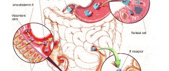
Iron absorption is regulated in accordance with iron reserves in the body and the intensity of erythropoiesis. It has been proven that in case of depletion of iron stores in the body, intestinal absorption of iron increases [18]. In addition, increased erythropoietin-induced erythropoiesis in the second and third trimester [19] stimulates iron absorption.
This combined stimulation of iron absorption has been confirmed in studies showing that with increasing gestational age there is an increase in iron absorption, and this is most pronounced after 20 weeks of pregnancy. The ability to absorb a significant amount of iron through the gastrointestinal tract is favorable for the treatment of IDA with tableted iron supplements in pregnant women. In this regard, the administration of oral iron supplements is first-line therapy, especially during the first and second trimesters of pregnancy [20]. For mild anemia with hemoglobin 90-105 g/l, the recommended dose is 100-200 mg of elemental iron per day. After oral treatment with iron supplements for 2 weeks. the effect should be assessed. If hemoglobin increases by more than 10 g/l, therapy should be continued during the remaining period of pregnancy with subsequent monitoring of hemoglobin and ferritin [13].
Refractoriness of anemia to therapy may be a consequence of non-compliance with the drug due to subjective reasons, gastrointestinal side effects, impaired iron absorption due to achlorhydria or inflammatory bowel disease, or hidden ongoing bleeding with iron loss. Gastrointestinal disorders, such as colic, nausea, vomiting, and diarrhea, occur in approximately 6–12% of patients taking iron supplements [21]. Until recently, iron salts were most widely used internally. However, their use is limited by low and uneven absorption, in particular dependent on food products [22]. Ferric compounds were created to avoid these problems. In the first trimester of pregnancy, the severity of iron deficiency, on the one hand, and a sufficient supply of time, on the other, allow for a smooth correction of iron deficiency conditions using the safest oral iron preparations.
The iron-polymaltose complex was developed as a molecule that dissolves at neutral pH. The drug contains iron in the form of a polymaltose complex of iron (III) hydroxide (PKH), for example Maltofer. This complex is stable and does not release iron in the form of free ions into the intestines. The structure of the drug is similar to ferritin. Due to this similarity, iron (III) moves from the intestine into the blood through active absorption. Iron, which is part of PKZh, does not have the pro-oxidant properties inherent in simple iron salts. Studies to evaluate the effectiveness and safety of polymaltose drugs in comparison with ferrous sulfate in pregnant women have shown their undoubted advantage. Polymaltose iron complex has been formulated so that the elemental form of iron is in a non-ionic state. This ensures that its use does not cause irritation to the gastric mucosa. Additionally, the high iron content eliminates the need for frequent dosing and therefore improves compliance. The polymaltose complex (Maltofer) should be taken during or immediately after meals, which also increases ease of use. Interesting data were obtained in an experiment using iron salts and PLC in pregnant rats. All treatment methods were effective in correcting anemia. However, as a result of the use of iron salts, signs of liver damage and oxidative stress were noted in the study of the fetus and placenta. PLC restored normal expression of TNF-α and IL-6 in the placenta, while the highest levels of cytokines were observed with ferrous sulfate, suggesting a local inflammatory response. Most of the negative effects associated with IDA were resolved by the administration of PKZh. Pregnancy outcomes with the use of iron salts were worse when PCZ was prescribed [23].
The use of various iron preparations in adults has been studied many times. A study by Badhwar et al [24] in both women and men with IDA demonstrated equivalent efficacy and better bioavailability of PCZH compared to ferrous fumarate. However, this study was conducted in non-pregnant women. A study by Pakar et al [25] also demonstrated the effectiveness and safety of PLC in both pregnant and non-pregnant women.
Important results were obtained in a randomized, double-blind, controlled trial of the use of PCZ during pregnancy [26].
The incidence of side effects was significantly higher in the iron salts group than in the PCZ group (78 vs. 31%, p < 0.001). The increased incidence of adverse effects of iron salts may be due to the release of free radicals, leading to cell damage and death [27], whereas PLC does not release free radicals. Reducing the incidence of side effects improves patient compliance and ensures regular treatment. In addition, better tolerability is essential to ensure long-term therapy during pregnancy. PLC is an effective therapeutic approach in the treatment of IDA in pregnant women. The improved tolerability profile compared to iron salt preparations and the equivalent efficacy profile strongly suggest that PKZh is the preferred form of oral iron for the treatment of IDA during pregnancy. Significantly better tolerability of PCZH in comparison with quickly satiating, but less tolerated preparations of inorganic iron salts ensures higher compliance of patients, contributing to the formation of a positive stereotype of daily drug use until the end of pregnancy, which is fundamentally necessary.
Iron preparations for intravenous administration
Treatment with intravenous iron is superior to oral iron in terms of the speed of restoration of hemoglobin and replenishment of iron reserves in the body. However, the safety of such drugs in the first trimester has not been sufficiently proven, so they can be recommended for use from the second or third trimester of pregnancy. Intravenous iron reduces the need for blood transfusions and is an alternative to transfusions in severe IDA. Currently, the most effective therapeutic approach, allowing to safely obtain the maximum effect in the shortest possible time, is the use of iron carboxymaltose [28]. This is a dextran-free complex that can be used in maximum doses (up to 1000 mg per intravenous injection) in a short period of time (15-30 minutes per infusion). Repeated infusions are carried out weekly at a rate of 15 mg of iron per kg of body weight. According to the Cochrane Database [29], among drugs for intravenous use, iron carboxymaltose is the drug of choice for the treatment of IDA during pregnancy. Intravenous iron supplements are considered safe in the second and third trimesters of pregnancy [30], although intravenous iron infusions should be administered by health care personnel to avoid possible allergic or other reactions. Treatment with intravenous iron preparations is indicated in cases of ineffectiveness of iron-containing drugs for oral administration (no increase in hemoglobin by 10 g/l within 2 weeks), with severe IDA (hemoglobin <90 g/l during pregnancy more than 14 weeks, and also as first-line treatment for IDA in the third trimester. At this stage there is insufficient time for oral therapy to be effective. This is important to reduce the manifestations of IDA and replenish iron stores before delivery to prevent anemia in labor and avoid blood transfusions The dose of intravenous iron should be sufficient to achieve a hemoglobin level of more than 105 g/l. For most women, a total dose of 1,000-1,250 mg of intravenous iron is adequate. When the hemoglobin level reaches 105 g/l, a transfer to maintenance therapy with an oral iron preparation is carried out 100 mg/day until the end of pregnancy.
Conclusion
Iron deficiency anemia is the most common form of anemia in pregnant women (up to 95%). The diagnosis of IDA is based on identifying a decrease in blood hemoglobin and serum ferritin levels. Among non-pregnant women of reproductive age, up to 40% have insufficient iron reserves in relation to the upcoming pregnancy, therefore, issues of timely correction of iron deficiency before the development of anemia are extremely important when planning pregnancy. For this purpose, complex preparations are used containing from 30 to 80 mg of iron, depending on the values of serum ferritin. Given the increasing iron requirements during pregnancy, dietary measures are insufficient to correct iron deficiency. Treatment of IDA during pregnancy should be carried out using drugs for oral administration, the most preferred of which is polymaltose complex of iron (III) hydroxide and iron preparations for intravenous administration. For IDA with a hemoglobin level of more than 90 g/l, the first line of therapy is PKH at a dose of 100-200 mg/day. Indications for prescribing an intravenous iron supplement are anemia with a hemoglobin level of less than 90 g/l, insufficient effect of therapy with oral medications (hemoglobin less than 100 g/l for 2 weeks) or poor tolerability. Timely detection and effective therapy, safe for pregnant women, allows you to quickly normalize hemoglobin levels and improve iron reserves, which, in turn, improves the quality of life of women and prevents the development of pregnancy complications. Taking into account the inevitability of the development of one or another degree of iron deficiency during gestation in the vast majority of pregnant women and taking into account the negative long-term consequences of iron deficiency on the ante- and postnatal development of the child, the tactics of early initiation of anemia treatment should be considered the most justified.
Literature
1. WHO Iron Deficiency Anaemia: Assessment, Prevention and Control. WHO/NHD/01.3, World Health Organization, 2001, Geneva, Switzerland. 2. Sifakis S and Pharmakides G. Anemia in pregnancy. Ann. NY Acad. Sci., 2000, 900: 125–36. 3. Ramsey M, James D & Steer P. Normal Values in Pregnancy, 2nd edn. WB Saunders, London, 2000. 4. Coad J, Conlon C. Iron deficiency in women: assessment, causes and consequences. Current opinion in clinical nutrition and metabolic care. 2011, 14, 625-634. 5. Friedman AJ et al. Iron deficiency anemia in women across the life span. Journal of women's health, 2012, 21: 1282-1289. 6. The Obstetric Hematology Manual edited by Sue Pavord, Beverley Hunt. Cambridge University Press 2010. pp. 13-27. 7. Ren A, Wang J, Ye RW, Li S, Liu JM, Li Z (2007). Low first trimester hemoglobin and low birth weight, preterm birth and small for gestational age newborns. Int J Gynaecol Obstet 98: 124–128. 8. Koller O (1982). The clinical significance of hemodilution during pregnancy. Obstet Gynecol Surv 37:649–652. 9. Breymann Ch et al. Diagnosis and treatment of iron-deficiency anemia during pregnancy and postpartum. Arch Gynecol Obstet 2010, 282:577-580. 10. Milman N (2006). Iron and pregnancy - a delicate balance. Ann Hematol 85:559–565. 11. Bothwell TH (2000). Iron requirements in pregnancy and strategies to meet them. Am J Clin Nutr 72:257–264. 12. Milman N, Byg KE, Ovesen L (2000). Iron status in Danes updated 1994. II. Prevalence of iron deficiency and iron overload on 1319 women aged 40–70 years. Influence of blood donation, alcohol intake, and iron supplementation. Ann Hematol 79: 612–621. 13. Milman N. Prepartum anemia: prevention and treatment. Ann Hematol 2008, 87:949–959. 14. Milman N (2006). Iron prophylaxis in pregnancy—general or individual and in which dose? Ann Hematol, 85: 821–828 doi:10.1007/s00277-006-0145-x. 15. Milman N, Bergholdt T, Byg KE, Eriksen L, Hvas AM (2007). Reference intervals for haematological variables during normal pregnancy and postpartum in 433 healthy Danish women. Eur J Haematol 79: 39–46. 16. Makrides M, Crowther CA, Gibson RA, Gibson RS, Skeaff CM (2003). Efficacy and tolerance of low-dose iron supplements during pregnancy: a randomized controlled trial. Am J Clin Nutr 78: 145–153. 17. Milman N, Bergholt T, Eriksen L, Byg KE, Graudal N, Pedersen P, Hertz J (2005). Iron prophylaxis during pregnancy — how much iron is needed? A randomized, controlled study of 20 to 80 mg ferrous iron daily to pregnant women. Acta Obstet Gynecol Scand 84: 238–247. 18. Skikne B, Baynes RD (1994). Iron absorption. In: Brock JH, Halliday JW, Pippard MJ, Powell LW (eds) Iron metabolism in health and disease. Saunders, London, pp. 151–187. 19. Milman N, Graudal N, Nielsen OJ, Agger AO (1997). Serum erythropoietin during normal pregnancy: relationship to hemoglobin and iron status markers and impact of iron supplementation in a longitudinal, placebo-controlled study on 118 women. Int J Hematol 66: 159–168. 20. Beris P, Maniatis A on behalf of the NATA working group on intravenous iron therapy (2007). Guidelines on intravenous iron supplementation in surgery and obstetrics/gynecology. TATM transfus Altern Transfus Med 9(Suppl 1): 29. 21. Adamson JW. Fauci AS. Kasper DL, et al. Iron deficiency and other hypoproliferative anaemias. In: Braunwald E, editor; Harrison's Principles of Internal Medicine.15th edition. Mc Graw Hill; 2001. pp. 660–66. 22. Sharma N. Iron absorption: IPC therapy is superior to conventional iron salts. Obstet Gynecol., 2001: 515–19. 23. Toblli JE, Cao G, Oliveri L, Angerosa M. Effects of iron polymaltose complex, ferrous fumarate and ferrous sulfate treatments in anemic pregnant rats, their fetuses and placentas. Inflamm Allergy Drug Targets, 2013, 12(3): 190-8. 24. Badhwar VR, Rao S, Fonseca MM. Comparative efficacy and safety of iron polymaltose+folic acid and oral ferrous fumarate in the treatment of adult patients with iron deficiency anemia. Indian Med Gazette 2003, 136: 296–301. 25. Patkar VD, Patkar S, Khandeparker PS, Dingankar NS, Shetty RS. Evaluation of efficacy and tolerability of iron (III) – hydroxide polymaltose complex tablets in the treatment of iron deficiency anemia in women. Indian Med Gazette 2001, 135: 306–309. 26. Saha L, Pandhi P, Gopalan S, Malhotra S and Saha PK. Comparison of Efficacy, Tolerability, and Cost of Iron Polymaltose Complex With Ferrous Sulphate in the Treatment of Iron Deficiency Anemia in Pregnant Women. MedGenMed, 2007, 9(1): 1. 27. McCord JM. Iron, free radicals, and oxidative injury. Semin Hematol., 1998, 35: 5–12. 28. Christoph P, Schuller C, Studer H et al. Intravenous iron treatment in pregnancy: comparison of high-dose ferric carboxymaltose vs. iron sucrose. J Perinat Med, 2012, 13, 40(5), 469-474. 29. Reveiz L, Gyte GML, Cuervo LG, Casasbuenas A. Treatments for iron-deficiency anemia in pregnancy (Review), Cochrane library, 2011. 30. Beris P, Maniatis A, on behalf of the NATA working group on intravenous iron therapy (2007) Guidelines on intravenous iron supplementation in surgery and obstetrics/gynecology. TATM transfus Altern Transfus Med 9(Suppl 1):29.
Source:
Medical Council, No. 9, 2015
Material under study
Deoxygenated blood.
Interpretation of research results contains information for the attending physician and is not a diagnosis. The information in this section should not be used for self-diagnosis or self-treatment. The doctor makes an accurate diagnosis using both the results of this examination and the necessary information from other sources: medical history, results of other examinations, etc.
Reference values:
- The level of ferritin in the blood in the first 2 months reaches 600 mcg/l.
- At the age of 2 months to six months, normal values range from 55 to 210 mcg/l.
- From six months to 15 years from 8 to 143 µg/l.
- After the onset of puberty in a young man, the reference values are: 21 - 250 mcg/l.
- In women under 15 years of age, the norm is from 7 to 120 mcg/l.
- Normal values for girls over 15 years old are from 10 to 125 mcg/l.
- According to information from the Obstetric Hematology Center, the ferritin norm during pregnancy is: in the 1st trimester - about 50% of the norm for a non-pregnant woman;
- in the 2nd trimester - about 30%;
- and in the 3rd trimester - at least 12%.
Indications for the purpose of the study
- Hereditary hemochromatosis. In this disease, too much iron is absorbed from food and deposited in various organs, causing them to become damaged.
- Multiple blood transfusions, intramuscular iron administration, administration of iron tablets.
- Inflammations, such as upper respiratory tract infections, urinary tract infections, autoimmune diseases. Moreover, an increase in ferritin in the acute phase of inflammation can mask the existing iron deficiency.
- Acute or chronic liver diseases.
- Alcoholism.
- Hemolytic anemia: associated with the destruction of red blood cells, B12 deficiency anemia, thalassemia.
- Hyperthyroidism is an increase in thyroid function.
- Oncological diseases of the bone marrow, breast cancer, Hodgkin's disease - a malignant neoplasm of lymphoid tissue. Ferritin levels will increase significantly.
Prices
| Ferritin | 360 ₽ |
All prices
Name in English: ferritin.
Ferritin is a complex protein complex (iron protein) that serves as the main intracellular iron depot in humans and animals. Structurally, it consists of the protein apoferritin and a ferric atom in the composition of phosphate hydroxide. One ferritin molecule can contain up to 4000 iron atoms. Contained in almost all organs and tissues and is a donor of iron to cells that need it.
In a situation where iron begins to be scarce (frequent blood loss or lack of its supply with food), the human body begins to use its reserve from the tissue. Ferritin levels begin to decline. A long-term lack of iron intake can lead to anemia. Ferritin levels can be reduced long before the symptoms of iron deficiency appear and allow anemia to be diagnosed in time.
Along with this, hemoglobin levels also decrease. The condition is accompanied by insufficient oxygen supply to the cells and tissues of organs throughout the body. The central and peripheral nervous system suffers to a greater extent.
Ferritin levels during pregnancy
Ferritin testing is not included in the compulsory medical insurance standards, so the expectant mother may begin to have problems with iron deficiency. If a woman has low ferritin, then a course of iron supplements is prescribed before conception. This will not only protect the expectant mother herself from feeling unwell during pregnancy, but will also help the baby develop normally in the first year of life.
By all international standards, a ferritin test is a simple diagnostic test for identifying iron deficiency,” says Anastasia Arseneva. — It answers the question: is there iron deficiency or not. But this analysis is not included in the compulsory medical insurance standards; a gynecologist can prescribe it, or he can limit himself to OAC. Most often, it happens that in the KBC hemoglobin does not cause cause for alarm, but iron reserves are depleted. In the 2nd trimester, iron begins to be sharply lacking because the body needs to make more blood cells, providing for the growing placenta (so that the baby receives more oxygen). And then the body takes iron from ferritin reserves. Food cannot provide the required amount of iron, even if a woman eats for two. If a pregnant woman is prescribed iron supplements when hemoglobin has decreased, this is already a missed opportunity. If you prescribe medications in advance, the expectant mother will waste the iron she receives from tablets, and not from her reserves. In this case, ferritin will not decrease and hemoglobin will remain normal. Yes, all women have physiological anemia, when hemoglobin decreases. But it cannot decrease indefinitely, the limit is 110 g/l. If hemoglobin drops below, it is necessary to check whether physiological anemia has become pathological.
In the latent stage of iron deficiency there are not always symptoms, but it is necessary to correct this condition. When a woman is preparing for pregnancy, ferritin levels also need to be known. If it is at the level of 30-40 mcg/l, then most likely she needs prophylactic medication, since ferritin will decrease during pregnancy. The ideal ferritin for a woman is from 50 to 150 mcg/l. With such indicators, the woman feels good, she has enough energy and there is no chronic fatigue.