Ringworm (microsporia) is a disease manifested as a fungal infection of the skin, nail plates and hair follicles. The pathogen is a mold fungus of the genus Microsporum. Its colonies form in keratinized substrates. Microsporia remains a relatively common disease - dermatologists identify 60-75 cases for every hundred thousand Moscow residents. The pathology has a pronounced seasonality. The peak incidence occurs at the end of summer and beginning of autumn - the period of breeding of offspring in cats and other animals.
Routes of infection
The causative agent of microsporia enters the body when a healthy person comes into contact with a carrier of the disease. An alternative way is to interact with objects covered with fungal spores. Ringworm is most often detected in children aged 5-10 years; in boys, microsporia is diagnosed five times more often than in girls. The pathology almost does not affect adults due to the presence of organic acids in their hair structure, which suppress the growth of fungal mycelium.
The reasons for the development of ringworm are microtraumas of the skin and its dryness. Spores get into cracks, scratches or open calluses. Healthy skin becomes an insurmountable barrier to fungus. The pathogen does not survive contact with personal hygiene products - thorough hand washing after contact with spore carriers eliminates the possibility of infection.
The risk group includes people who regularly come into contact with the ground and wild animals. The active growth of the fungus is facilitated by disturbances in the functioning of the sebaceous glands due to changes in the chemical composition of their secretions. Microsporum spores can remain viable for three months when left in open ground.
Causative agents of ringworm
Today, 12 species of fungi of the genus Microsporum have been studied and described. Pathogens of microsporia that are of interest to doctors are represented by 3 groups:
- The zoophilic group is represented by Microsporum canis and Microsporum distortum. Mostly animals - cats and dogs - are infected. The infection from them is transmitted to humans.
- The anthropophilic group is represented by Microsporum audouinii (more often) and Microsporum ferrugineum (less often). Mostly people get sick, rarely animals.
- The geophilic group is represented by Microsporum gypseum and Microsporum nanum. They live in the soil. The disease is rarely caused.
Rice. 7. Ringworm in dogs and children.
Microsporum canis (furry, feline)
Cat microsporum is the most common in the Russian Federation.
Type of colonies (macroscopic picture):
- When growing on nutrient media, fungal colonies are powerful, mealy in the center, loose and fluffy along the periphery.
- The reverse side of the colonies is red-brown in color, giving the overall appearance of the colonies a salmon tint.
Type of pathogens under a microscope:
- The mycelium is bamboo-shaped and consists of rocket-shaped cells. The hyphae are septate and produce numerous macroconidia.
- On the filaments of the mycelium there are large exospores (macroconidia) of a spindle-shaped form, they have a jagged-villous shell, spinous, multi-chambered, the shell is 2-contour.
- On the sides of the branches of the mycelium, microconidia are formed, which are single-celled formations of a round or pear shape, located singly or in groups.
- The organs of vegetative reproduction are represented by chlamydospores - round-shaped cells.
Rice. 8. Microscopic picture of microsporum fluffy. The mycelium (photo on the left) and numerous exospores (photo on the right) are clearly visible.
Rice. 9. The photo shows exospores of the pathogen. They are large, spindle-shaped, located on mycelial filaments.
Rice. 10. Colonies of microsporum fluffy fungi.
Microsporum ferrugineum (rusty microsporum)
Rusty microsporum rarely causes disease.
Type of colonies (macroscopic picture):
- Colonies of pathogens are wide, lumpy or flat, leathery, dome-shaped in the center, divided into convex sectors by grooves.
- They have a brownish, yellowish or reddish color.
- Some colonies are waxy, yellowish, line-like, folded, or whitish-powdery, finely lumpy.
Type of pathogens under a microscope:
- The mycelium of mushrooms is wide.
- Chlamydospores are 30 microns in diameter.
- Macroconidia are absent.
- Microconidia are rarely present.
Rice. 11. Microsporum ferrugineum colonies.
Rice. 12. Rusty microsporum under a microscope.
Microsporum gypseum
Microsporum gypseum is distributed throughout the world. They live in the soil. People cultivating the soil are getting sick. The disease affects hair and smooth skin. In addition to microsporia, they cause skin infections in the form of herpes zoster Corporis and Capitis. There is information about nail damage. The pathogen has been isolated by some researchers from dogs, horses, cats and rodents.
Type of colonies (macroscopic picture):
- Fungal colonies grow quickly.
- Flat and powdery in appearance, later a velvety-looking raised area forms in the center.
- They have a yellow-pink color and yellow on the reverse side.
Type of pathogens under a microscope:
- Macroconidia (exospores) are numerous, spindle-shaped, blunt-pointed, wide, smooth.
- Microconidia are numerous and oval or pear-shaped.
Rice. 13. Colonies of Micrsporum gypseum.
Rice. 14. View of pathogens under a microscope. Numerous spindle-shaped exospores are visible.
Symptoms of pathology
Symptoms of ringworm appear 4-6 weeks after the patient becomes infected. A red spot appears on a smooth area of skin of a child or adult. It rises above the surface and has smooth boundaries. Over time, the size of the lesion increases. The surface becomes covered with nodules, blisters and scabs. The spots develop into rings that can intersect or merge with each other. The diameter of skin formations ranges from 5 to 30 millimeters.
Signs of ringworm include an acute inflammatory reaction. It often develops in children and girls. The lesions begin to peel off intensively. Patients suffering from dermatitis do not immediately identify symptoms of microsporia. Areas of active fungal growth may appear as inflammatory skin lesions.
A common manifestation of ringworm in a child is damage to the scalp. This symptom affects patients aged 5-12 years. Older children experience changes in the chemical composition of sebum. Its elements become dangerous for the microsporia pathogen.
The suppurative type of ringworm is characterized by the appearance of soft nodules on the patient’s skin. The nodes are dotted with numerous abscesses. When they are compressed, purulent contents are released.
Are you experiencing symptoms of ringworm?
Only a doctor can accurately diagnose the disease. Don't delay your consultation - call
State Autonomous Institution "Regional Clinical Dermatovenerological Dispensary"
Definition.
Microsporia is a fungal disease affecting hair, smooth skin with or without involvement of vellus hair, and extremely rarely nails, caused by fungi of the genus Microsporum.
In addition to its medical name, this fungal disease has another common name - ringworm.
The term “ringworm” is the traditional designation for a group of diseases of the skin and scalp in which the hair is affected and breaks off, resulting in the formation of bald spots. And since 100 years ago doctors were not able to identify infectious agents due to the lack of appropriate techniques, all diseases were classified, described and named mainly by external manifestations. That is why microsporia was called ringworm. However, with the development of science and technological progress, doctors were able to identify not only signs of diseases, but also isolate their causative agents, which was literally a breakthrough. During this period, it was possible to establish that the disease, which has always been called ringworm, can be caused by two types of pathogenic fungi - Trichophyton and Microsporum. And then the type of ringworm caused by fungi of the genus Trichophyton began to be called trichophytosis, and Microsporum - accordingly, microsporia. But since the external signs and course of trichophytosis and microsporia are very similar, these two infections retain the same common name - ringworm. Thus, according to modern concepts, microsporia is a fungal infection (mycosis) that affects the skin, hair, and very rarely nails, and at the same time is considered one of the types of ringworm.
Relevance.
Currently, microsporia is the most common mycosis in children.
The causative agent of infection. Among the fungi of the genus Microsporum there are about 20 species that can cause disease. Fungi of the genus Microsporum are resistant to damaging environmental factors, as well as to various antifungal drugs, which is due to their structural features. This type of fungus has an ultrastructurally dense thick wall, consisting of 6 layers, reinforced by costal protrusions. These fungi can remain viable in hair for up to 10 years, and in skin scales for up to 7 years.
Epidemiology.
Sources of microsporia can be animals, people and soil.
The main animals involved in the preservation and transmission of infection are cats, especially kittens (70-80%), less often dogs (4%). Light-colored cats and brindle cats are especially often affected by microsporia, apparently this is due to reduced resistance to fungi in these cat species.
Contact with stray cats and dogs, which often turn out to be sick with microsporia, is extremely dangerous.
Infection with this mycosis is also possible from a person with microsporia, more often from children (3-10%), and extremely rarely from soil (0.7%).
Rare animals that suffer from microsporia and can be a source of infection for people include monkeys, tigers, lions, wild and domestic pigs (especially piglets), horses, sheep, silver-black foxes, rabbits, rats, mice, hamsters, guinea pigs and others small rodents, as well as poultry.
Mechanism of infection
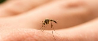
Most often, microsporia is infected directly from a sick animal, when they play with it, wash it, warm it under a shirt, let it into bed, and also through environmental objects infected by it. And since kittens are most often carriers of microsporia, there are two seasonal peaks in the incidence of this infection - in mid-summer and autumn, when cats give birth. To become infected with microsporia, it is enough to pet a cat or dog that has the infection or is an asymptomatic carrier.
Infection with microsporia is possible not only through direct contact with a sick animal, but also through touching objects that have fur and skin flakes of an infected animal.
The fact is that cats and dogs that suffer from microsporia or are carriers of the fungus can leave small and unnoticeable pieces of hair on various household items (furniture, carpets, beds, sofas, armchairs, clothes, shoes, etc.), in which contains fungal spores. A person touching such pieces of wool containing fungal spores also becomes infected with microsporia.
Transmission of infection from a sick person to a healthy person through direct close contact or through the use of various objects on which there are skin flakes of an infected person (for example, when using a comb, hat, hair scissors belonging to a person with microsporia).
At home, the subject of infection can be bed linen, towels, clothing, bedding for animals and animal care items.
In the entrances of houses and courtyards, the infection can be transmitted through door mats, dust from staircases, sand from children's sandboxes, and water from puddles.
The source of infection for newborns can be a baby stroller left overnight in the entrance of a house and favored by cats.
In hairdressing salons, if sterilization is improper, you can become infected through hair clippers, scissors, combs, negligees, curlers, hair dryers, and wash brushes.
If a fungus simply gets on the skin, a person will not get sick with microsporia, since the pathogenic fungus will be destroyed by normal microflora and the immune system or simply washed away during hygiene measures.
Thus, for the disease microsporia it is necessary not only for the fungus to get on the skin, but also for the presence of certain predisposing factors that will allow it to penetrate the skin and provoke an infection. These include: 1. Traumatic skin injuries; 2. Skin maceration; 3. Reduced immunity 4. Failure to comply with hygiene measures.
Interesting Facts
An increase in the incidence of microsporia is observed from June to November, then decreases to a minimum by April.
Microsporia is more often recorded in cities with multi-storey buildings, where stray animals come into contact with domestic ones. The incidence among stray animals reaches 50%.
Microsporia in children is much more common in children than in adults, which is explained by two main factors. Firstly, children are more likely to come into contact with sick animals, and accordingly, they have a higher risk of contracting an infection. And secondly, the sebaceous glands of the skin of children do not produce acids that have a destructive effect on fungi. That is, a fungus that gets on the skin of a child is much more likely to cause microsporia than in an adult in a similar situation, since after puberty the glands begin to produce acids that have a detrimental effect on microsporia pathogens. It is important that children have less developed hygiene skills than adults.
Clinic
The incubation period is in most cases 1-2 weeks.
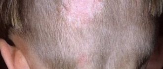
With microsporia of the scalp, single 1 or 2 lesions up to 3-5 cm in diameter appear, with a regular round-oval shape with clear boundaries. The hair in the lesions becomes dull, grayish, broken off at the same level at a height of 4-6 mm, as if trimmed. At a quick glance, the lesions resemble the appearance of “gray spots.” The surface of the lesions is covered with grayish scales, as if lightly sprinkled with flour.
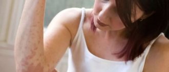
With microsporia of smooth skin, lesions are most often observed on the face, neck, hands, forearms, shoulders, but can also be on the torso. Typical are round or oval erythematosquamous edematous spots from 0.5 to 2-3 cm in diameter, surrounded by a continuous elevated peripheral ridge on which blisters and quickly drying crusts are visible. Microsporia can also affect vellus hair.
Diagnosis of microsporia.
The clinical diagnosis of microsporia must be confirmed by laboratory data.
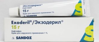
Treatment principles include:
- Isolation from a sick animal;
- Taking systemic antifungal drugs by mouth
- Local ointment antifungal treatment.
Principles of microsporia prevention
- The main measures to prevent microsporia in children are to teach the child how to communicate properly with animals: the child should know that such contacts may be unsafe. The child should know the external signs of microsporia in animals and not allow contact with sick animals. After petting a cat or dog, a child should wash their hands.
- It is unacceptable to use other people's objects - such as a comb, handkerchief, etc. You cannot wear someone else's clothes.
- You should not allow animals, especially cats and dogs, into bed or warm them under your shirt. After playing with them, you should immediately wash your hands with warm water and soap.
- You should avoid contact with stray animals, examine domestic animals, including those that do not come into contact with wild ones, to reduce the risk of infection with microsporia.
- Examinations of children in organized groups (kindergartens, pioneer camps) should be carried out at least 2 times a year, using a Wood lamp
- If a disease is detected, the child should be isolated from other children.
- By decision of the doctor, the patient may be sent for treatment to a specialized hospital. Indications for hospitalization are:
- severe course of the disease and the presence of concomitant pathologies
— lack of effect from outpatient treatment;
- according to epidemiological indications: patients from organized groups in the absence of the possibility of isolating them from healthy individuals (for example, in the presence of microsporia in persons living in boarding schools, orphanages, dormitories, children from large and asocial families).
- All things that belonged to a sick child must be disinfected in a steam-air chamber, and in its absence, boiled and ironed.
- In the apartment where the sick kitten was, all furniture, equipment, floors, carpets, and animal bedding should be treated with a disinfectant that has an antifungal effect. After treating items with a disinfectant, they are washed with hot water and soap.
- Children must have individual bedding, underwear, towels, combs, hairpins, bows, etc.
- Contact with stray, and often sick with microsporia, cats and dogs is especially dangerous. Sometimes a monkey, hamster, or guinea pig given by parents can infect a child, which, although rarely, also suffer from this mycosis. Therefore, pets should be periodically observed by a veterinarian.
- Pets sick with microsporia are subject to full treatment with veterinary supervision to ensure they are cured.
- It is prohibited to keep any animals in children's institutions, especially kindergartens and nurseries.
- Hair clippers, scissors, and combs in hairdressing salons must be disinfected after each client.
- A significant role in the fight against microsporia belongs to health education. In special lectures, wall newspapers, and mass printed publications, it is advisable to talk about ways of infection with microsporia, preventive measures, and the consequences of the disease.
Diagnostic measures
Diagnosis and treatment of ringworm are carried out by a dermatologist. The doctor examines the patient and identifies typical manifestations of microsporia. Examination of skin scrapings under a microscope reveals fungal mycelium and changes in the structure of hair and skin. Differential diagnosis makes it possible to exclude trichophytosis from the patient’s history, which has similar manifestations upon microscopy of the patient’s biomaterials.
Microflora culture appears to be a more informative diagnostic technique. Laboratory staff determine the type and genus of fungi. Based on the laboratory report, the dermatologist selects drugs that will cure the patient.
Luminescent examination makes it possible to identify pathological lesions on the skin of the patient and those living with him. This diagnostic method is based on the green glow of the fungal mycelium under the influence of a gas-discharge light source.
Definition of microsporia
The name microsporia was given by fungi of the genus Microsporum, which provoke pathological changes. There are an average of 60-70 cases of the disease per 100 thousand people. Of the entire group of dermatophytosis (skin infectious diseases), this pathology is the most contagious (infectious). As a rule, a person becomes infected through contact with infected animals or objects with their fur on them. Transmission of microsporia in the active phase from person to person is possible, although it is observed extremely rarely - in only 2% of cases of their total number.
First actions after detection of microsporia
“In the first 3 days after identifying the patient, a dermatologist examines people who have been in contact with him. Next, observation is carried out with mandatory examination 1-2 times a week for 21 days. In an apartment where there was a sick animal (cat, dog), all furniture, floors, carpets, and animal bedding are treated with a disinfectant with an antifungal effect. After treatment, the sick person’s belongings are washed with hot water and soap. The animals are sent to a veterinary hospital for examination and treatment.”
Kuldibaeva Alina Talgatovna
expert
Pulmonologist, Allergist-immunologist, Pediatrician
Treatment
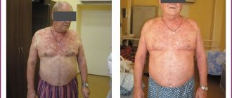
The combination of drugs prescribed to the patient depends on the severity of damage to the skin, nail plates and hair follicles. Antifungal therapy may be local or general. In the first case, a child or adult needs to use creams and ointments that suppress the activity of the microsporia pathogen. Drugs in this group are not recommended for use by girls during pregnancy and lactation. Topical sprays are effective in treating large areas of ringworm. Modern formulations do not leave stains on the skin and are not absorbed into the fabric of clothing.
A severe inflammatory reaction is treated with a combination of antifungal and hormonal drugs. Patients will have to apply ointment applications to the affected areas. After this, the skin is treated with iodine solutions. The oral tablet intake schedule is formed by a dermatologist based on the clinical picture of the pathology.
The addition of a secondary infection to microsporia involves the use of drugs based on betamethasone, gentamicin or clotrimazole.
Diagnosis of microsporia
The clinical picture and history of contact with animals provide the doctor with grounds for making a preliminary diagnosis. To confirm it, the following diagnostic studies are prescribed:
- Inspection using a fluorescent filter. This method is based on the ability of fungi of the genus Microsporum to fluoresce when using ultraviolet radiation. For this purpose, a device known in medicine as a Wood's lamp is used. This lamp emits ultraviolet light, which, when passed through a darkened filter, helps identify pathogenic fungi: when examined, they glow pale yellow or greenish.
- Histological examination. An area of skin is scraped for microscopic examination, which makes it possible to identify the fact of infection and the degree of inflammation. However, microscopy does not determine the type of pathogen.
- Cultural research. To determine the species of the pathogen and its sensitivity to antifungal drugs, the fungus is sown and grown on a nutrient medium. The disadvantage of the cultural method is that you have to wait at least a week to get a reliable result.
- Additionally, clinical tests of blood and urine are performed. If the results deviate from the norm, the tests are repeated once every 10 days. A biochemical study of blood serum is done before the start of the therapeutic course and 3 weeks after its completion.
Preventive measures
Prevention of ringworm is based on regular medical examinations of children attending preschool educational institutions. Parents should talk to their children about the inadmissibility of contact with stray animals. An important preventive measure is compliance with personal hygiene rules by patients of all age groups.
When purchasing pets, you must visit a veterinarian. The doctor will examine the cat or dog and give recommendations on how to eliminate any health problems with the pet. Following your veterinarian's advice will help prevent microscopic outbreaks within your family.
Prevention and prognosis
Prevention of fungal skin infections comes down to maintaining hand and skin hygiene, and avoiding contact with other people's animals. Your own pets should be checked for various infections at least 2 times a year. This applies to families with small children. When a disease is detected, the patient must be isolated for adequate treatment and to prevent spread to other family members and surroundings. Household items and other personal belongings of the patient are subjected to antiseptic treatment and disinfection. With prompt consultation with a doctor and proper therapy, the prognosis for patients is favorable.
The disease does not pose any threat to the lives of patients. Proper treatment, organized on time, helps prevent irreversible damage to the structure of the skin (scars, scars). An untreated fungus on the scalp can cause baldness in the affected area and cause inferiority of the scalp. In addition to aesthetic defects, the disease can cause deep emotional distress, including depression and an inferiority complex. Timely therapeutic treatment can eliminate the risk of developing such negative complications.