All tissues of the oral cavity, both teeth and gums, are interconnected and naturally have a complex structure. Gums, just like teeth, require careful and thorough care. Periodontology is a separate science in dentistry that deals with the study of gum disease. Inflammation from the teeth can spread to periodontal tissue, and vice versa. Therefore, it is so important to maintain careful hygiene of the entire oral cavity and promptly treat any dental diseases.
What it is
Periodontium is periodontal tissue, the main function of which is to hold the tooth in the alveolus. All periodontal tissues are interconnected, so any changes in the functioning of one or another element inevitably affect the functioning of other elements. The periodontal structure includes periodontium, gums, alveolar processes and cementum. Some dental scientists also include tooth enamel, dentin and pulp in its composition.
The term “periodontium” appeared in dentistry a little over a hundred years ago and has since firmly taken its place in modern dentistry, although in Russia the term “took root” a little later, around the mid-30s of the last century. of periodontology deals with a thorough study of the periodontium, its main functions, structure, and possible diseases .
Anatomy, physiology
The periodontium
is a complex organ of the oral cavity surrounding the teeth, formed from specialized tissues. It is located on the jaws and its main functions are to support (support) the teeth and their nutrition and innervation.
Nutrition (trophism) is provided by blood and lymphatic vessels that supply oxygen and metabolic products to the periodontal tissue.
There are four main anatomical and functional elements of the periodontium:
- gum,
- periodontium,
- alveolar bone with periosteum,
- root cement
The gum is a soft tissue - a mucous membrane covering the alveolar processes, with an underlying connective tissue layer.
It contains lymphatic vessels (they are involved in regulating fluid pressure in tissues and protecting against microbial agents).
Through the periodontium - the ligament of the tooth, the latter is fixed in the jaw bone.
Periodontium
(otherwise known as desmodont) is a connective tissue formation surrounding the tooth and located between the root cement and the inner cortical plate of the alveolar bone.
This ligament, consisting of the main substance, fibers and cellular elements, like an elastic gasket, absorbs the load on the tooth, giving it minimal mobility. When periodontal disease and death occur, fusion of the bone with the tooth – ankylosis – can occur.
The next element of the periodontium is cement, which is also a hard tissue covering the root of the tooth. It is formed by collagen fibers directed along the root and an adhesive substance.
Bone
The alveolar process, in which the tooth is strengthened by periodontium, is composed of outer and inner dense plates and a spongy part between them.
Spongy bone consists of trabecular septa, the direction and shape of which is determined by the load on the jaws and teeth, and bone marrow.
Bone tissue contains mainly crystals of hydroxylapatite (inorganic) and small amounts of water and organic substances.
The cellular elements of bone - osteoblasts, osteoclasts and osteocytes, under the influence of the nervous and humoral system, determine changes in the shape, volume and quality of this tissue.
The bone tissue of the alveolar process is covered with a dense connective tissue structure - the periosteum; it, with the vessels located in it, plays a significant role in the blood supply to the bone.
The blood supply provides tissues and anatomical formations with organic and mineral substances.
At the level of the enamel-cement junction (neck of the tooth), an anatomical formation stands out in the marginal part of the gum - a vascular cuff, which, like an elastic band, ensures a tight fit of the gum to the tooth due to hydrostatic pressure.
The main source of blood supply to periodontal tissue is the external carotid artery with the maxillary and mandibular arteries branching from it.
Venules and veins collect blood into the internal jugular vein. The periodontium is innervated by the trigeminal nerve and the superior cervical sympathetic ganglion.
Among others, there is an anatomical formation - the dentogingival junction
. This is the connection between the gum epithelium and the neck of the tooth.
It is physicochemical: molecules of epithelial cells adhere to cement structures through gingival fluid cells.
Gingival fluid produced by periodontal tissue plays the role of a protective barrier against microorganisms through the activity of phagocytes and its chemical properties. It should be noted that the periodontium is washed by saliva. It is secreted by the major and minor salivary glands.
In addition to water (99.42%), saliva contains organic substances, salts and trace elements. Organic substances enter the oral fluid from blood serum and are secreted by the salivary glands and microorganisms present in the mouth.
Of the inorganic substances, calcium phosphate and calcium bicarbonate (they take part in the formation of tartar), phosphate and sodium chloride are important.
Diagram 1: periodontium
. a – submucosal layer with lymphatic vessels; b — mucous membrane of the attached gum; c - spongy bone of the alveolar process between the plates of the compact substance; d – periodontal fibers are woven into the root cement; e – gingival margin with vessels of the gingival cuff; e – dentogingival junction; g – tooth enamel; h – dental pulp; and – root cement; j – root dentin.
Age-related changes in periodontium
In the periodontium, with age, there is a decrease in the number of collagen fibers and a decrease in their quality. The epithelial layer of the mucosa becomes thinner, and keratinization is disrupted.
The bone tissue of the alveoli becomes less dense, the cortical layer atrophies.
Structure and functions
The periodontal structure includes:
- Desna . Soft tissues that cover part of the tooth root, protecting it from the external environment. The gums are based on collagen fibers, which take an active part in the functionality of the dentofacial apparatus. The soft tissue of the gums is covered on top with epithelium, which has excellent regenerative properties.
- Alveolar process of the jaw . Bone bed of the tooth. It consists of two bone plates, has a spongy structure and is filled with vessels and nerves.
- Periodont . A special connective tissue that fills the space between the alveolar process and the tooth. Consists of special connective fibers, blood and lymphatic vessels, and nerve fibers.
- Cement . Refers to the tissues of the tooth and covers the root of the tooth. Its structure resembles bone tissue.
- Tooth enamel . The hardest part of the tooth, it covers the surface of the crown of the tooth. It is thanks to the hardness of tooth enamel that we can bite and chew food.
- Dentin . Refers to the tissues of the tooth, it is covered with cement and enamel. Dentin is less hard than tooth enamel, it has a huge number of tubules, as well as a cavity filled with pulp.
- Dental pulp . The softest dental tissue, which is responsible for the innervation and nutrition of the tooth. The pulp consists of connective tissue, nerves and blood vessels.
Functions of periodontium:
- Support-retaining . Fixation of the tooth in the alveolus. Thanks to the ligamentous apparatus of the periodontium, alveolar process and gums, the tooth is securely fixed inside the alveolus in a suspended state and does not fall out of its place even under fairly heavy loads.
- Shock-absorbing . Evenly distributes pressure on the teeth and jaw while chewing food. This is facilitated by the presence of connective tissue and tissue fluid, which acts as a natural shock absorber.
- Trophic . It is provided due to the presence of blood and lymphatic vessels, as well as a large number of different nerve receptors.
- Barrier or protective . It is carried out due to the protective properties of the gum epithelium, the presence of lymphoid, plasma and mast cells, the presence of enzymes and other active substances.
- Reflex . It is carried out using the oral mucosa and the presence of nerve receptors in periodontal tissues. Responsible for the force of chewing pressure during eating.
- Plastic . High ability of periodontal tissues to regenerate due to the presence of fibroblasts and osteoblasts.
Periodontal functions
In a healthy state, the periodontium performs a number of functions assigned to it:
- Support. The main function is due to which the tooth is held between bone plates.
- Shock-absorbing function. Correctly distributes pressure over the entire dentition.
- Trophic. A function responsible for nutrition and ensuring metabolism of the tissue complex.
- A protective function that helps create a barrier against the effects of bacteria.
- Reflex – affects the correct distribution of the chewing load.
- The plastic function is responsible for the elasticity of periodontal tissues.
Etiology and pathogenesis of periodontal diseases
The pathogenesis of periodontal diseases has not been fully established. It is known that at different stages of the development of periodontology, the causes of periodontal diseases such as
- general diseases of the body;
- presence of dental plaque;
- the presence of a large number of aggressive harmful bacteria in the patient’s mouth.
The etiology of periodontal diseases lies in the presence of dental plaque, without which the occurrence of diseases is simply impossible. It is the presence of dental plaque that is the primary factor in the occurrence of periodontal diseases.
Secondary factors include:
- presence of tartar;
- traumatic occlusion;
- the presence of low-quality fillings or dentures in the patient’s mouth;
- anomalies in the position of teeth and bite;
- structural features of soft tissues;
- features of saliva composition;
- genetic predisposition;
- frequent stress;
- hormonal imbalance;
- smoking.
Classification of periodontal diseases.
Gingivitis
Gingivitis is an inflammatory superficial disease of the gum tissue.
With gingivitis, there is no disruption of the periodontal junction and bone. Based on the form of manifestation of periodontal disease, the following are distinguished:
- catarrhal gingivitis,
- hypertrophic gingivitis,
- ulcerative gingivitis.
According to the type of course, gingivitis is divided into acute, chronic, aggravated and remission. According to the distribution, gingivitis can be localized and generalized. Without active treatment, gingivitis progresses to periodontitis.
Periodontitis.
Periodontitis is a deep inflammatory disease of the periodontium with destruction of the periodontal junction and bone tissue of the alveoli.
There are acute, chronic course of periodontitis, exacerbation of the disease and remission. Periodontitis can be mild, moderate or severe.
According to the prevalence, this gum disease is divided into generalized and localized periodontitis.
Photo 1. X-ray picture of severe periodontitis. The roots of the teeth are exposed by two-thirds.
2. 1.
Additionally, there is a condition known as
an aggressive form of periodontitis.
This diagnosis can be made by a doctor based on the presence of a set of signs of gum disease. In this case, periodontal destruction is generalized and rapid.
Such periodontitis is typical for ages from adolescence to 35 years. Often the aggressive form of periodontitis is accompanied by gum growth and granulation. During the period of remission, inflammatory manifestations may subside and even disappear.
Periodontal disease
Periodontal disease is a chronic degenerative periodontal disease.
In European and American classification as a separate gum disease
doesn't stand out. Periodontal disease is characterized by a long-term, benign course and almost does not lead to loosening and loss of teeth.
Periodontal diseases - as symptoms of other diseases
There are idiopathic periodontal diseases with progressive destruction of periodontal tissue (periodontal syndrome).
Such conditions can occur when exposed to physical factors (radiation), poisoning, immunodeficiency, diabetes mellitus, eosinophilic granuloma, Papillon-Lefevre syndrome, neutropenia, etc.
Periodontomas. Epulis (epulid) and gingival fibromatosis
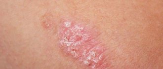
Periodontomas are tumors and tumor-like conditions of the gums. The most common of them are epulis (epulid) and gingival fibromatosis.
Gingival fibromatosis is characterized by the proliferation of periodontal tissues, which causes tumor-like damage to the gum tissue. Gingival fibromatosis is most common in adults, but is sometimes diagnosed in children.
The diagnosis of gingival fibromatosis is made by a specialist based on the patient’s complaints, examination data, radiography, and histological examination.
The main reason for the growth of gum tissue, as a rule, is a violation of metabolic processes; drug-induced fibromatosis of the gums is also identified. Treatment of the disease is surgical and consists of excision of the overgrown gum to the periosteum.
Photo 2. Gingival fibromatosis
Differential diagnosis of diseases
The doctor makes a diagnosis based on the results of examining the patient's oral cavity using dental instruments, as well as on the results of an x-ray examination. It is also important to ask the patient in detail about the symptoms, their intensity, and nature. It is very important to conduct a detailed clinical examination of the patient to exclude the presence of other diseases.
Differential diagnosis of periodontal diseases is based on the analysis of radiographic data. With gingivitis, there are no changes in the bone base of the periodontium.
When diagnosing periodontal diseases, so-called indices are often used, which make it possible to determine the degree of the inflammatory process and changes in bone tissue, which allows for the most accurate diagnosis.
Local factors:
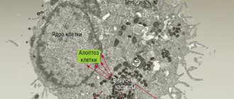
Local factors include, first of all, dental plaque, which is a consequence of improper and untimely oral hygiene. It is a conglomerate of food debris and the bacteria that live in them.
Figure 3. Plaque under a microscope
At first, the plaque is soft and can be easily removed with a toothbrush, but over time, bacteria firmly bind to the tooth enamel and the plaque gradually turns into tartar (1), which constantly injures the gums (2) and is a constant source of infection of the tissues surrounding it ( 3-6).
Figure 4. Local causes of periodontal disease development
In addition to dental plaque, local factors also include overload of periodontal tissues, insufficient chewing of food, and underload of certain groups of teeth.
Vector device in periodontology
The Vector device allows you to quickly and reliably cure patients of many symptoms. It not only helps to get rid of the disease, but also activates the reserve forces of the periodontium, which allows you to avoid many problems in the future. With the invention of the Vector device, periodontics has reached a qualitatively new level of disease treatment. Literally in one visit to the doctor you can get rid of such unpleasant symptoms as bleeding gums, inflammation and soreness of the gums. Moreover, the treatment is almost painless.
The Vector periodontal device was invented in Germany and is most often used to remove dental plaque, which is the main cause of periodontal disease. Using the device, you can also treat the surface of the teeth with ultrasound before fixing the dentures. However, its main purpose is the treatment of periodontal diseases.
If you have suffered greatly from an inflammatory disease, Vector will help replace curettage, which is why the device is often used for osteoplasty and gingivoplasty.
Treatment of periodontal diseases
The general treatment program for periodontal diseases includes:
- removal of microbial plaque and mineralized deposits from the surface of teeth;
- caries therapy;
- preventing functional overload of certain areas of the dentition;
- rejection of bad habits;
- carrying out orthodontic treatment (if indicated);
- anti-inflammatory therapy;
- combating systemic diseases;
- the use of drugs that enhance immunity, stimulate osteogenesis and activate the adaptive and protective functions of the body;
- carrying out general hygiene measures (monitoring the regime of work, nutrition, rest, hygiene).
The treatment regimen for periodontal diseases is drawn up taking into account all manifestations of the disease and the results of a comprehensive examination of the patient. Timely seeking of professional help by a patient significantly improves the prognosis of the disease, shortens the duration of treatment and avoids the development of complications.
Possible signs
Based on the nature of the flow, the following options are distinguished:
Acute catarrhal pericoronitis
During the process of teething, the patient experiences unpleasant sensations, which intensify when eating. The mucous membrane in the affected area will be red, swollen and painful when touched. Regional lymph nodes increase in size. Their palpation becomes painful.
Acute purulent pericoronitis
With this form of the disease, pain increases. Purulent discharge appears from under the hood. Pain when eating is accompanied by discomfort when swallowing. Mouth opening is limited.
The patient's general condition changes. There is an increase in body temperature to 37.5-38 degrees. Sleep is also disturbed and appetite is lost.
Chronic pericoronitis
The chronic course of the disease occurs if the necessary treatment is not carried out in a timely manner. Painful sensations decrease in intensity and appear only periodically. Since the hood remains, food continues to get trapped under it. This leads to bad breath. Regional lymph nodes are slightly enlarged and painful only on palpation. Swallowing and mouth opening are not impaired.
What are the possible complications?
Improper teething, accompanied by pericoronitis without treatment, can lead to the following serious complications:
- Formation of necrotic ulcers along the gingival margin. Subsequently, necrosis can spread to other tissues of the oral cavity;
- Damage to healthy neighboring teeth;
- Phlegmon of the perimaxillary spaces;
- Periostitis, which easily spreads to nearby areas;
- Osteomyelitis;
- Abscess formation.
Any of these pathologies can become life-threatening, so treatment of inflammation of the mucous membrane must be timely.
Treatment of pericoronitis is not difficult. A timely visit to the dentist and diagnostics will allow you to immediately carry out the correct treatment. For example, if a tooth initially erupts incorrectly, then removing it at the very beginning of the process will avoid further discomfort.
If for one reason or another it was not possible to visit a doctor on time, then you should come for a consultation when the first signs of inflammation appear. The purulent form is much more difficult to treat and causes more inconvenience. In addition, pus is the main cause of complications.
The post Treatment of pericoronitis appeared first on.
]]>