Emphysema is a lung disease in which the lung tissue becomes “overinflated” due to the gradual destruction of the alveoli. There is a lot of air, but this air does not participate in gas exchange. In bullous emphysema, air bubbles form in the lungs. These bubbles are called bullae and are filled with air.
- Bulla is an air bubble with a diameter of at least 1 cm and having a thin wall (no more than 1 mm).
- Single or multiple bullae constitute bullous emphysema and bullous pulmonary disease.
- Bullous emphysema and bullous pulmonary disease are different.
- 12% of people over 30 years of age have various bullous changes in the lungs.
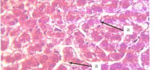
Bullae arise when the structure of the alveoli and the partitions between them is destroyed. Alveoli are tiny sacs made up of a thin membrane. The process of respiration occurs in the alveoli - oxygen enters the blood, and carbon dioxide from the blood enters the exhaled air.
The process of forming bullae is similar to blowing soap bubbles, when small bubbles combine into large ones, and then even larger ones. The cause of destruction of the alveoli and interalveolar septa is COPD or congenital lung pathology.
Bullous emphysema is more common in patients with COPD against the background of panacinar and centrilobular emphysema of both lungs.
In bullous disease, there is lung tissue without emphysema, and there may be several or one bulla. In congenital syndromes such as Marfan and Ehlers–Danlos syndromes, primary bullous pulmonary disease is isolated
Classification
For convenience of description and classification, bullae can be single or multiple.
In case of multiple distribution, the following options are determined:
- several bullae within one (focal) lobe
- several bullae in several lobes (multifocal).
- bullae are observed in all lobes of the lungs and are called diffuse.
Bullae located near the pleura or scars in the lung tissue can reach large sizes. Such bullae occur in paraseptal emphysema; they are devoid of vessels and interalveolar septa. Another type of bulla is located superficially and may contain vessels and fragments of lung tissue. Such bullae lead to pneumothorax.
The third type of bullae is located deep in the lungs. These bullae are rich in blood vessels and contain a lot of lung tissue.
A special place is occupied by giant bullae - this is a super bulla that occupies more than 1/3 of the lung and compresses the surrounding lung tissue. Giant bullae are characteristic of the lungs of a person who smokes tobacco or marijuana.
Patients with bullous disease are younger than patients with bullous emphysema. This is mainly due to congenital deficiency of α1-antitrypsin, as well as congenital genetic diseases (Marfan syndrome, Ehlers–Danlos syndrome).
Symptoms of subcutaneous pneumothorax - how to recognize air under the skin?
Patients with pneumothorax complain of the sensation of air bubbles under the skin. Feeling of severe discomfort in the neck and chest. Additionally there may be:
- breathing problems,
- difficulty swallowing and speaking
- change in voice timbre,
- feeling of fullness in the neck.
Patients complain of sore throat, wheezing, or neck pain. The tissue around the pneumothorax may be slightly swollen. On examination, a squeaking sound of the skin when touched is characteristic of subcutaneous pneumothorax.
About
Symptoms of bullous emphysema
Bullous pulmonary disease can be asymptomatic or have severe and life-threatening consequences.
The asymptomatic course of bullous pulmonary disease is a characteristic feature of the disease. Even giant bullae may not cause shortness of breath. After all, there is no pulmonary emphysema in this condition and gas exchange may not be disrupted.
Shortness of breath and cough are characteristic symptoms of COPD, against which bullous emphysema occurs as a complication of COPD. Bullae arise in this case against the background of panacinar or centrilobular emphysema.
The size of the bullae may increase or remain unchanged. Cases of their disappearance or reduction are also described.
Bullae in the lungs can lead to respiratory failure, pneumothorax, suppuration of bullae, empyema, and hemoptysis.
Bullous deformity may predispose to the development of cancer. According to the literature, lung cancer develops four times in the lung tissue adjacent to the bullae. Therefore, it is necessary to carefully evaluate the lung tissue around the bullae.
What does a bulla in the lung mean?
According to the definition given at the international medical CIBA symposium, a bulla should be considered an air cavity in the lung, the diameter of which exceeds 1 cm, while the alveolar wall thins to 1 mm or less.
In medicine, it is customary to distinguish between bullous emphysema and bullous disease. They are distinguished depending on the causes of occurrence, etiology, and also depending on the health consequences. Bullous emphysema is usually diagnosed in patients with signs of COPD (chronic obstructive pulmonary disease) against the background of bilateral emphysema of the panacinar or centrilobular type. With bullous disease, emphysema is not observed, and the cavity may be one or slightly larger. Bullous disease can occur with congenital Marfan syndrome. Bullous emphysema, in the absence of treatment and adequate actions to curb its growth (quitting smoking, working in hazardous industries, treating chronic bronchitis, etc.) over time can lead to respiratory or heart failure, lung collapse, severe coughing with blood, and physical intolerance. loads
Bullae are formed according to the principle of soap bubbles: first small ones appear, then they combine to form large bullae, and so on. With bullous emphysema, the lung tissue becomes like an old sponge - as it wears out, the pores become larger, and the material loses its elasticity and tears.
Large bullae (diameter can reach up to 10 cm) are formed when the alveolar septa are destroyed. Alveoli are vesicles that make up normal air lung tissue. Air storage, oxygen transportation, air exchange are carried out in the alveoli, and the process itself requires a structure of dosed air supply. Bullae, even very small ones, introduce an element of chaos and entropy into the work of the most important respiratory organ.
Diagnosis of bullous disease
Bullae are detected on CT scan of the lungs. This test measures the number of bullae, their size and shape. CT will also allow you to evaluate changes in lung tissue, the presence or absence of emphysema, bronchiectasis or other cavities.
CT scan of the lungs can exclude cystic diffuse lung diseases. They may resemble bullous deformity.
When diagnosing bullous changes in the lungs, breathing tests are used - respiratory function, body plethysmography and diffusion test.
For example, bullous emphysema is characterized by obstructive pulmonary ventilation disorders. And for bullous disease, restrictive disorders are more expected. The exact answer to restriction and obstruction is given by the body plethysmography test.
A diffusion test will assess respiratory failure.
For all patients with giant bullae, measurement of α1-antitrypsin levels is mandatory.
All diagnostic tests are necessary to solve the following problems:
- Exclude other lung diseases similar to bullous disease
- Determine contraindications for surgical treatment
- Identify the cause-and-effect relationship of the occurrence of bullae: smoking, marijuana, connective tissue diseases.
Bullae in the lungs on CT
Bullae in the lungs on CT are visualized as distinct, relatively darker, bubble-like areas of lung tissue, resembling holes or large pores of a sponge. The denser the tissue, the lighter it appears on CT scans—so, for example, bones are white, but airy lung tissue is a relatively uniform graphite gray color.
Using a CT scan of the lungs, a radiologist can accurately determine the diameter of the bullae, their number, and find out whether there are signs of emphysema, bronchiectasis or other diffuse pulmonary diseases.
Treatment of bullous emphysema
Conservative treatment
If bullous lung disease is detected, a person must stop all types of smoking.
If bullous changes are part of COPD, then standard COPD therapy is used. There are no pills or inhalers for bullae.
During life, bullae may increase in size and number. The patient should be aware of this possibility and understand what symptoms to look out for. The patient should know what symptoms indicate immediate medical attention.
Since bullae can progress over time, patients need to be closely monitored and should be informed of alarming symptoms that may require seeking medical help. There is no conservative treatment for bullous changes.
Surgery
Surgical treatment is called bullectomy. If a person is diagnosed with a giant bulla, then this is an indication for surgical treatment. Small bullae are difficult to treat surgically and the effect of the operation is insignificant. Shortness of breath rarely improves.
Bullae that are not accompanied by clinical symptoms (shortness of breath) or complications (pneumothorax, suppuration) are not recommended to be removed.
Contraindications to bullectomy include continued smoking, severe cardiovascular disease, and diffuse emphysema with low compression of the surrounding lung tissue.
The indication for surgical treatment may be recurrent pneumothorax against the background of bullous changes. In this case, bullectomy and pleurodesis are performed simultaneously.
How to treat bullae in the lungs
The patient needs to quit smoking and quit working in harmful production activities, otherwise all therapeutic measures will be meaningless. In uncomplicated cases and in the early stages of emphysema (or bullous disease), treatment is conservative and combined. Therapy is prescribed by a pulmonologist and may include:
- Medicines (diuretics, bronchodilators, hormonal drugs);
- A special course of exercise therapy and breathing exercises;
- Regular oxygen therapy using an oxygen concentrator.
- Regular diagnostic measures (CT of the lungs, spirometry, consultation with a pulmonologist).
The positive aspect is that complete and lifelong cessation of smoking in most patients stops the development of bullous emphysema, and the use of oxygen therapy in seriously ill patients can prolong life by 5-10 years.
Surgical treatment of bullae (endoscopic “suturing”) may be indicated for recurrent pneumothorax against the background of growing cavities. The patient undergoes bullectomy and pleurodesis operations. However, the absolute success of such treatment cannot be guaranteed.
Recommendations for air travel
Pneumothorax is a complication of bullous transformation of the lungs. Air travel poses a certain risk for these patients because at an altitude of 2.5 km, the cavities in the lungs containing air can increase by 38-40%. This expansion can lead to bulla rupture and pneumothorax.
To be fair, episodes of pneumothorax during air travel are rare and there are no categorical bans on flying patients with bullae in the lungs.
If pneumothorax occurs 7-14 days before the flight, air travel should be postponed.
Subcutaneous emphysema - causes, symptoms, treatment of air bubbles under the skin
Subcutaneous emphysema is a condition in which air bubbles are visible under the skin. Typically, subcutaneous emphysema is located on the chest, neck or face. The cause is trauma in these areas, medical procedures (for example, after laparoscopy, tracheotomy) or dental procedures (for example, after tooth extraction). Symptoms of pneumothorax include pain and fullness in the neck and difficulty breathing. Treatment for subcutaneous pneumothorax is usually not required because the air is self-absorbed.
Fotolia
Diagnosis of emphysema in Israel
Examination of a patient with pulmonary emphysema in Israel is carried out using laboratory and instrumental methods.
Laboratory diagnostics
- General and biochemical blood tests;
- Study of gases (oxygen and carbon dioxide) in arterial blood;
- Tank. sputum analysis;
- General urine analysis.
Instrumental diagnostics
- X-ray or computed tomography of the chest - to determine the severity of the pathology and detect other causes of shortness of breath (pulmonary fibrosis);
- Functional pulmonary tests (spirometry) - measurement of vital capacity of the lungs, inhalation/exhalation volumes, lung reserve volume, Tiffno index, etc.;
After diagnostic procedures, the patient is examined by a pulmonologist and gets acquainted with the results of the examination. Then the patient’s condition is assessed, an accurate diagnosis is determined and a plan for further treatment is developed.
Causes of emphysema
Emphysema develops as a result of exposure to external and internal factors on the human body.
External (exogenous)
- Work in hazardous working conditions, involving regular/continuous inhalation of various impurities - pollutants (coal, silicate, textile, silicon, etc.);
- Bad habits – smoking (inhalation of nicotine and toxic substances);
- Unfavorable environmental conditions in the area of residence (large industrial cities);
- Low socio-economic status (inability to maintain one’s health and lead a healthy lifestyle).
Internal (endogenous)
- Hereditary predisposition – congenital genetic mutations leading to a deficiency of alpha-1-antitrypsin protein in the body;
- Increased sensitivity (hyperreactivity) of the respiratory tract to allergic factors - especially in children, the risk of developing emphysema in the future increases 3 times;
- The presence in the body of a chronic recurrent respiratory infection.
Risk group
The disease risk group is compiled taking into account the most common causes of its development. According to WHO, centrilobular emphysema occurs in 78% of cases in people over 50 years of age. More often, the pathology is observed in heavy smokers and people who have suffered from chronic diseases of the respiratory system for a long time. Paraseptal and centrilobular emphysema are common pathologies in people with chronic lung diseases.
People whose activities involve contact with chemicals often get sick. Builders and workers in hazardous industries are susceptible to the development of pathology.
Due to the abnormal development of connective and pulmonary tissue, patients suffering from autoimmune diseases and pathologies of the endocrine system are also at risk.
Periods and features of the course of emphysema
- Non-infectious – the leading factors in the development of the disease are exogenous.
- Infectious - the accumulation and reduction of sputum discharge in the central bronchi contributes to the spread of infection to the distal parts of the bronchopulmonary system.
Obstruction (narrowing of the lumen) of the bronchi can be reversible and irreversible.
Reversible processes include:
- inflammatory disorder of bronchial patency due to swelling of the mucous and submucosal layer;
- obstruction by excessive mucus production;
- bronchial hyperreactivity, accompanied by bronchospasm.
Irreversible mechanisms:
- stenosis (narrowing), deformation and obliteration (complete blockage) of bronchioles;
- fibroplastic changes (remodeling) of the bronchial walls;
- collapse of the bronchial wall during exhalation.