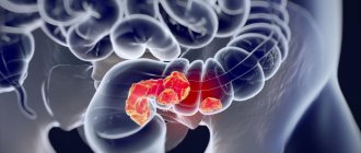
Carcinoid is a slow-growing tumor that develops from cells of the neuroendocrine system, mainly in the gastrointestinal tract (cecum, stomach, small intestine, colon, rectum) or lungs. It produces a hormone (serotonin) that causes symptoms such as diarrhea or facial flushing (carcinoid syndrome).
In 50-60% of cases, carcinoids are located in the appendix area. The tumor is usually benign. Only when the size is more than one centimeter does the carcinoid become malignant. In order not to miss the moment of tumor growth, it is recommended to undergo detailed diagnostics in our clinic in Germany.
Symptoms of carcinoid tumor of the appendix
Increased serotonin production due to carcinoid causes a decrease in tryptophan and leads to niacin deficiency (pellagra), dermatitis, dementia and diarrhea.
Serotonin is a neurohormone of the nervous system. Especially when metastases form, the so-called carcinoid syndrome occurs, which is expressed by rapid redness of the facial skin and accompanying manifestations:
- rapid heartbeat;
- sweating;
- breathing problems;
- cramping pain in the abdomen;
- diarrhea.
Diagnosis of the disease
Most carcinoid tumors of the appendix are discovered incidentally during surgery performed for other reasons. To diagnose carcinoid, a blood test is performed to determine the level of free hormones. When hormones break down, increased concentrations of certain substances are formed, for which a urine test is performed.
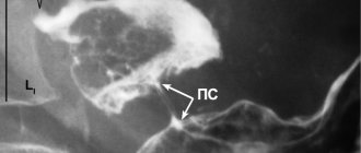
To determine the exact position and size of a neuroendocrine tumor of the appendix, imaging methods are used:
- Computed tomography (CT)
- Magnetic resonance imaging (MRI)
- PET-CT
- Ultrasound examination
- Somatostatin receptor scintigraphy
- X-ray
- in some cases, colonoscopy.
To confirm the diagnosis, a tumor biopsy is performed.
Treatment
Surgical treatment The main treatment method for NET is surgery. Possible options for surgical intervention are determined by the location of the tumor, the presence or absence of metastases and complications of the tumor process. However, symptom control during surgical treatment is not always achieved !
Non-drug treatment Monitoring the manifestations of carcinoid syndrome: avoiding stress, alcohol, following a diet.
Drug treatment
Biotherapy:
- synthetic analogues of somatostatin: solution for injection Octreotide and lyophilisate for the preparation of a suspension for intramuscular administration of prolonged action Octreotide-depot.
- interferon-?, prolonged form of INF-? (PEG-interferon).
Chemotherapy:
- monochemotherapy: streptozocin, doxirubicin, fluorouracil, dactinomycin, etoposide, cisplatin, dacarbazine.
- PCT: combinations of streptozocin, fluorouracil and doxirubicin, cisplatin with etoposide.
Combination chemotherapy:
biotherapy + chemotherapy.
Symptomatic therapy:
- antihypertensive drugs
- antihistamines
- diuretics
- bronchodilators
- antidiarrheal drugs.
Mechanism of action of OCTREOTIDE-DEPO
- The drug has a symptomatic effect, reducing the secretion of hormones and peptides excreted in VIPomas in 89%, in glucoganomas in 75%, in insulinomas in 65%.
- After using Octreotide depot for various NETs, diarrhea stops in 40-60% of patients.
- With VIPomas, Octreotide-depot completely stops diarrhea in 38% of cases, and in another 38% of patients it significantly reduces it.
- Improves general condition in 75-85% of cases.
- Octreotide depot controls hypoglycemia in insulinomas and necrolytic skin lesions in glucanomas, which significantly improves the quality of life of patients.
For carcinoid tumors, the use of Octreotide Depot can lead to a decrease in the severity of symptoms of the disease, primarily such as hot flashes and diarrhea. In many cases, clinical improvement is accompanied by a decrease in plasma serotonin concentrations and urinary 5-hydroxyindoleacetic acid excretion.
For tumors characterized by overproduction of vasoactive intestinal peptide (VIP), the use of Octreotide Depot leads in most patients to a reduction in severe secretory diarrhea, which is characteristic of this condition, which, in turn, leads to an improvement in the patient’s quality of life. At the same time, there is a decrease in concomitant electrolyte imbalances, for example, hypokalemia, which makes it possible to cancel enteral and parenteral administration of fluids and electrolytes. According to computed tomography data, in some patients the progression of the tumor slows down or stops, and even reduces its size, especially metastases to the liver. Clinical improvement is usually accompanied by a decrease (down to normal values) in the plasma concentration of vasoactive intestinal peptide (VIP).
In case of glucagonomas, the use of Octreotide depot in most cases leads to a noticeable reduction in the necrotizing migratory rash that is characteristic of this condition. In patients suffering from diarrhea, Octreotide Depot causes its reduction, which is accompanied by an increase in body weight. When using Octreotide depot, a rapid decrease in plasma glucagon concentration is often observed, but with long-term treatment this effect does not persist. At the same time, symptomatic improvement remains stable for a long time.
For gastrinomas/Zollinger-Ellison syndrome, Octreotide depot, used as monotherapy or in combination with H2 receptor blockers and proton pump inhibitors, can reduce the formation of hydrochloric acid in the stomach and lead to clinical improvement, incl. and regarding diarrhea. It is also possible to reduce the severity of other symptoms, probably associated with the synthesis of peptides by the tumor, incl. tides In some cases, there is a decrease in plasma gastrin concentration.
In patients with insulinomas, Octreotide depot reduces the level of immunoreactive insulin in the blood. In patients with operable tumors, Octreotide depot can ensure the restoration and maintenance of normoglycemia in the preoperative period. In patients with inoperable benign and malignant tumors, glycemic control may improve without a simultaneous prolonged decrease in blood insulin levels.
In patients with rare tumors that overproduce growth hormone releasing factor (somatoliberinomas) , Octreotide Depot reduces the severity of symptoms of acromegaly. This appears to be due to suppression of the secretion of growth hormone releasing factor and growth hormone itself. In the future, it is possible to reduce the size of the pituitary gland, which was enlarged before treatment.
It was found that the use of Octreotide depot in doses of 30 mg and above has an antiproliferative effect associated with the direct cytotoxic effect of the drug on the tumor when interacting with somatostatin receptors, as well as with the inhibition of the formation of blood vessels feeding the tumor.
Dosage regimen
For endocrine tumors of the gastrointestinal tract and pancreas for patients in whom subcutaneous administration of Octreotide provides adequate control of the manifestations of the disease, the recommended initial dose of Octreotide depot is 20 mg every 4 weeks. Subcutaneous administration of Octreotide should be continued for another 2 weeks after the first administration of Octreotide depot. For patients who have not previously received subcutaneous Octreotide, it is recommended to begin treatment with subcutaneous administration of Octreotide at a dose of 0.1 mg 3 times a day. for a relatively short period of time (approximately 2 weeks) to assess its effectiveness and overall tolerability. Only after this is Octreotide depot prescribed according to the above scheme. In the case when therapy with Octreotide depot for 3 months. provides adequate control of clinical manifestations and biological markers of the disease, it is possible to reduce the dose of Octreotide depot to 10 mg, prescribed every 4 weeks. In cases where after 3 months. Treatment with Octreotide depot only achieved partial improvement; the dose of the drug can be increased to 30 mg every 4 weeks. During treatment with Octreotide Depot, on some days there may be an increase in clinical manifestations characteristic of endocrine tumors of the gastrointestinal tract and pancreas. In these cases, additional subcutaneous administration of Octreotide is recommended at the dose used before the start of treatment with Octreotide depot. This may occur mainly in the first 2 months. treatment.
Octreotide depot is a first-line drug for monotherapy or combination therapy of highly differentiated neuroendocrine tumors of the gastrointestinal tract and pancreas, effectively relieving hormonal syndromes by suppressing the hypersecretion of hormones by these tumors, increasing the quality of life and survival of patients.
Treatment of carcinoid tumor of the appendix in Germany
Treatment for carcinoid depends on several factors:
- its location;
- presence and extent of metastasis;
- general condition of the patient;
- stage of the disease.
The main directions of treatment for carcinoid tumor of the appendix are:
- Surgery – the most complete removal of a malignant tumor
- Radiation therapy
- Chemotherapy
- Targeted therapy
- Radioreceptor therapy
When metastases form, other forms of treatment are used, such as Octreotide or the radiopharmacone 131I-MIBG (meta-iodine-bencyl guanidine) to stop the growth of cancer cells, as well as embolization techniques.
Carcinoid syndrome
Carcinoid tumors can appear where there are enterochromaffin cells, basically throughout the body. A larger proportion of carcinoid tumors (65%) develop in the gastrointestinal tract. In most cases, carcinoid tumors develop in the small intestine, appendix, and rectum. Carcinoid tumors develop most rarely in the stomach and colon; The pancreas, gallbladder, and liver are least likely to develop carcinoid tumors (even though carcinoid tumors usually metastasize to the liver).
Approximately 25% of carcinoid tumors affect the airways and lungs. The remaining 10% can be found anywhere. In some cases, doctors cannot locate the carcinoid tumor despite symptoms of carcinoid syndrome. Carcinoid tumor of the small intestine
In general, tumors of the small intestine (benign or malignant) are rare, much less common than tumors of the colon or stomach. Small carcinoid tumors of the small intestine may not cause any symptoms other than mild abdominal pain. For this reason, it is difficult to determine the presence of a small bowel carcinoid tumor at an early stage, at least until the patient undergoes surgery. It is possible to detect only a small proportion of small intestinal carcinoid tumors in the early stages, and even then this occurs unexpectedly on x-ray. Typically, carcinoid tumors of the small intestine are diagnosed in late stages, when the symptoms of the disease have become apparent and usually after metastases have appeared.
Approximately 10% of carcinoid tumors of the small intestine cause carcinoid syndrome. Typically, the development of carcinoid syndrome means that the tumor is malignant and has reached the liver.
Carcinoid tumors often obstruct the small intestine when they become large. Symptoms of small intestinal obstruction include paroxysmal abdominal pain, nausea and vomiting, and sometimes diarrhea. Obstruction can be caused by two different mechanisms. The first mechanism is tumor enlargement within the small intestine. The second mechanism is twisting of the small intestine due to fibrosing mesenteritis, a condition caused by a tumor in which extensive scarring occurs in the tissues located in close proximity to the small intestine. Fibrosing mesenteritis sometimes obstructs the arteries that carry blood to the intestines, which can result in the death of part of the intestine (necrosis). In this case, the intestines can rupture, which is a serious threat to life. Appendiceal carcinoid tumor
Although tumors in the appendix are quite rare, carcinoid tumors are the most common tumors in the appendix (about half of all appendiceal tumors). In fact, carcinoid tumors are found in 0.3% of removed appendixes, but most of them do not grow larger than 1 cm and do not cause any symptoms. In most cases, they are found in appendices that have been removed for reasons other than tumors. Many institutions believe that appendectomy is the most appropriate treatment for these small appendiceal carcinoid tumors. The chances of the tumor returning after an appendectomy are very low. Appendiceal carcinoid tumors larger than 2 cm in 30% can be malignant and form local metastases. Therefore, larger carcinoid tumors should be removed. A simple appendectomy will not help in this case. Fortunately, large carcinoid tumors are quite rare. Carcinoid tumors in the appendix, even with metastases to local tissues, usually do not cause carcinoid syndrome. Rectal carcinoid tumors
Rectal carcinoid tumors are often diagnosed incidentally during plastic sigmoidoscopy or colonoscopy. Carcinoid syndrome is rare in rectal carcinoid tumors. The likelihood of metastases is related to tumor size; 60-80% chance of metastases for tumors larger than 2 cm. For carcinoid tumors smaller than 1 cm, 2% chance of metastases. In summary, small rectal carcinoid tumors are usually successfully removed, but larger tumors (greater than 2 cm) require extensive surgery, which can even lead to partial removal of the rectum in some cases. Gastric (stomach) carcinoid tumors
There are 3 types of gastric (stomach) carcinoid tumors: type I, type II and type III.
Type 1 gastric carcinoid tumors are usually less than 1 cm in size and are benign. There are complex tumors that spread throughout the stomach. They usually appear in patients with pernicious anemia or chronic atrophic gastritis (a condition in which the stomach stops producing acid). The lack of acid causes the cells in the stomach, which produce the hormone gastrin, to release large amounts of gastrin, which enters the blood. (Gastrin is a hormone secreted by the body to enhance the activity of stomach acid. Acid in the stomach blocks the production of gastrin. In pernicious anemia or chronic atrophic gastritis, the lack of acid is the result of an increase in the amount of gastrin). In addition, gastrin also influences the transformation of enterochromaffin cells in the stomach into a malignant carcinoid tumor. Treatment for type 1 carcinoid tumors includes treatments such as somatostatin-containing drugs that stop gastrin production or surgical removal of the part of the stomach that produces gastrin.
The second type of gastric carcinoid tumor is less common. Such tumors grow very slowly and the likelihood of them becoming malignant is very low. They appear in patients with a rare genetic disorder called MEN (multiple endocrine neoplasia) type I. In these patients, tumors arise in other endocrine glands, such as the pineal gland, parathyroid gland and pancreas.
The third type of gastric carcinoid tumor is a tumor larger than 3 cm that appears separately (appearing one or more at a time) in a healthy stomach. Tumors of the third type are usually malignant and there is a high probability of their deep penetration into the walls of the stomach and the formation of metastases. Type 3 tumors can cause abdominal pain and bleeding, as well as symptoms due to carcinoid syndrome. Type 3 gastric carcinoid tumors usually require surgery to remove the stomach and nearby lymph nodes. Carcinoid tumors of the colon
Colon carcinoid tumors usually form on the right side of the colon. Like carcinoid tumors of the small intestine, carcinoid tumors of the colon are often discovered at advanced stages. Thus, the average tumor size at diagnosis is 5 cm, and metastases are present in 2/3 of patients. Carcinoid syndrome is rare in carcinoid tumors of the colon.
Where is the best place to treat appendix carcinoid?
Carcinoid tumor of the appendix belongs to neuroendocrine tumors, the treatment of which involves the interaction of doctors from different fields: surgeons, endocrinologists, radiologists, oncologists, which is successfully provided at the Nordwest Clinic. The treatment regimen for neuroendocrine tumors directly depends on their type and stage of development, and therefore accurate diagnosis of the disease is of particular importance. The diagnostic equipment of the Nordwest Clinic provides the most modern capabilities for determining and confirming all types and stages of cancer. When you contact us, you can be sure that the therapy will be tailored individually, taking into account the characteristics of the development of the disease.
INSTRUMENTAL DIAGNOSTICS
X-ray methods
- chest x-ray
- X-ray of the esophagus and stomach
- irrigoscopy
Endoscopic methods
- EGDS
- sigmoidoscopy
- colonoscopy
- bronchoscopy
Ultrasound methods
- Ultrasound
- Ultrasound Dopplerography of blood vessels
- Endosonography
- Intraoperative ultrasound
- Laparoscopic ultrasound
Contrast-enhanced spiral CT and MRI
RADIOISOTOPIC DIAGNOSTICS
- Skeletal bone scintigraphy
- Scintigraphy with Octreotide labeled with 111In
NETs on the cell surface have receptors with high affinity for the hormone somatostatin. In 87% of cases they are present both in the primary tumor and in metastases. In this regard, in recent years, a radioisotope method with 111In-labeled Octreotide has been used to determine the location of the tumor and metastases. Octreotide administered intravenously, 111In after 24-48 hours is determined on somatostatin receptors and allows you to visualize a somatostatin-positive tumor, as well as determine the possibility of using somatostatin analogues for treatment.
The method using Octreotide, 111In has a sensitivity of 87%, a specificity of 75% and a diagnosis agreement rate of 87%.