Every year there is an increasing increase in oncological pathology. Lung cancer occupies the first place in the structure of oncological diseases. This is due to both environmental pollution and smoking, alcoholism, genetic predisposition and other factors. The statistics of deaths from lung cancer are also frightening. Everything is connected with the fact that a person does not notice symptoms at all for a long time, and even when he notices them, he easily finds a simpler explanation for them. This is followed by a period of symptomatic treatment, and only when things get really bad do patients seek help. Unfortunately, more than half of patients already have numerous metastases when a pronounced clinical picture appears.
Depending on the location, central and peripheral lung cancer are divided, and if the entire lung is involved in the process, it is called massive. Central lung cancer is localized in large bronchi, and peripheral lung cancer is located in bronchi with a smaller diameter down to the alveoli. But scientists argue that the differences are not only in localization, but also in structure, origin, pathogenesis, clinic, etc. For example, it is believed that smoking and prolonged inhalation of polluted air are the etiological factors for central, and for peripheral lung cancer, lymphogenous and hematogenous are characteristic ingress of carcinogens.
How long you live with peripheral lung cancer depends on many factors, including treatment. The prognosis for peripheral cancer is significantly more favorable with timely diagnosis and effective specific therapy.
About seventy percent of cases of peripheral cancer of the right lung are localized in the upper lobes, about twenty in the lower lobes, and less than ten in the middle lobes. Histologically, peripheral cancer is most often represented by either adenocarcinoma or squamous cell carcinoma.
It is the peripheral form of cancer that is practically asymptomatic. Diagnosis at an early stage is often associated with preventive medical examinations.
The Yusupov Hospital is equipped with all the modern equipment necessary to diagnose lung cancer. Comfortable rooms, polite staff, highly qualified doctors - these are all the keys to successful treatment.
Forms of the disease in question
Tumor processes in the lungs are diverse. Peripheral lung cancer has several types. The cortico-pleural appearance is an oval-shaped formation that grows in more than one node. This neoplasm grows directly into the chest, after which it is located in the so-called subpleural space. The resulting pathology belongs to the squamous cell type of cancer. The structure of the tumor is homogeneous; it has a lumpy inner surface and poorly distinguishable contours. The tumor can grow both into adjacent ribs and into the bodies of adjacent thoracic vertebrae.
The so-called cavitary form of peripheral lung cancer is characterized by the absence of symptoms. It is detected at later stages, when pathological processes have become irreversible. Cavity formations are formed inside the pulmonary sections, having the shape of a ball, a lumpy outer surface and poorly distinguishable contours. The growth of the tumor leads to the proliferation of formations with parallel thickening of their walls.
Epidemiology
90% of all pulmonary neoplasms are lung cancer. It is the most malignant tumor in men (35% of all tumors) and women (30%) aged 45-70 years. Recently, statistics indicate an increase in the incidence of primary lung cancer in females. The disease occurs in men at a later age than in women.
According to statistics, in the United States, tumors are detected with a frequency of 70 per 100,000 population. And among the patients there are more African Americans than Caucasians. As for the countries of the world, pulmonary neoplasms are most often recorded in Poland and England. There are 100 diseases per 100 thousand people. The most minimal statistics are in Senegal and Nigeria. There is less than one case of lung tumors per 100 thousand inhabitants.
Oncology of the upper lobe of the left lung
In people with this type of cancer, X-rays clearly show blurred contours, an irregular shape of the formation and its heterogeneous structure. The pulmonary roots are expanded due to the influence of the vascular trunks. In most cases, the lymph nodes remain the same size (in the case of lower lobe cancer, the situation is the opposite).
As for oncology of the upper lobe of the right lung, it is in many ways similar to the previous pathology, but it is diagnosed much more often. The formation of the nodular form occurs in the terminal bronchioles. The disease makes itself felt after the soft lung tissues germinate. X-ray examination allows us to examine a formation that has a nodular shape, clear contours and a bumpy surface. Sometimes the edge of the tumor contains a small depression. The presence of such a depression indicates that a large vessel has entered the node.
Treatment
While the person is being examined, symptomatic therapy is prescribed. Emergency therapeutic and diagnostic bronchoscopy is prescribed for severe hemoptysis or pulmonary hemorrhage. For pneumonia in the atelectatic lobe of the lung, broad-spectrum antibiotics are prescribed. In some cases, doctors prescribe medications to relieve pain.
Treatment for small and non-small cell lung cancer is different. For peripheral small cell cancer without regional metastases, surgery is prescribed, followed by chemotherapy and radiation therapy. For central small cell cancer, only this therapy is needed, because at the time of diagnosis it is often not subject to surgery due to the number and location of metastases. Non-small cell cancer is treated only with surgery.
Surgery
Benign tumors are removed because they can only be accurately diagnosed after removal. Radical therapy for lung cancer consists only of removing a portion of the lung or the entire lung. A stage IV tumor is considered inoperable.
Other treatments and prognosis
Radiation therapy can be used as an independent method. Sometimes it is combined with surgery and chemotherapy. Radiation therapy is most effective for Pancoast tumors. Chemotherapy is effective only when combined with radiation therapy for small cell lung cancer. The best results were obtained with lobectomy in patients with peripheral tumors. The prognosis for lung cancer is mainly related to the histological type of the tumor and the stage of the disease at the time of detection.
For bronchoalveolar cancer, 5-year survival is recorded in 30-35% of cases, and for squamous cell cancer - 8-16%. Even less for adenocarcinoma and small cell cancer.
Tumor collapse and centralization syndrome
When the pathogenic formation reaches a large size, the pulmonary blood supply deteriorates, and this leads to tumor disintegration. The decay occurs gradually with the formation of cavities inside the tumor node. Due to the unevenness of the process, small tumor masses remain on the walls of the cavity. When small cavities merge into one, the stage of major decay begins. At the end, the phase of the so-called central decay begins. Thanks to the examination, it is possible to identify the presence of a cavity formation with thick walls; There is a horizontal liquid level inside it. The course of the described pathology is similar to the course of an abscess. A person’s temperature rises, the expectoration becomes purulent (sometimes there is blood in the secreted mucus).
In which bronchi do malignant tumors occur?
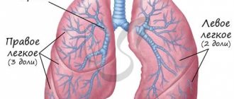
The air that a person inhales passes through the nose, pharynx, larynx, and trachea. At the level of the upper edge of the fifth thoracic vertebra, the trachea ends and divides into two main bronchi . This location is called the tracheal bifurcation . The main bronchi are the first order bronchi , they are divided into lobar (second order) , then into segmental (third order), subsegmental (fourth order), lobular, and finally into terminal (end) bronchioles.
All these branches together make up the bronchial tree . The walls of large and small bronchi are structured in the same way: on the inside they are lined with mucous membrane , under it there is a framework - fibrocartilaginous membrane , on the outside - the adventitial membrane.
Malignant tumors that originate from the mucous membrane are called cancer. They can occur in any part of the lung, but are most often found in the hilum - where the main bronchus enters the lung. In two thirds of cases, cancer develops in the bronchi of the first, second and third order.
Symptoms of the described deviation
In the early stages of peripheral lung cancer, a person may suffer from shortness of breath and chest pain. There is often expectoration of blood. The progression of the pathology leads to an increase in temperature and copious sputum production. After a certain time, secondary symptoms are added to the main symptoms, which appear after the pathogenic formation grows into adjacent tissues, as well as nearby organs.
Atelectasis - forms after the tumor grows inside the bronchus. This germination disrupts the pneumatization of lung tissue.
Syndrome of neurological abnormalities - progresses when metastases enter the brain. The syndrome is characterized by signs of paralysis of the phrenic and recurrent nerves.
Perifocal inflammation - forms in situations where a focus of pneumonia forms around a tumor node. Symptoms include hyperthermia, cough with sputum production.
Pleural effusion is a deviation that cannot be treated by pleural puncture. After a certain time after removal, the pathology develops again.
Pancoast syndrome - manifests itself in the form of atrophy of the muscles of the upper extremities; There is often pain in the shoulder girdle area. Progresses in the presence of apical oncology, affecting the nerves and vessels of the shoulder girdle.
The so-called mediastinal compression syndrome makes itself felt by difficulty swallowing and pain in the chest area.
Etiology (causes)
The vast majority of lung cancer cases in men and most cases in women are due to the carcinogenic effects of cigarette smoke from smoking. Active smokers have a 13 times higher risk of developing lung cancer, and one and a half times the risk of passive smoke inhalation, than those who do not smoke. Occupational factors also play a role in the development of lung tumors, but in less than 15% of cancer cases in men, and in less than 5% of cases in women. Industrial poisons are suspected to be carcinogens. The role of hereditary factors in the development of some forms of lung cancer is also being investigated.
Differential diagnosis
In accurately identifying the pathology in question, bronchographic as well as radiation examination is of enormous importance. Thanks to such procedures, it is possible not only to diagnose the presence of cancer, but also to determine its form. If a person suffers from central cancer, x-rays will show insufficient ventilation of the lungs, atelectasis of lung tissue, narrowing of the central bronchi, and abnormal sizes of mediastinal lymph nodes. The tumor will have poorly distinguishable contours, as well as a heterogeneous structure. An x-ray will be able to show decay cavities with poorly visible contours; in addition, it will allow you to examine the node against the background of lung tissue. Bronchographic examination will provide consideration of multiple bronchial amputations; in addition, it will show narrowing of the small bronchi.
| To main |
(Radiation Diagnostic News 2001 1-2: 11-15)
Central lung cancer.
Dzyuban V. P.
Research Institute of Oncology and Medical Radiology named after. N.N.Alexandrova, Minsk.
Lung cancer firmly occupies first place in the structure of morbidity and mortality from malignant neoplasms in most developed countries of the world.
In the Republic of Belarus, the incidence rate is steadily increasing from year to year, in 1998. it amounted to 44 people per 100,000 population. It is predominantly the male population that is affected; lung cancer accounts for 23% of the cancer incidence. The ratio of sick men and women in different age groups is from 7:1 to 13:1. The incidence figures look much more ominous when we look at age-specific incidence. In the age group of men 70-75 years old, the incidence of lung cancer reaches 500 people per 100,000, and the overall incidence of men aged 50 to 75 years is 317 people per 100,000. This means that in the specified age interval, 8 out of 100 men, or every twelfth, will develop lung cancer. It should also be remembered that in rural areas the incidence is 1.3-1.4 times higher than in the city.
In the structure of cancer mortality of the entire population, lung cancer constantly and firmly holds first place. Due to the fact that only 37% of patients are diagnosed in stages I and II of the disease, the one-year mortality rate from lung cancer remains very high, exceeding 60%. These figures emphasize the special social and medical significance of the problem of lung cancer.
An internationally recognized synonym for lung cancer is the term “bronchogenic cancer,” which indicates the source of the tumor – the bronchial epithelium, regardless of the caliber of the bronchus.
Currently, we call central cancer a tumor that affects the proximal, i.e. central sections of the bronchial tree, including subsegmental branches. The priority research method for central cancer is bronchoscopy: it allows you to directly visualize the tumor, determine the proximal limit of its spread and, most importantly, obtain material for morphological verification of the diagnosis. In connection with the development of bronchoscopic technology in recent decades, not only segmental bronchi, but also their subsegmental branches, i.e., have become accessible to inspection. bronchi of the 4th order. Accordingly, the distal border of the central cancer has shifted: it is a tumor that affects the main, intermediate, lobar, segmental and subsegmental bronchi. A tumor, the source of which is an even smaller bronchus, in the presence of a pronounced peribronchial node, is considered as peripheral cancer.
Among the variety of histological forms of lung cancer, from the point of view of a radiologist, it is advisable to distinguish 3 types:
1) Squamous cell carcinoma
- the most typical histological form in our republic (about 70% of all lung cancer), affecting mainly men, is a tumor with moderate growth rates, the usual stages of metastasis to the lymph nodes, with a tendency for distant metastasis to target organs, typical of lung cancer. It is squamous cell carcinoma that we are talking about when we consider the “classical” traditional X-ray diagnosis of lung cancer.
2) Adenocarcinoma
– a tumor “similar” in appearance to squamous cell carcinoma, which often has clinical and biological features that allow it to be called an “unpredictable” or “insidious” tumor. These features are as follows: a) The primary tumor is prone to infiltrative, branched growth, and therefore quickly spreads to the proximal parts of the bronchial tree, trachea, and the opposite side. Growing along the pulmonary vessels, the tumor quickly reaches the pericardium and heart chambers. In the case of peripheral cancer, there is a tendency to rapid invasion into the pleura and dissemination throughout the pleura. b) Early onset lymph node metastasis is often combined with normal size or slight enlargement of the affected lymph nodes. There is a violation of the typical phasing of damage to groups of lymph nodes; sometimes there are “jumps” of metastases from one group of lymph nodes to another, bypassing the intermediate ones between them. c) Adenocarcinoma is characterized by the rapid development of distant metastases, especially dissemination in the lungs and pleura. In this case, typical types of metastases may occur; a subtle picture of lymphangitis often develops in the lungs, and the first symptom of pleural damage is often effusion in the pleural cavity.
Thus, it is in cases with lung adenocarcinoma that radiologists most often make mistakes in diagnosing the locoregional state of the tumor. When establishing this histological form, one should be especially careful and purposefully look for minimal signs of local spread, lymphogenous and distant metastasis of cancer.
3). Undifferentiated cancer.
The majority of this group is small cell cancer, one of the most malignant tumors of the human body. From the point of view of radiodiagnosis, this form of cancer manifests itself with rapidly progressing lymphogenous metastasis. Against the background of massive conglomerates of lymph nodes, it is difficult to detect a primary tumor; due to the rapid development of the lesion, narrowing of the bronchi and impaired ventilation often do not have time to develop, and therefore difficulties arise in the differential diagnosis of undifferentiated cancer and mediastinal lymphoma. This tumor is also prone to rapid distant hematogenous metastasis to the brain, liver, kidneys, bones, and adrenal glands.
The radiological diagnostic algorithm for establishing lung cancer, determining its regional spread, lymphogenous and distant metastasis includes the following methods: 1. Fluorography 2. Radiography in 2 projections, fluoroscopy, radiopaque examination of the esophagus 3. Digital methods of X-ray diagnostics, primarily DFR -tomography 4. CT scan of the chest, if necessary CT scan of the abdominal cavity and brain 5. Ultrasound of the abdominal organs 6. Additionally, angiography (aortography, venocavagraphy, angiopulmonography), bone radiography, osteoscintigraphy, pulmonary perfusion scintigraphy, echocardioscopy can be used. 7. Bronchoscopy with biopsy and morphological methods for verifying the diagnosis are mandatory.
X-ray diagnosis of central lung cancer is based on the detection of a combination of three X-ray syndromes: 1. Syndrome of nodular formation in the root of the lung. 2. Syndrome of bronchial lumen obstruction. 3. Syndrome of impaired ventilation in the area drained by the affected bronchus.
Along with the three “classical” syndromes, we always strive to identify the fourth – regional lymphadenopathy syndrome, which characterizes metastatic lesions of regional lymph nodes. This syndrome is of fundamental importance not only for determining regional metastasis, but also from the point of view of differential diagnosis. In cases where the first three syndromes do not manifest themselves convincingly, or two of them are not expressed at all, the diagnosis cannot be formulated definitely. But when these “insufficient” signs are combined with regional enlargement of the lymph nodes, the diagnosis becomes almost absolute, since their combination practically does not occur in other diseases.
Before considering each syndrome separately, it is advisable to recall the forms of growth of central lung cancer. We adhere to a classification partially borrowed from bronchologists, which is based on the relationship of the tumor with the bronchial wall, that is, with the cartilaginous framework of the bronchus.
A tumor growing outside the bronchus is called peribronchial, while a tumor growing inside is called endobronchial. In turn, each of these forms can manifest itself as nodular or infiltrative growth. In the case of endobronchial cancer, nodular growth within the lumen of the bronchus is called exophytic, and infiltration of the mucosa and submucosal layer is called endophytic. Nodular growth of a tumor outside the bronchus is called so, and peribronchial tumor infiltration is called the creeping form of growth. An extreme expression of infiltrative growth with endobronchial damage to the bronchial tree over a large area, often with transition to the trachea and the opposite side, is considered as a separate form. This type of tumor is called branched.
In principle, most often we encounter a mixed form of growth of central cancer, when both endo- and peribronchial components are represented to varying degrees. In these cases, the peribronchial component always predominates (due to the larger space for its development than the small endobronchial volume), which forms the first of the radiological syndromes under consideration - the shadow of a tumor in the root of the lung.
The basis for diagnosing a node is fundamentally comparing the latter with the opposite side - for this it is enough to have a plain chest x-ray or even a good quality fluorogram.
The second thing that confirms or refutes the idea of a node at the root is a comparison with previous radiographs or fluorograms. Sometimes just a quick glance at the “old” and “fresh” fluorograms is enough to confidently make a diagnosis of lung cancer. This is why oncologists insist so persistently on providing a fluorographic archive when referring patients with suspected lung cancer. Conventional tomography helps clarify the shape, size, outline and structure of the tumor. CT, in addition, indicates the relationship of the tumor with the pulmonary vessels and mediastinal structures.
Signs of a nodule in the root of the lung are loss of structure and an increase in the intensity of the shadow of the root, expansion of its borders outward. The tumor node usually has large or small-lumpy outlines; as a rule, it is possible to identify areas with a radiant contour, which is especially characteristic of cancer. The tumor is located locally around the affected bronchi, it is necessary to differentiate it from the enlarged lymph nodes located in the immediate vicinity, which have clear outlines and convex, wavy contours, and the tumor is more or less blurry, blurry boundaries. However, in most cases, a single conglomerate of the tumor and metastatically affected enlarged lymph nodes is formed in the root of the lung, in which only by the appearance of the external outlines one can judge where the primary tumor is localized and where its metastases are. It should be noted that the tumor node with central cancer does not receive such a clear x-ray image as with peripheral cancer. This is due to the large number of shadows of the pulmonary vessels in the root, which veil and hide the shadow of the tumor itself. At the same time, when determining the size of the tumor, one should remember the contribution of normal anatomical elements to shadow formation and subtract their size from the total size of the root shadow. It is especially important to observe this principle when analyzing CT images without vascular contrast. We emphasize that without sufficient knowledge of the anatomy of the lung root and pulmonary vessels, one should not attempt to assess the extent of lung cancer based on CT data, which is always indicated when radiologically detected contact of the tumor with the mediastinal shadow.
Increased attention is paid to analyzing the structure of the tumor shadow in order to identify areas of decay in it. Tumor disintegration in central cancer is a relatively rare phenomenon, but potentially very dangerous for the patient due to the likelihood of developing fatal profuse pulmonary hemorrhage. Such patients should be considered as a surgical emergency, evaluated as quickly as possible, and urgently referred to thoracic oncological surgeons. Decay cavities in a tumor node are of two types: a) located on the periphery of the large peribronchial node; b) located centrally, being, as it were, a continuation of the affected bronchus and drained by it. It is this type of decay that is potentially most threatening with regard to bleeding.
In conclusion, we note that almost exclusively central squamous cell carcinomas undergo decay. The analysis of the following syndrome in central cancer—the study of the bronchial lumen—should be approached especially carefully. Detection of narrowing and breakage of the bronchi puts lung cancer in first place in the differential series, since in other lung diseases such changes are the exception and not the rule. Radiography usually does not give an idea of the state of the bronchial tree; one can rather guess than confidently judge the patency of the bronchi. The use of supervoltage modes and modern special screen-film systems (for example, Insight Thoracic) improves visualization of the bronchi, but does not replace our traditional method of longitudinal tomography. Only in cases of complete atelectasis of the lung against its background is the affected main bronchus so clearly visible that additional methods to the plain radiograph are not required. CT also allows reliable visualization of the bronchi, but the axial plane is not optimal for studying them, and subsequent multiplanar reconstruction leads to a significant deterioration in resolution along the longitudinal axis. The solution is the introduction of multi-slice spiral scanning, a feature of which is the isotropy of images (i.e., equal quality in all planes), but there are no such devices in the republic today.
In addition to traditional tomography in frontal and lateral projections, radiologists now have a new digital tomography technique in their arsenal. It made it possible to perform tomograms with the patient in an upright position and to select arbitrary oblique tomography projections for optimal alignment of the slice with the required bronchial plane. At the Research Institute of OMR, this technique has become the main one for x-ray visualization of the bronchi.
The nature of changes in the lumen of the bronchi provides important information not only for establishing the diagnosis of central cancer, but also for determining the form of its growth.
Amputation of the bronchial tube may occur - a complete absence of lumen in the usual place. In this case, there is often a protrusion into the lumen of a larger bronchus from the mouth of the amputated fragment of the exophytic component of the tumor in the form of a semicircular, semi-oval additional shadow. Most often we have to deal with a bronchial stump of various shapes - that is, a partially preserved bronchial lumen is found that has no further continuation.
The following stump shapes can be distinguished: semicircular, rectangular, trapezoidal, irregular, meniscoid.
All the changes in the bronchial lumen described above indicate the presence of an exophytic component of the tumor, which is very typical for lung cancer. Only the meniscoid shape of the stump requires serious differential diagnosis - it is more typical for benign bronchial tumors and special forms of cancer (mucoepidermoid and adenocystic or cylindroma) than for central bronchogenic cancer. A picture similar to exophytic cancer can be created by foreign bodies of the bronchi with granulations developing around them. If the foreign body is not calcified and there is no history of possible aspiration, only bronchoscopy can make the correct diagnosis.
Peribronchial tumor growth is indicated by conical or circular narrowing of the bronchial lumen. It is important that the clarity and evenness of the walls of the bronchi is maintained; sometimes their contour seems emphasized. A conical bronchial stump indicates the presence of a peribronchial component of the tumor, while an endobronchial component is not excluded. The most difficult thing to diagnose is infiltrative tumor growth along the walls of the bronchi - it manifests itself as thickening of the walls, increasing the intensity of their shadow, which is difficult to interpret against the background of the complex pattern of the vessels of the root. Endophytic growth is accompanied not only by a circular or conical narrowing of the bronchial lumen, but also by its deformation and unevenness; the walls appear finely lumpy, and in some places their clarity is lost. With peribronchial creeping growth, as already noted, the clarity and evenness of the walls, the uniformity of narrowing of the lumen must be documented, otherwise the nature of the growth should be considered mixed.
Impaired ventilation is the most common and pronounced syndrome, its manifestations predominant over other signs of central cancer. Its symptoms have been studied in the most detail, widely covered in the literature, and the priority of a number of scientific developments on this topic belongs to the Soviet radiology school.
According to classical concepts, a violation of ventilation in the parenchyma drained by a bronchus obstructed by a tumor goes through 3 stages - hypoventilation, valvular emphysema and atelectasis.
Valve emphysema, which develops when the bronchus is incompletely obstructed by an exophytic tumor, the elasticity of the walls of which is partially preserved, has the least clinical and diagnostic significance. On inhalation, air enters the lung tissue, and on exhalation, when the bronchus collapses, it cannot escape out. According to our observations, this short-lived condition, quickly replaced by atelectasis, is detected mainly retrospectively.
The term “hypoventilation” is used less and less by radiologists; it is more suitable for assessing the functional state of the lungs (for example, with ventilation pulmonary scintigraphy). It was replaced by the internationally recognized equivalent “dyselectasis”. This term denotes changes in a subsegment or larger anatomical unit of the lung, including a triad of signs: 1) volumetric decrease 2) decrease in pneumatization 3) thickening of the vascular pattern
Dyselectasis is characterized, on the one hand, by easy detection on survey photographs and nonspecificity, on the other. This condition often accompanies severe somatic and surgical diseases in bedridden patients. When identifying signs of dystelectasis at the stage of primary diagnosis, especially in the risk population - men who smoke after 40 years of age, the radiologist is obliged to tomographically examine the bronchus draining the altered zone of the lung, and if there is no clear image of its lumen, recommend bronchoscopy to exclude central cancer.
Note that distelectasis is not accompanied by inflammatory changes in the parenchyma. If the latter develops, this condition is called obstructive pneumonia or obstructive pulmonitis. X-ray, in addition to signs of dystelectasis, reveals peribronchovascular infiltration in the form of blurred and widened vascular shadows, as well as uneven infiltration of the pulmonary parenchyma. Against the background of such darkening, small or large foci of clearing are often observed due to abscess formation. The type of obstructive pneumonia is even less specific than distelectasis, and requires serious differential diagnosis, primarily with a large group of nonspecific pneumonia. Recently, it has become increasingly common to observe a protracted course and slow resolution of segmental and polysegmental pneumonia, in the late stage of which fibrous changes form and a volumetric decrease in the inflamed area develops. This kind of pneumonia, especially those occurring cyclically, with relapses, requires mandatory x-ray visualization of the corresponding bronchus and bronchoscopy. It is this group of protracted, recurrent “pneumonias” that is the source of advanced forms of lung cancer. Neither the development of abscess formation nor good positive dynamics during antibacterial therapy should be taken as reliable diagnostic criteria for rejecting the diagnosis of lung cancer.
Atelectasis develops when the bronchus is completely obstructed by a tumor and is characterized by complete resorption of air from the lung tissue and a sharp decrease in its volume. In addition to these direct signs, as with dyselectasis, secondary symptoms, to one degree or another accompanying atelectasis, are of great diagnostic importance. This is, first of all, a displacement of the interlobar pleura and its concavity towards a volumetric decrease. Displacement of the root of the lung towards atelectasis, the mediastinum towards the affected side, elevation of the dome of the diaphragm and narrowing of the intercostal spaces on the affected side, vicarious emphysema of the adjacent areas of the lung are also revealed. These signs are also nonspecific and indicate volumetric reduction of the lung. Inflammatory and destructive changes also often develop in atelectasis. It is not always possible to diagnose them; only in some cases cavities with gas and liquid levels develop. A sign of severe, extensive purulent destruction in atelectasis is the normal size of the lobe or even its volumetric increase - such cases require emergency surgical care.
When forming atelectasis of segments and lobes, certain rules are followed, knowledge of which facilitates topical orientation and shortens the diagnostic search. Firstly, any atelectatic area is displaced medially and is necessarily associated with the root of the lung. Secondly, the shadow of atelectasis shifts to where the segment or lobe was normally located before. Thirdly, all lobes and segments bordering the interlobar pleura necessarily retain clear outlines in certain projections. The areas that are not in contact with the pleura have a blurred outline in any projection. In addition to the medial direction, the lobes fall as follows: on the right, the upper one is up and forward, the middle one is forward, the lower one is downward and backward; on the left, the upper one is forward, the lower one is down and backward. Let us recall the segments bordering the interlobar pleura: on the right in the upper lobe - S2 and S3, in the lower - S6, S7, S8, on the left in the upper - S1-2, S4 and S5, in the lower - S6 and S8. Both segments of the middle lobe are limited by the interlobar pleura. During the formation of atelectasis, the interlobar pleura often significantly changes its position and is revealed in projections perpendicular to those in which it is normally visible. For example, with atelectasis of the middle lobe, the horizontal interlobar pleura is visible only in the lateral projection; with atelectasis S6 on both sides (especially often on the left), the oblique interlobar pleura is clearly visible in the direct projection.
The combination of atelectatic lobes and segments indicates an affected bronchus, but this relationship is not always fully observed. With severe stenosis of the bronchial lumen, there may be no signs of impaired ventilation of the lung tissue. Conversely, with a clearly visible lumen, distelectasis of the parenchyma can form. This option is especially typical for cancer B1-2 on the left, which often has peribronchial creeping growth and is accompanied by the symptom of “bronchus elongation.”
Some types of atelectasis are “hidden” in nature and their identification requires attentiveness, experience and compliance with examination techniques. First of all, this is atelectasis of the lower lobe on the left, which is “hidden” retrocardially and appears in the direct projection only as an additional paracardial contour. Sometimes the atelectatic middle lobe is not visible in the direct projection, but it is impossible not to notice it on the lateral image.
There are cases when, against the background of atelectasis, distelectasis or obstructive pneumonia, the lumens of small bronchi are determined over a large area. Detection of such an air bronchogram can be the cause of a fatal diagnostic error. This symptom is considered pathognomonic for inflammatory processes in the lungs, and is especially popular in CT diagnostics. However, for inflammatory processes, the following rule is strictly observed - the air bronchogram must be traced throughout, from the main, lobar bronchi to the small branches. If there are areas of interruption of the bronchial lumen, then first of all you should think about central cancer.
Complete atelectasis of a lobe or lung often does not allow one to determine the true size of the tumor node in the root. Due to their similar density, even non-contrast CT will not help in such cases, and only bolus-enhanced CT can sometimes differentiate atelectasis from tumor. Against the background of atelectasis, one should not try to look for the outlines of a tumor or lymph nodes, since the true contours in the lungs are formed not at the border of two tissues - air and airless (that is, soft tissue density).
When looking for enlarged intrathoracic lymph nodes, it is important to compare previous and subsequent X-ray and fluorograms. If you suspect the presence of lymphadenopathy in the roots or mediastinum, it is necessary to resort to median tomography in a direct projection. Its information content in relation to the lymph nodes of the roots of the lungs is especially great: the most important diagnostic criteria in this case are the expansion of the borders of the roots, their compaction, lack of structure and convexity of the outer borders; expansion, rounding of the spurs of large bronchi. These features are often more convincing than non-vascular CT findings. Let us recall the features of the anatomy of the right and left roots: on the right, the vessels are located outside and in front of the bronchi, on the left - outside and behind. The lymph nodes are also localized according to the course of the vessels, so in the lateral projection they should be looked for on the right - in front, and on the left - behind the main bronchi.
The Naruke map of intrathoracic lymph nodes used by surgeons includes 14 groups, but for a practical radiologist in a general medical network it is enough to remember 5 groups of mediastinal lymph nodes - the upper and lower paratracheal (or tracheobronchial) on the right, the aortic windows on the left, bifurcation and prevascular (on both sides), which usually manifest as darkening of the retrosternal space. An increase in the latter is relatively rare in metastatic lesions and requires differential diagnosis with malignant lymphomas.
When looking for enlarged lymph nodes, you need to have an idea of the stages of metastasis of lung cancer in various locations, despite frequent deviations from the given rules.
Cancer of the upper lobe on the right: root lymph nodes (upper pole) - right lower paratracheal - right upper paratracheal.
Cancer of the lower lobe on the right: root lymph nodes (middle, caudal part) - bifurcation - right lower paratracheal - right upper paratracheal.
In case of cancer of the middle lobe, the second stage after the root of the lung can be both bifurcation and lower paratracheal lymph nodes.
Cancer of the upper lobe on the left: lymph nodes of the root on the left (upper pole) - lymph nodes of the aortic window - right lower paratracheal - right upper paratracheal.
Cancer of the lower lobe on the left: lymph nodes of the entire left root - bifurcation - right lower paratracheal - right upper paratracheal.
Thus, there is a tendency for left-sided cancer to metastasize to the right side of the mediastinum and a general tendency for cancer of any localization to metastasize to the right paratracheal lymph nodes.
Like the lymph nodes in the roots of the lungs, metastatically affected mediastinal lymph nodes appear as homogeneous semicircular, semi-oval shadows of various sizes with clear, smooth wavy or coarsely tuberous contours, while the mediastinal shadow is expanded. CT is of leading importance in the diagnosis of metastatic lesions of the mediastinal lymph nodes, in which all groups of lymph nodes are visible according to the Naruke map; the size, fusion of individual lymph nodes into conglomerates, the condition of the surrounding tissue, and invasion of metastases into the structures of the mediastinum are determined. For the diagnosis of bifurcation metastases, contrast esophagography remains important, making it possible to identify a rigid depression of various shapes and depths in the subbronchial segment of the esophagus on an anterior-right, anterior or anterior-left (for left-sided cancer) radiograph.
With the development of the material and technical base of radiation diagnostics, one should expect a decrease in the role of traditional methods of X-ray examinations in lung cancer and their replacement with spiral computed tomography with bolus contrast enhancement, as has already happened in economically developed countries.
Surgery
• removal of the pulmonary lobe (the most common procedure);
• tumor removal - this type of operation is usually performed on elderly people or patients with concomitant disease that prohibits extensive abdominal surgery (due to danger to life);
• complete removal of the lung - such an intervention is carried out in the 2nd and 3rd stages of cancer development;
• combined surgery – involves removal of the tumor, as well as affected neighboring organs and tissues.
Establishing diagnosis
Determining the type of malignant tumor in a timely manner is not easy. Most often, the formation is mistaken for the development of pneumonia, tuberculosis, retention cyst, etc.
Among the most informative diagnostic methods, the following should be highlighted:
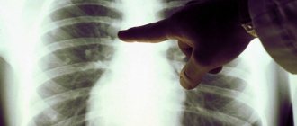
- X-ray is the main test for identifying lung cancer.
- MRI, CT, as well as PET-CT and PET-MRI are techniques that allow you to visualize the pulmonary area and study the tumor (type, size, etc.).
- Biopsy – extraction of a biopsy sample from a tumor allows, using histological examination, to obtain the most accurate information and prescribe effective therapy.
Stages of bronchial cancer
Bronchopulmonary cancer is divided into stages depending on how far the tumor has spread in the body. In this case, they are guided by the generally accepted TNM classification. The letters in it mean:
- T—size of the primary lesion, degree of invasion into adjacent tissues.
- N - damage to regional (close to the primary tumor) lymph nodes.
- M—presence of distant metastases.
Depending on these indicators, the stage of the tumor is determined, which is designated by Roman numerals I–IV.
Diagnostic methods
The examination usually begins with a chest x-ray. Computed tomography helps to obtain more detailed information about the number, size and location of tumor foci.
A biopsy is performed to confirm the diagnosis. It can be done in different ways:
- Using a needle inserted into the tumor through the chest wall.
- During bronchoscopy - endoscopic examination of the bronchi.
- During thoracoscopy - endoscopic examination of the pleural cavity (the space between the lungs and the walls of the chest, limited by a thin film of connective tissue - the pleura ).
- During thoracentesis , a puncture is made in the chest wall to remove accumulated fluid.
- During mediastinoscopy - endoscopic examination of the space between the lungs - the mediastinum .
A cytological examination of sputum is performed. In order to assess the extent of cancer spread and identify distant metastases, the doctor may prescribe an MRI, PET scan, radiography and radioisotope bone examination, and ultrasound.
Treatment tactics depend on the stage of cancer, general health and age of the patient.