Pulmonary embolism (PE) is a serious, potentially life-threatening condition. Pulmonary embolism occurs due to blockage of blood vessels by thrombotic masses in the lungs. Pulmonary embolism (PE) can cause symptoms such as chest pain and/or shortness of breath. Also, in some cases, PE may have no symptoms at all, and it is difficult to detect without specialized examination. Massive pulmonary embolism can cause collapse and death. PE usually occurs due to the formation of a thrombosis in the lower extremity - deep vein thrombosis (DVT).
Pulmonary embolism (PE)
Prompt treatment is important and can save lives. Pregnancy, various medical conditions and medications, immobility, and major surgery all increase the risk of PE. Anticoagulant therapy, including heparin drugs, direct oral anticoagulants, or warfarin, is the usual treatment for PE.
General information
Thrombosis is the formation of a blood clot in a blood vessel that interferes with blood flow.
Its danger lies in the fact that there is a danger of a blood clot or its fragment moving through the bloodstream - an embolism and blockage of another vessel occurs. For example, supplying the brain or heart. Pulmonary embolism often occurs, which leads to severe organ dysfunction or death. What is pulmonary embolism in medicine? Pulmonary embolism is an acute occlusion (blockage) of the pulmonary artery trunk or its branches (main, lobar or segmental). Occlusion occurs more often due to embolization of a thrombus from the right half of the heart or veins of the lower extremities. The incidence of this disease increases with age and the age of such patients is 62 years.
Pulmonary artery thrombosis is an emergency in cardiac resuscitation and is often the cause of patient death. Seriously ill patients with a high risk of pulmonary embolism and death are patients with cardiac arrhythmias, cancer pathology , and thrombosis of the veins of the lower extremities . Diagnosis is often difficult, so the disease is often not recognized. Early diagnosis and initiation of intensive treatment are important in the prognosis of this disease. The code for pulmonary embolism according to ICD-10 is I 26.
What causes pulmonary embolism?
The usual cause of pulmonary embolism is deep vein thrombosis (DVT).
In almost all cases, the cause is a blood clot (thrombosis) that originally formed in a deep vein of the lower limb (known as deep vein thrombosis). This clot travels through the circulation and eventually becomes lodged in one of the lung's blood vessels. The broken thrombus is now called an embolus (and therefore can cause an embolism). Most deep vein thromboses arise from the veins of the legs or pelvis. Sometimes PE can occur from a blood clot in a vein in the arm or from a blood clot that forms in the heart. However, these situations are quite rare.
Other causes of pulmonary embolism
In rare cases, a blockage of a blood vessel in the lung may be caused by an embolus, which is not a blood clot. It could be:
- Fatty tissue from the bone marrow of a broken bone (if a large long bone such as a femur is broken).
- Foreign material from unsterile injection - for example, due to drug abuse.
- Amniotic fluid during pregnancy or childbirth (rare).
- Entry of a significant volume of air into the vein (rare).
- A small fragment of cancerous material (tumor) that has broken off from a larger tumor in a different location.
- Mycotic emboli are material from the focus of a fungal infection.
Pathogenesis
The main thing in the pathogenesis of pulmonary embolism is occlusion of the pulmonary artery and its branches by embolism, which leads to disruption of hemodynamics and gas exchange. During occlusion, the pressure in the pulmonary artery increases by almost 50% due to vasoconstriction associated with the release of serotonin and thromboxane A2 . A sharp increase in pulmonary resistance causes expansion of the right ventricle, stretching of myocytes and tension of the ventricular walls. Lengthening the time of its contraction towards diastole causes protrusion of the septum into the left ventricle. Therefore, left ventricular filling becomes more difficult, cardiac output decreases, and systemic hypotension .
Massive infiltrates are found in the ventricular myocardium, which further destabilizes hemodynamics. Right ventricular failure due to pressure overload is a leading cause of death. Respiratory failure is also associated with hemodynamic disturbances. A decrease in cardiac output causes desaturation of venous blood. The appearance of zones of reduced blood flow causes a mismatch between perfusion and ventilation. Small emboli in the periphery do not affect hemodynamics, but cause pulmonary hemorrhage with hemoptysis, pleurisy and pleural effusion (“pulmonary infarction”).
Classification of pulmonary embolism
The 2008 classification of the European Society of Cardiology identifies:
- Low risk pulmonary embolism.
- Intermediate.
- Tall.
Clinical classification takes into account the caliber of the pulmonary arteries and the percentage of pulmonary involvement:
- Massive (cessation of blood flow in less than 50% of the pulmonary bed). It is of great clinical importance because it is accompanied by shock or decreased pressure, pulmonary hypertension. With occlusion of the trunk, pronounced cardiorespiratory disorders develop. Under such conditions, the right ventricle cannot perform the function of a pump and its cavities quickly expand, and tricuspid valve insufficiency develops. The septum between the ventricles shifts to the left (towards the left ventricle), which is accompanied by poor filling in diastole. Due to the cessation of blood flow, the pulmonary parenchyma is not supplied with blood, but is ventilated. In the affected area, obstruction of the bronchi occurs, the alveoli collapse and surfactant is not formed in them, which contributes to the development of atelectasis (collapse) of the lungs on days 1-2 of embolism. Such hemodynamic disturbances and impaired pulmonary function often lead to the death of the patient.
- PE of small branches of the pulmonary artery (cessation of blood flow is noted in less than 30% of the pulmonary bed). In this case, there is occlusion of lobar and segmental small branches. The disease is not severe without hemodynamic disturbances and patients only need anticoagulant therapy. The pulmonary circulation has compensatory capabilities and there is a possibility of independent dissolution of blood clots when fibrinolysis .
- Submassive—thromboembolism of the branches of the pulmonary artery (cessation of blood flow in less than 30-50% of the bed). It manifests itself as right ventricular failure, and hemorrhagic infarctions form in the lungs.
What are the prospects for pulmonary embolism?
This depends on the type of pulmonary embolism and the presence of concomitant diseases.
If PE is treated correctly and promptly, the prognosis is good and most patients can make a full recovery.
The prognosis is worse if there is a serious disease that contributed to the embolism, such as advanced cancer. Massive PE is more difficult to treat and is life-threatening. PE is a serious condition and can have a high risk of death, but this is greatly reduced with early treatment.
The most dangerous time for complications or death is the first few hours after an embolism. In addition, there is a high risk of developing a new pulmonary embolism within six weeks of the first one. Therefore, treatment must be started immediately and continued for at least six months.
Causes of pulmonary embolism
Considering the causes of pulmonary embolism, it is necessary to highlight a number of diseases and conditions that are accompanied by the formation of blood clots, which are the basis of embolism:
- Varicose veins and phlebothrombosis of the deep veins of the lower extremities. This pathology causes thromboembolism in 90% of cases. The threat is posed by floating blood clots, which are freely located in the lumen of the vessel and are connected to the vein wall in only one part.
- Congestive heart failure , overstretching of the right ventricle, which creates conditions for the formation of blood clots in the cavity of the right ventricle.
- The use of oral contraceptives and pregnancy , which are associated with an increased risk of blood clots.
- Antiphospholipid syndrome (autoimmune thrombotic vasculopathy ), manifested by venous and arterial thrombosis . Vessels of different sizes may be affected - capillaries and large arterial trunks. Deep vein thrombosis of the lower extremities is a typical manifestation of antiphospholipid syndrome. Repeated pulmonary embolisms are typical.
- Central venous catheterization.
- The presence of malignant diseases.
- Carrying out chemotherapy .
- Spinal cord injury.
- Forced immobilization after surgery, during a stroke , fractures of the pelvis and limbs.
- Hip replacement.
- Atrial fibrillation.
- Polytrauma and extensive surgery.
- Hereditary predisposition to thrombosis ( thrombophilia ), which is caused by mutations of certain genes, deficiency of antithrombin III, deficiency of protein C and S.
Considering pulmonary thrombosis , it is necessary to note the risk factors for this condition:
- Damage to the venous endothelium.
- Obesity.
- Age.
- Hypercoagulation.
- Slowing of venous blood flow.
- Infection.
- Blood transfusions.
- Migraine.
- The combination of several factors is accompanied by a high risk of thrombus embolism .
Venous thromboembolic complications
Pulmonary embolism (PE) is part of a group of clinical problems collectively known as venous thromboembolic events (VTE).
Thrombosis is the blockage of a blood vessel by a blood clot (thrombus). Embolisms occur when part or all of a blood clot displaces from where it formed. The thrombotic masses then move with the bloodstream until they get stuck in narrower blood vessels in other parts of the body. In this case, the blood clot is called an embolus. Deep vein thrombosis (DVT) is the cause of venous thromboembolism, including pulmonary embolism (PE). Thrombosis most often occurs in the veins of the lower extremities.
Symptoms of pulmonary embolism (PE)
Making a diagnosis in the early stages is difficult, only in the acute course the symptoms of pulmonary artery thrombosis appear manifestly and suddenly: shortness of breath , chest pain , tachycardia , decreased blood pressure. In this case, the patient has risk factors for thromboembolism - varicose veins and phlebothrombosis .
Hypotension and shock indicate central thromboembolism . Severe acute shortness of breath also develops with central pulmonary embolism. With the same localization, chest pain is angina-like in nature . Massive thromboembolism is accompanied by signs of right ventricular overload - this is the bulging of the jugular veins in the neck and the right ventricular gallop rhythm.
Signs of PE in subacute cases are not very specific. Respiratory and right ventricular failure progresses, infarction pneumonia and hemoptysis appears. With a recurrent course, repeated attacks of shortness of breath , there are signs of pneumonia and periodic fainting occurs. If the thrombus blocks small arteries in the periphery, shortness of breath is minor and transient. Only in the case of chronic lung pathology or heart failure does shortness of breath worsen. Sometimes PE is asymptomatic.
The clinical picture depends on the caliber of the blocked vessel and, accordingly, the degree of involvement of the pulmonary vascular bed. Massive pulmonary embolism (most often thrombosis of the main branch ) occurs with symptoms of shock or a decrease in pressure by 40 mm Hg. Art. for a short time that is not associated with arrhythmia , sepsis , or decreased blood volume. Characteristic shortness of breath , cyanosis , and sometimes fainting .
Submassive pulmonary embolism (obstruction of lobar or segmental branches) is manifested by right ventricular failure (swelling of the neck veins, pallor, cyanosis), but without arterial hypotension. The patient develops shortness of breath , tachycardia , pulmonary infarction (fever, cough, pulmonary-pleural pain, sputum streaked with blood). In non-massive cases, there are no signs of right ventricular failure, and the pressure is normal.
How is pulmonary embolism diagnosed?
The diagnosis is based on clinical symptoms and medical history. For example, if a bedridden patient undergoing major surgery in a hospital develops shortness of breath, it is likely to be a pulmonary embolism.
Pregnant women who experience symptoms and/or signs suggestive of PE should be hospitalized immediately, as pulmonary embolism during pregnancy is a very serious condition and requires prompt diagnosis and treatment. For other patients, the Wells score is used to decide whether hospitalization is necessary. This allows you to assess the risk of developing pulmonary embolism, taking into account the following clinical signs:
- Clinical features of deep vein thrombosis.
- Heart rate more than 100 beats per minute.
- Immobilization for more than three days or surgery in the previous four weeks.
- Previous deep vein thrombosis and pulmonary embolism.
- Coughing up blood (hemoptysis).
- Treatment of cancer over the past six months.
- The alternative diagnosis is less likely than PE. Example: pneumothorax, pneumonia, myocardial infarction or gastroesophageal reflux.
If the likelihood of pulmonary embolism is high, emergency hospitalization of the patient is necessary. Various tests will be used to confirm the diagnosis. These may include the following activities:
Duplex ultrasound scanning of the veins of the lower extremities
This study is used to determine blood flow disorders in the veins of the lower extremities. Lower extremity ultrasound is a simple, non-invasive test that can detect deep vein thrombosis. If DVT is detected, it can be assumed that the cause of other symptoms (such as shortness of breath or chest pain) was a pulmonary embolism. It is better to start treatment with anticoagulants immediately both in cases of deep vein thrombosis and in cases of suspected pulmonary embolism. If thrombotic masses are detected in the veins of the lower extremities, further studies may not be required. However, if the ultrasound results are negative, both deep vein thrombosis and pulmonary embolism cannot always be excluded. Further clinical studies will be required.
D-dimer blood test
This test detects fragments of the breakdown products of a thrombotic blood clot. The higher the level, the greater the likelihood of a blood clot forming in the vein. Unfortunately, the test can be positive in a number of other situations, such as if you have recently had surgery or are pregnant. Therefore, a positive result does not diagnose DVT or PE. However, in a complex of studies, this test can show how likely the presence of thrombosis is. To some extent, this may help decide whether further research is needed. A negative D-dimer result with a low risk of thrombosis means that the likelihood of a blood clot is extremely low. However, if your risk of venous thromboembolism is high, the D-dimer test cannot rule out the possibility of a blood clot and you will need further testing.
Ultrasound scan of the heart (echocardiography)
Echocardiography is useful for patients who may have massive PE, as it can show the effect on the heart. If there is massive PE, this puts stress on the right side of the heart. This test can be performed at the patient's bedside.
Computed tomography of the lungs with blood flow contrast
This is a specialized test that examines blood circulation in the lungs. This technique is the most accurate for detecting pulmonary embolism.
General studies
These studies are focused on diagnosing the condition of the heart, lungs and blood. This may help with diagnosis or identify other conditions:
- An electrocardiogram (ECG) is performed on all patients. This is necessary to identify any signs of cardiac strain that may occur with PE. Any heart rhythm abnormalities, such as atrial fibrillation, that may result from thromboembolism are identified.
- Blood tests are used to look for signs of heart attack, infection, or inflammation. An arterial blood gas test may also be done, which involves taking a blood sample from an artery rather than a vein. This is necessary to check the oxygen level in the blood.
- A chest x-ray is used to detect pneumonia or other chest conditions.
- Treatment of pulmonary embolism
Tests and diagnosis of pulmonary embolism
Instrumental diagnostics of this condition includes:
- Chest X-ray . In the lungs, discoid atelectasis , a raised dome of the diaphragm, or pleural effusion . All these signs are nonspecific, but exclude various causes of pain and shortness of breath.
- ECG.
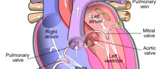
- Echocardiography . It is of key importance in diagnosis, especially in patients with unstable hemodynamics. This test directly detects blood clots in the right side of the heart, and blood clots in the large arteries of the lungs (trunk and main branches). An indirect sign of pulmonary embolism detected by echocardiography is overload of the right chambers. An indirect sign of pulmonary artery thrombosis is identified - overload of the right ventricle: expansion of the ventricular cavity, unusual movement of the septum between the ventricles and a D-shaped left ventricle.
- Ultrasound of the deep veins of the extremities.
- A special research method is ventilation-perfusion scintigraphy . This is a safe study that involves the intravenous administration of albumin microspheres that are labeled with technetium. Albumin microspheres block pulmonary capillaries and lung perfusion is assessed based on this feature. This study is complemented by a ventilation study, which increases the specificity of the examination (in case of thrombosis, ventilation in segments of the lungs in which there is poor blood supply remains normal). That is, a discrepancy between ventilation and perfusion is detected. Radioactive radiation during scintigraphy is lower than during CT angiography. Perfusion scintigraphy alone can be performed in patients with a normal radiograph.
- CT angiography . This study has become the method of choice for diagnosing pulmonary vascular pathology. The pulmonary arteries can be viewed down to the segmental branches. The specificity of the method is 96%. The technique is optimal for diagnosing a patient whose condition is stable. In another case, the patient cannot be transported from intensive care.
- Pulmonary angiography is considered the gold standard for diagnosing PE. However, it is rarely performed since a less invasive test, CT angiography, has become available. Diagnosis is based on identifying a blood clot that causes a defect in the filling of the pulmonary artery branch or is completely absent. The method allows you to obtain images of peripheral pulmonary arteries and detect blood clots of 1-2 mm in the smallest arteries. A contrast agent is injected for the study.
ECG signs of PE and radiographic signs
Blood tests:
- D-dimer is a fibrin degradation product. Its increase in plasma is observed during acute thrombosis - this is explained by the activation of fibrinolysis and coagulation. A normal D-dimer level makes the diagnosis of PE unlikely. Also, this indicator is nonspecific, since fibrin overproduction is observed during inflammation, bleeding, myocardial infarction , aortic aneurysm , oncological processes, trauma, and surgery. The specificity of D-dimer for thrombosis decreases with age. This indicator is paid attention to during treatment, since an increase in its level after the end of anticoagulant treatment indicates a risk of relapse.
- of brain natriuretic peptide increases , which is associated with distension of the right ventricle. The degree of increase is proportional to the severity of the patient. However, an increase in this marker is nonspecific, since it can be observed with myocardial ischemia , left ventricular hypertrophy , sepsis and tachycardia . However, the absence of a significant increase indicates a favorable prognosis for pulmonary embolism.
Basic methods of treating pulmonary embolism
Anticoagulant therapy
Anticoagulant therapy is often referred to as blood thinning. However, it does not actually thin the blood. Anticoagulant therapy changes certain chemicals in the blood to prevent thrombotic clots from forming. This therapy does not dissolve the clot (as some people mistakenly think). Anticoagulation prevents the formation of new clots, and therefore thromboembolism. The body’s own mechanisms in the blood destroy the clot. Anticoagulant treatment is usually started immediately (if PE is suspected) to prevent worsening of the thrombosis while awaiting test results. Anticoagulation medications come in two forms: injections and tablets (or syrup for those who cannot swallow tablets). The first include heparin and fondaparinux. Tablet forms include warfarin and direct oral anticoagulants.
Supportive treatment is also used in the treatment of pulmonary embolism.
This therapy helps the body cope with the consequences of pulmonary embolism.
- Oxygen to reduce shortness of breath.
- In some cases, intravenous fluids are given to support circulation.
- If the patient is unwell or has a massive pulmonary embolism, careful monitoring and possibly intensive care are necessary.
- Additional procedures
They are usually used to treat severe or massive PE when the patient is very unwell or when treatment with anticoagulants is not possible.
Thrombolysis is an injection of drugs that is used to dissolve a blood clot. The drugs commonly used are alteplase, streptokinase, or urokinase. They are more powerful than the anticoagulants heparin and warfarin described above. However, there is a greater risk of side effects such as unwanted bleeding. Unwanted bleeding may even include bleeding into the brain (intracerebral hemorrhage).
Kava filters . They can be used to prevent blood clots from reaching the lungs. The filter is placed in a large vein called the inferior vena cava. The filter is inserted through a thin tube, which is inserted into a large vein and then inserted along the vein into the correct position. This procedure does not require anesthesia and can be performed at the patient's bedside. Filters may be useful when anticoagulant treatment alone is not sufficient or for patients who for some reason cannot undergo anticoagulant treatment. Currently, the indications for the use of vena cava filters are narrowing.
Surgical embolectomy. In some cases, it is possible to remove the embolus surgically. This is called an embolectomy. This is a major operation because it involves surgery on the organs of the chest, near the heart. A specialized medical facility and a highly specialized surgical team are required. This is usually considered a last resort for seriously ill patients. The operation carries a significant risk of death. This intervention will only be considered as an option if there is a massive PE, limiting other treatment options.
Catheter thrombolysis . This type of treatment is called catheter embolectomy or catheter clot fragmentation. It involves advancing a catheter through the blood vessels until it reaches the clot in the lung. Once the clot is reached, it can be removed or broken up (fragmented) with treatment given through the tube. This is a highly specialized treatment, so it is only available in certain treatment facilities.
In children
Thrombosis in children occurs much less frequently. For their occurrence, a combination of acquired and congenital factors is necessary. Of the hereditary ones, it is worth noting the hereditary decrease in the activity of antithrombin III and proteins C, S, which are natural anticoagulants. Acquired factors:
- severe somatic diseases
- infectious diseases;
- systemic connective tissue diseases;
- malignant tumors;
- connective tissue dysplasia ;
- hemolytic anemia;
- antiphospholipid syndrome.
The most important factor in thrombosis is the use of an intravascular catheter. Up to 80% of thromboses are associated with catheterization. In children, pulmonary embolism is often associated with deep vein thrombosis. Venous thrombosis in children is preceded by infection, thrombocytopenic purpura , antiphospholipid syndrome , and in some, excessive insolation. Recurrent pulmonary embolism also occurs, but the source of thrombosis cannot be identified. PE in children is often mistakenly regarded as pneumonia .
How to prevent pulmonary embolism?
To avoid pulmonary embolism, it is necessary to prevent and promptly treat chronic diseases. One of the causes of venous thrombosis is varicose veins. Any person who suspects he has this disease should visit a phlebologist and have the venous system of the lower extremities diagnosed. After diagnosis, you need to follow the doctor's recommendations. Also, patients planning major surgery should be assessed for the risk of deep vein thrombosis, and people at high risk of DVT may require prophylactic doses of anticoagulants before and after surgery.
Prevention of pulmonary embolism
- Primary prevention of embolism after surgery on the pelvic organs and orthopedics.
- If the risk of thromboembolism is low, prevention involves the use of compression stockings or elastic compression with an elastic bandage of the lower extremities.
- enoxaparin or fondaparinux or dabigatran until discharge .
- If the risk is high, these recommendations should be followed for a month.
- New oral anticoagulants are registered for primary prevention after orthopedic surgery.
There are also non-surgical patients (chronic heart failure class III-IV), who are also at risk for pulmonary embolism. Their choice of anticoagulant is the same. The standard of treatment is enoxaparin .
Secondary prevention is the prevention of recurrent thromboembolism. Without prophylaxis, recurrence of pulmonary embolism within the first three months is observed in 20-45% of patients. With prevention, the number of relapses is reduced to 1%. Warfarin is used for prevention , and for the first six months, cancer patients are prescribed low molecular weight heparins ( Fraxiparin , Fraxiparin Forte , Clexane , Enixum , Fragmin , Daltep ), then they are transferred to oral medications ( vitamin K antagonists - Warfarin , Warfarex ).
How long should I take them? To do this, the risk of bleeding is assessed based on risk factors (age 65 years or older, history stroke malignant tumor , renal and liver failure , diabetes mellitus , low platelet levels, anemia and others). The risk of bleeding is high if more than two risk factors combine.
It is also possible to use “new” anticoagulants for secondary prevention, which are included in the American Guidelines for the prevention of pulmonary embolism. Rivaroxaban ( Xarelto ) is superior in long-term treatment and can be prescribed for 6-12 months with a low risk of bleeding, providing protection against relapse.
Who develops pulmonary embolism?
Almost all cases of pulmonary embolism are caused by deep vein thrombosis. Thus, people who are more likely to get pulmonary embolism are people who are prone to deep vein thrombosis. Let's look at some risk factors for pulmonary embolism. Important factors are immobility, severe somatic pathologies and major surgical intervention (especially gynecological operations, operations on the pelvis and lower extremities). The risk of developing deep vein thrombosis or PE in hospital can be significantly reduced by activating the patient to walk as soon as possible. Medicines to help prevent deep vein thrombosis and PE are also prescribed to those patients who are at high risk.
Consequences and complications
- Hemodynamic disturbances (increased vascular resistance in the pulmonary artery system and increased afterload on the right ventricle).
- Respiratory failure.
- Hypoxia.
- Pulmonary-pleural complications ( atelectasis , pleurisy , lung abscess , pulmonary infarction , pneumonia , pyopneumothorax , pleural empyema ).
- Post-embolic pulmonary hypertension .
List of sources
- Vatutin N.T., Sklyannaya E.V., Eshchenko E.V. /Pulmonary embolism. Review of recommendations of the European Society of Cardiology for diagnosis and treatment (2014 // Practical Angiology. - 2015. - No. 1 (68). - P. 5-18.
- Russian clinical recommendations for the diagnosis, treatment and prevention of venous thromboembolic complications // Phlebology. — 2015.— No. 4, issue 2. 46 p.
- Vatutin N.T. Emergency cardiology. - Donetsk, 2011. - 236 p.
- Vatutin N.T., Kalinkina N.V., Perueva I.A. /Pulmonary embolism // Practical angiology. – 2011. – No. 2. – P. 32-40.
- Vatutin N.T., Kalinkina N.V., Eshchenko E.V. and others. Pulmonary embolism (basic information and own observation) // Heart and Vessels. – 2013. – No. 1. – P. 120-123.
Frequently asked patient questions about pulmonary embolism (PE)
What are the symptoms of pulmonary embolism?
The main symptoms to suspect pulmonary embolism are cough, shortness of breath, chest pain and hemoptysis.
How to avoid pulmonary embolism?
In order to prevent the development of pulmonary embolism, it is necessary to prevent venous thrombosis: promptly treat varicose veins and other vein pathologies, avoid prolonged immobilization for various diseases and surgical treatment, use anticoagulants in case of increased risks of thrombosis.
Who is more likely to develop pulmonary embolism?
Pulmonary embolism, as a rule, affects patients with severe cardiac and pulmonary pathology, cancer patients, and patients in various surgical hospitals after long-term surgical interventions.
What should you do if you suspect pulmonary embolism?
At the slightest suspicion of pulmonary embolism, you must call an ambulance. Before the arrival of medical personnel, the patient must be laid on a flat surface and ensure free access of air.