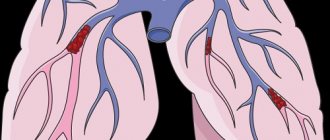
An artery in the pulmonary circulation that carries deoxygenated blood from the heart to the lungs
| Pulmonary artery | |
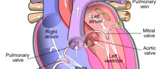
Anterior (frontal) view of an open heart. White arrows indicate normal blood flow. (The pulmonary artery is indicated at the top right.) | |
| Details | |
| Predecessor | truncus arteriosus |
| System | Cardiovascular, Respiratory |
| Source | Right ventricle |
| Identifiers | |
| Latin | pulmonary artery |
| MeSH | D011651 |
| TA98 | A12.2.01.101 A12.2.01.201 |
| TA2 | 4077, 4091 |
| F.M.A. | 66326 |
| Anatomical terminology [edit in Wikidata] | |
And the pulmonary artery
is an artery in the pulmonary circulation that carries deoxygenated blood from the right side of the heart to the lungs.
The largest pulmonary artery is the main pulmonary artery
or
pulmonary trunk
from the heart, and the smallest are the arterioles, which lead to capillaries that surround the pulmonary alveoli.
Structure
The pulmonary arteries are blood vessels that carry blood from the right side of the heart to the capillaries of the lungs. The transported blood, unlike other arteries, does not contain oxygen (“deoxygenated”). The main pulmonary arteries emerge from the right side of the heart, and they divide into smaller arteries that gradually divide and become smaller until they become arterioles and eventually capillaries.
Main pulmonary arteries
Volumetric rendering of a high resolution computed tomography scan of the chest.
The anterior chest wall, airways, and pulmonary vessels anterior to the root of the lung were digitally removed to visualize different levels of pulmonary circulation. In order of blood flow, the pulmonary arteries begin as the pulmonary trunk
or
the main pulmonary artery
. The main pulmonary artery begins at the base of the right ventricle. It is short and wide - approximately 5 centimeters (2.0 in) long and 3 centimeters (1.2 in) in diameter.
The main pulmonary artery divides into true
and
the left main pulmonary artery
.[1] The left main pulmonary artery is shorter and somewhat smaller than the right, passing horizontally in front of the descending aorta and left bronchus to the root of the left lung. The superior left main pulmonary artery is connected to the concavity of the proximal descending aorta by means of the ligamentum arteriosus.[2]
The opening of the pulmonary artery (or pulmonary trunk) is round in shape and located at the apex of the conus arteriosus, close to the interventricular septum.
It is located above and to the left of the atrioventricular orifice, and is guarded by the pulmonary semilunar valves.
Pulmonary arterial tree
It then divides into two lobar arteries, one for each lobe of the left lung. The right main pulmonary artery has a longer and more horizontal course as it crosses the mediastinum. It passes under the aortic arch, behind the ascending aorta and in front of the descending aorta. It passes posterior to the superior vena cava and in front of the right bronchus. Upon reaching the hilum of the right lung, the right main pulmonary artery divides into two branches:
- truncus anterior - supplies blood to the right upper lobe
- interlobar artery - the lower and greater branch, supplies blood to the middle and lower lobes of the lung
At the far end, the pulmonary arteries (labeled below) become capillaries to the pulmonary alveoli.
The right and left main pulmonary arteries arise from branches that roughly correspond to the lobes of the lungs and in such cases may be called lobar arteries
.
The lobar arteries branch into segmental arteries
(approximately 1 per lobular segment), which in turn branch into
subsegmental pulmonary arteries
.[3] They eventually form
the intralobular arteries
.[4]
Complications of pulmonary embolism
Most people with PE are treated successfully and have no serious complications. However, serious complications are possible, including:
- Collapse - due to the effect of a blood clot on the heart and circulation. This can cause cardiac arrest and can be fatal. PE can cause heart strain. This can lead to a condition called heart failure, where the heart pumps less than normal.
- Recurrence of pulmonary embolism. Blood clots may occur again later (called recurrent PE). Treatment with anticoagulants helps prevent this.
- Bleeding. Complications due to treatment. Treatment with anticoagulants may have side effects. The main one is bleeding in various locations, for example, from a stomach ulcer. Approximately 3 in 100 patients will have significant bleeding due to treatment of PE with anticoagulants. This type of bleeding can usually be treated successfully. However, it is almost always safer to take anticoagulants to prevent recurrent PE, which can be even more severe.
- Chronic pulmonary hypertension. If small PEs recur, they may contribute to the development of a condition in which there is increased pressure in the blood vessels of the lungs (pulmonary hypertension).
Development
The pulmonary arteries originate from the truncus arteriosus and the sixth pharyngeal arch. Truncal arteriosis is a structure that forms during the development of the heart as a successor to the conus arteriosus.[5]
By the third week of development, the endocardial tubes developed swelling in the area closest to the heart. The swelling is known as Bulbus cordis and the superior portion of this swelling extends into the truncus arteriosus.[6] The structure is ultimately mesodermal in origin.[5] During cardiac development, the heart tissue undergoes folding and the truncus arteriosus is exposed to the left and right ventricles. As the septum develops between the two ventricles of the heart, two bulges form on either side of the truncus arteriosus. They gradually increase in size until the trunk divides into the aorta and pulmonary arteries. [7]
During early development, the ductus arteriosus connects the pulmonary trunk and aortic arch, allowing blood to bypass the lungs.[8]
Function
The pulmonary artery carries deoxygenated blood from the right ventricle to the lungs.[9] Here the blood passes through the capillaries next to the alveoli and becomes oxygenated as part of the breathing process.[10]
Unlike the pulmonary arteries
, then the bronchial arteries supply nutrition to the lungs themselves.[11]
Pressure
B pulmonary artery pressure
(
PA pressure
) is a measure of the blood pressure found in the main pulmonary artery. This is measured by inserting a catheter into the main pulmonary artery.[12]:190–191 The mean pressure is usually 9–18 mm Hg. Art.[13] and the wedge pressure measured in the left atrium can be 6-12 mmHg. Wedge pressure may be increased in left-sided heart failure,[12]:190–191 mitral valve stenosis, and other conditions such as sickle cell disease.[14]
Clinical significance
The pulmonary artery is relevant in a number of clinical conditions. Pulmonary hypertension is used to describe an increase in pulmonary artery pressure and can be defined as a mean pulmonary artery pressure greater than 25 mmHg.[12]:720 As measured on a CT scan, a diameter greater than 29 mm is often used as a cutoff to indicate pulmonary hypertension.[15] This may occur as a result of a heart problem such as heart failure, a lung or airway disease such as COPD or scleroderma, or a thromboembolic disease such as a pulmonary embolism or the emboli seen in sickle cell disease.[12]:720 –721
A pulmonary embolism is an embolus that becomes lodged in the pulmonary circulation. This may occur due to deep vein thrombosis, especially after a period of immobility. Pulmonary embolism is a common cause of death in patients with cancer and stroke.[12]:720–721 A large pulmonary embolus that lodges in the bifurcation of the pulmonary trunk with extensions into the left and right main pulmonary arteries is called a saddle embolus
.[16]
Causes and symptoms of pulmonary embolism
The development of pulmonary embolism has several main causes:
- Acute deep vein thrombosis of the ileofemoral segment.
- Deep vein thrombosis of the leg.
- Thrombosis of the venous plexuses of the pelvis.
- Blood clots in the right cavities of the heart.
- Thrombophlebitis of the superficial veins (a fairly rare cause).
- Septic generalized process.
- Cardiovascular diseases that predispose to the formation of blood clots (ischemic heart disease, rheumatism with the presence of mitral stenosis, endocarditis, cardiomyopathy, etc.).
- Oncological diseases.
- Thrombophilia (impaired vascular hemostasis, changes in blood clotting properties).
- Antiphopholipid syndrome (formation of antibodies to phospholipids of platelets, endothelial cells and nervous system).
The greatest danger to the patient’s health is posed by so-called “floating” blood clots. They have a single small point of fixation, so they can be damaged quite easily and move into the pulmonary circulation.
Risk factors for the development of pulmonary embolism
When diagnosing an acute condition, the presence of risk factors that could provoke thrombus formation must be taken into account. The most significant are:
- fracture of the upper femoral neck;
- hip or knee replacement;
- undergone major operations;
- history of serious injuries;
- brain damage.
The following should also cause caution regarding thrombosis:
- arthroscopy of the knee joint;
- installation of a central venous catheter;
- history of chemotherapy;
- oncological diseases;
- taking hormonal therapy (including combined oral contraceptives);
- history of strokes;
- pregnancy, childbirth and the postpartum period;
- thrombophilia.
The presence of cancer and concomitant chemotherapy significantly increases the risk of developing pulmonary embolism. About 15% of patients with a malignant neoplasm die due to blockage of the pulmonary artery by a thrombus or embolus. Chemotherapy treatment increases the risk of thrombosis by 47%.
In more rare cases, the development of thromboembolism is facilitated by:
- long-term immobilization of a limb or bed rest (more than 3 months);
- frequent air travel;
- elderly and senile age;
- phlebeurysm;
- history of laparoscopic surgery.
Also, risk factors characteristic of the formation of any thrombosis can lead to the development of pulmonary embolism:
- smoking;
- obesity;
- physical inactivity (sedentary lifestyle);
- diabetes;
- increased blood cholesterol and triglyceride levels (especially LDL and VLDL);
- regular psycho-emotional overload;
- poor nutrition.
With age, the risk of developing pulmonary embolism increases. A hereditary predisposition to the development of the disease has also been proven. Therefore, if close relatives have had episodes of pulmonary embolism, you need to carefully monitor your health.
Symptoms of pulmonary embolism
The course of the disease is multifaceted and varied. There are no specific symptoms characteristic only of PE. Therefore, for correct diagnosis, the professionalism of the doctor and modern medical equipment are important, which will help differentiate the pathology in the shortest possible time.
The first manifestations of PE (acute) may be chest pain (similar to myocardial infarction), shortness of breath, cough, hemoptysis, decreased blood pressure, cyanosis and loss of consciousness (fainting).
In most cases, the diagnosis of pulmonary embolism is made by exclusion. First of all, it is necessary to differentiate it from myocardial infarction.
A characteristic feature of PE is the suddenness of shortness of breath. For example, habitually climbing the stairs to the 2nd floor can provoke it.
If small pulmonary vessels are affected, the symptoms may be blurred and non-specific. Signs of pulmonary infarction appear only 3-5 days after the development of the disease: pain in the chest, cough and hemoptysis. Pleural effusion accumulates in the chest. The fever can last from 2 to 12 days.
The full range of symptoms can be found only in every seventh patient. Most people have only 1-2 nonspecific symptoms. Particular difficulties are caused by making a diagnosis in cases where small branches of the pulmonary artery are affected. The manifestations of the pathology are erased, so often the correct diagnosis can be made only on the 3-5th day.
For patients with pulmonary embolism, it is important to make a timely diagnosis. This helps reduce the symptoms of the disease, as well as improve overall well-being. To speed up diagnosis, special scales have been developed. For inpatient settings, the PSWells model is used, and for outpatients, the Geneva model is used.
In parallel with establishing the diagnosis of pulmonary embolism, it is important to identify the cause of thrombosis. This is also quite difficult to do, since blood clots formed in the veins tend to be asymptomatic.
How does pulmonary embolism develop?
The main link in pathogenesis is venous thrombosis. There are a number of factors that contribute to the development of thrombosis. Among them:
- decreased blood flow speed in the veins;
- increased blood viscosity;
- turning off passive contraction of the venous wall;
- compression of veins by space-occupying formations.
The main prerequisites for the development of venous thrombosis are:
- disorders of the blood coagulation system. May develop due to internal factors in the body, as well as when taking certain medications. Patients taking hormone replacement therapy, including combined oral contraceptives, should regularly take a blood coagulogram. The latter can cause the development of thrombosis and pulmonary embolism in young women.
- Damage to the vascular wall Can develop for many reasons: trauma, surgical interventions on the vascular wall, viral damage, thrombophlebitis, hypoxia, etc.
Blood clots are dangerous formations that can cause sudden death. Therefore, it is important to carry out high-quality diagnostic measures to identify them. Ultrasound is considered the main way to detect thrombosis.
Floating (moving) blood clots pose a particular health hazard. They are loosely attached to the wall of the vessel and are able to move in its lumen. The detachment of such formations occurs very easily, and they quickly move with the blood flow and are accompanied by impaired pulmonary circulation.
Changes in hemodynamic parameters during pulmonary embolism appear when 30-50% of the vascular bed is blocked. In the pulmonary circulation, pressure increases and the load on the right ventricle of the heart increases. As a consequence, acute right ventricular failure develops.
Oxygen transportation suffers, the concentration of carbon dioxide in the blood increases, and hypercapnia develops. Coronary blood flow is disrupted, blood pressure decreases and systemic hypotension develops. All this leads to fainting, collapse, cardiogenic shock, or even clinical death.
Stabilization of blood pressure during pulmonary embolism is a misleading indicator of hemodynamic stability. Normal numbers after 24–48 hours may again be significantly reduced due to repeated thromboembolism and ongoing thrombosis of the arterial vessels of the lungs. A so-called “vicious circle” is formed when some symptoms aggravate others.
Classification of pulmonary embolism
The classification of pulmonary embolism is varied: according to the volume of the lesion, the severity of the process, the speed of development, etc.
According to the volume of damage to the vascular bed
The following options for TELA are distinguished:
- Massive Localization of the thrombus - the main trunk of the pulmonary artery or large vessels adjacent to it. Damage to the vascular bed reaches 50-75%. The patient's condition is extremely serious and urgent medical attention is required. There is a significant decrease in blood pressure and increasing tachycardia. Cardiogenic shock and acute right ventricular failure develop. Patients with massive pulmonary embolism have a high mortality rate.
- Embolism of lobar, segmental pulmonary arteries Damage to the vascular bed varies between 25-50%. The patient's condition is moderate. All the main symptoms of the disease are expressed, however, blood pressure is reduced only slightly.
- Embolism of small branches of the pulmonary arteries Damage to small vessels is characterized by a blurred clinical picture and an asymptomatic course. Up to 25% of the vascular bed is affected. The condition has a relapsing course, and therefore also requires careful monitoring of the patient.
According to the clinical course
The following variants of the course of pulmonary embolism are distinguished:
- Acute (“lightning-fast” development of symptoms) Instant and complete blockage of the pulmonary arteries (main trunk). As a result, acute respiratory failure and respiratory arrest develop. The patient enters a state of collapse and ventricular fibrillation begins. With this course, death can occur in just a few minutes.
- Acute In this condition, obstruction of the lobar branches of the pulmonary artery, as well as its main trunk, rapidly increases. The disease begins suddenly and progresses rapidly. Symptoms of respiratory and cerebral insufficiency increase. In the absence of adequate treatment, death occurs within 3-5 days.
- Subacute (protracted course of symptoms) Subacute course is characterized by the development of multiple pulmonary infarctions. The large and medium branches of the pulmonary artery become blocked. May last for several weeks. Progression is slow, gradual. Symptoms of respiratory and right ventricular failure become more pronounced as the disease progresses. It is characterized by the risk of developing repeated thromboembolism and exacerbation of symptoms in the absence of treatment.
- Chronic (recurrent) Thrombosis affects the lobar and segmental branches of the pulmonary artery. The main manifestation of the pathology is repeated pulmonary infarctions, gradually increasing arterial hypertension of the pulmonary circulation and symptoms of right ventricular failure. However, most often patients with a chronic course first present with complaints to a pulmonologist, where a diagnosis of “bilateral pleurisy” is made. Also, this type of pathology often develops in the postoperative period or against the background of existing diseases (cardiovascular, cancer, etc.)
The main influence on the rate of development of disease symptoms is the percentage of damage to the vascular bed of the pulmonary circulation. For each type of course, treatment is selected individually. In the case of a fulminant and acute form, you need to seek emergency medical help, and in the case of a chronic course, together with your doctor, choose the most gentle treatment tactics that will not have a significant impact on your lifestyle.
By severity
Pulmonary embolism has several degrees of severity:
- mild severity of the disease develops in 15–27% of patients;
- moderate severity is observed in 45–57% of patients with pulmonary embolism;
- severe degree is recorded in 16–35% of patients.
The prognosis of the course of the disease in patients with pulmonary embolism is based on the use of special scales. They include 11 indicators that help identify the risk of 30-day mortality for a given condition.
Complications of pulmonary embolism
The main danger of the disease in its acute course is sudden death due to cardiac arrest. If the disease develops gradually, pulmonary hypertension appears.
Chronic thromboembolic pulmonary hypertension is a pathology in which blockage of small and medium branches occurs with thrombotic masses. This leads to an increase in the load on the heart (especially its right parts: the atrium and ventricle). This condition can be treated with both conservative and surgical methods. The most informative way to confirm the diagnosis is pulmonary artery catheterization. There is pressure in the pulmonary artery above 25 mm Hg, an increase in pulmonary vascular resistance above 2 Wood units, emboli during long-term anticoagulant therapy (3-5 months).
The most severe complication of chronic thromboembolic pulmonary hypertension is right ventricular failure. It is characterized by:
- general weakness;
- feeling of heartbeat;
- decreased exercise tolerance;
- accumulation of fluid in the abdominal cavity (ascites);
- accumulation of fluid in the chest (hydrothorax);
- accumulation of fluid in the heart sac (hydropericardium).
A feature of shortness of breath in right ventricular failure is its absence at rest in a horizontal position. Long-term circulatory disorders and hypoxia cause atrophy of internal organs, protein imbalance and weight loss. The prognosis for this condition is unfavorable. Drug therapy can only temporarily stabilize the condition. As symptoms progress, cardiac reserves become depleted. Life expectancy for right ventricular failure with poorly selected therapy does not exceed 2 years.
How is pulmonary embolism diagnosed?
Diagnosis of pulmonary embolism is carried out according to the following algorithm:
- Estimation of clinical probability (pre-test probability).
- Determination of D-dimer level (threshold values previously adjusted for age and level of clinical probability of pulmonary embolism are taken into account).
- Contrast-enhanced computed tomography of the pulmonary artery. It is a highly evidence-based method for diagnosing pulmonary embolism. It can be used to visualize both small and large branches of the pulmonary artery. It is not possible to perform the study during pregnancy, intolerance to iodine-containing contrast agents and some other conditions.
- Lung scintigraphy. A study of pulmonary blood flow is performed. To do this, a small amount of radioactive substance is injected into the body. And then, using a gamma camera, the distribution of this substance in organs and tissues is visualized. The method is effective and recommended for the vast majority of patients. However, lung scintigraphy is not readily available.
- Angiopulmonography. An invasive test that is also designed to evaluate pulmonary blood flow. To do this, an X-ray contrast agent is injected into the vessels of the lungs. Using this technique, it is possible not only to determine the fact of embolism, but also the extent of damage to the vascular bed.
- Magnetic resonance imaging.
- Echocardiographic examination (bedside, if there is a high probability of pulmonary embolism).
- Compression ultrasound examination of venous vessels.
To assess the clinical likelihood of pulmonary embolism, the following risk factors are taken into account:
- surgeries and fractures in the previous month;
- history of malignant neoplasms;
- age over 65 years;
- tachycardia (high heart rate);
- pain in one of the lower extremities;
- hemoptysis.
Tests to diagnose pulmonary embolism
To effectively diagnose this pathological condition, a number of studies are carried out:
- Determination of D-dimer One of the most effective and informative methods for diagnosing pulmonary embolism. However, even this laboratory test is not completely specific. Increased results may also occur in pregnant women, the elderly, cancer patients, and during atrial fibrillation.
- Troponin levels increase as ischemia of the right ventricle (in rare cases, the left) increases.
- H-FABR A cardiac protein capable of binding fatty acids. Provides information in acute pulmonary embolism.
- The level of sodium diuretic peptide increases with right ventricular failure.
Electrocardiographic study for pulmonary embolism
An ECG is a medical procedure that is mandatory for every patient with suspected pulmonary embolism. The popularity of the technique is associated with its availability in any medical institution, and any emergency medical team has a portable ECG device. On the film for pulmonary embolism the following is recorded:
- acute overload of the right atrium and ventricle;
- complex rhythm disturbances;
- signs of coronary blood flow insufficiency.
All this allows you to suspect the correct diagnosis, choose the appropriate treatment tactics and reduce the risk of developing severe complications.
EchoCG is also performed to diagnose PE. This study allows you to obtain important information about the state of blood flow and the level of pressure in the pulmonary artery. EchoCG allows one to differentiate PE from pericardial tamponade, aortic dissection and other cardiac pathologies. Despite its high information content, the technique is rarely used in emergency patients. This may be due to the impossibility of organizing a 24-hour ultrasound service, the lack of a transesophageal sensor, the patient’s obesity, etc.
Ultrasound for diagnosing pulmonary embolism
Ultrasound has important diagnostic value for pulmonary embolism. It can be used to examine the veins of the lower extremities. It is carried out at four points: in the groin and popliteal region on both sides. The larger the study area, the higher the diagnostic value of the technique.
How is pulmonary embolism treated?
Preserving life and maximum health is the main priority in the treatment of PE. Doctors’ efforts are also aimed at preventing the development of chronic pulmonary hypertension.
When pulmonary embolism is detected, it is first necessary to eliminate the thrombus or embolus that is preventing blood flow in the vessels of the pulmonary circulation.
Emergency condition: methods of treating pulmonary embolism in the acute phase
For the treatment of thromboembolism in the acute phase, the following are used:
- Drug therapy aimed at eliminating right ventricular failure.
- Life support. If necessary, patients are connected to a ventilator (artificial lung ventilation).
- Mechanical circulatory support.
- Extracorporeal membrane oxygenation (saturation of blood with oxygen).
- Anticoagulant therapy Injectable forms of anticoagulants are used. This is a group of drugs that prevents the formation of new blood clots. New generation anticoagulants (NOACs) are presented in tablet form. However, at present they are used quite rarely to relieve acute conditions.
- Oral vitamin K antagonists.
- Systemic thrombolysis. This procedure is aimed at dissolving existing thrombotic masses in the area of the systemic circulation. In each individual case, the doctor individually selects the drugs and the required dosages.
- Percutaneous catheter therapy.
- Use of compression orthopedic garments “for the entire leg.” Elastic bandages can be used as an alternative.
For each patient, treatment tactics are selected individually. The clinical severity of the condition, symptoms and medical history must be taken into account.
Surgical techniques for the treatment of pulmonary embolism
Surgical treatment of pulmonary embolism is not uncommon in modern medical practice. Surgical intervention is indicated for massive lesions, when it is necessary to restore the patency of almost completely blocked vessels. Thrombectomy is used to normalize hemodynamics.
To determine the degree of risk and select a treatment strategy in patients with stable hemodynamics, specialized scales are used.
| Options | PESl scale | sPESl scale |
| Age (in years) | Age in years | 1 (if age over 80 years) |
| Male | +10 | — |
| Presence of malignant neoplasms | +30 | 1 |
| CHF (chronic heart failure) | +10 | 1 |
| Chronic lung diseases | +10 | — |
| Heart rate more than 110 beats/min | +20 | 1 |
| Systolic pressure level is less than 110 mmHg. | +30 | 1 |
| Respiratory rate more than 30 per minute | +20 | — |
| Body temperature less than 36.0 degrees Celsius | +20 | — |
| Impaired consciousness | +60 | — |
| Blood oxygen saturation level less than 90% | +20 | — |
| 30-day mortality risk level | ||
| Class I (≤65 points) Very low risk 0–1.6% | 0 points – risk 1% Confidence interval: 0-2.1% | |
| Class II (66–85 points) Low risk 1.7–3.5% | ||
| Class III (86–105 points) Moderate risk 3.2–7.1% | ≥1 point – risk 10.9% Confidence interval: 8.5–13.2% | |
| Class IV (106–125 points) High risk 4.0–11.4% | ||
| Class V (≥126) Very high risk 10.0–24.5% | ||
Thrombolytic therapy in the treatment of pulmonary embolism
Thrombolytic therapy has been shown to be effective in 92% of patients with pulmonary embolism. With its help, it is possible to achieve an improvement in basic hemodynamic parameters. This method of treatment, when started in a timely manner, significantly improves the prognosis of the disease, with a small number of contraindications to the technique.
The main disadvantage of thrombolytic therapy is time limitations. It is effective only in the first 2 days after the development of pulmonary artery thrombosis. After this period, the effectiveness of the drugs decreases, and hemorrhagic complications make the treatment more dangerous than effective. In low-risk patients, thrombolysis is not indicated.
Implantation of venous filters
In cases where it is impossible to prescribe anticoagulants or they are ineffective, venous filters are installed. Special devices are installed in the inferior vena cava that trap thrombotic clots and emboli from peripheral veins.
Alternative Treatments
If the patient has contraindications to systemic fibrinolysis, then the transcatheter thrombus fragmentation technique can be used. During this manipulation of blood clots, they are destroyed, and their contents are immediately aspirated (removed).
If the patient has a thrombus or embolus in the central pulmonary vessels, surgical removal is recommended. Especially with the development of refractory cardiogenic shock to therapy, the presence of contraindications to fibrinolytic therapy or its low effectiveness.
Features of anticoagulant therapy
Anticoagulant therapy in patients with acute venous thrombosis continues for at least 3 months. All medications are taken strictly as prescribed by the attending physician in appropriate dosages. Dynamic monitoring of the state of the blood coagulation system is carried out.
Treatment of acute conditions begins with parenteral (intravenous) unfractionated heparin. With its help, it is necessary to increase the activated partial thromboplastin time by 1.5–2 times in comparison with the initial laboratory values.
After stabilization of the condition, a transition to maintenance anticoagulant therapy is necessary. Intravenous administration of the drug is abandoned in favor of subcutaneous injections. In parallel, warfarin is prescribed to achieve normal INR (international normalized ratio) values. In modern clinics, warfarin is replaced with more modern drugs: Pradaxa, Xarelto, etc. Compared to warfarin, they have an extremely important advantage: they do not require constant monitoring of the INR and can be used in oral forms even at home.
In some cases, an increase in the duration of anticoagulant therapy is required. This is especially often necessary for patients with repeated episodes of pulmonary embolism, right ventricular dysfunction, antiphospholipid syndrome, etc. The duration of taking medications is prescribed by the doctor individually. Self-medication is prohibited, as it can lead to the development of a number of serious complications (for example, bleeding from internal organs and varicose veins).
Additional recommendations for pulmonary embolism
PE is a serious condition that requires not only drug treatment, but also lifestyle adjustments.
Diet therapy for pulmonary embolism
With the development of pulmonary artery thrombosis and after treatment, strict adherence to the diet is required. It is necessary to exclude from the diet:
- spicy food;
- fatty foods;
- foods high in glucose.
It is also necessary to limit the consumption of foods containing vitamin K. These include: broccoli, milk, cottage cheese, cabbage, olive oil, etc. This is necessary in order to avoid stimulation of the blood coagulation system.
Pregnancy and pulmonary embolism
PE occurs in pregnant women with a frequency of 0.3–1 case per 1000 births. Diagnosis of this condition is complicated by the fact that complaints of shortness of breath can be caused not only by pathological processes, but also by physiological changes in the woman’s body. Thus, often an increase in the height of the diaphragm dome is one of the most common causes of shortness of breath in pregnant women. And in the absence of additional symptoms, it is most often accepted by doctors as a normal condition.
Also, for women carrying a child, indications for X-ray examinations, computed tomography and magnetic resonance imaging are strictly limited. This creates diagnostic difficulties.
The main method for diagnosing pulmonary embolism is ultrasound of the lower extremities. If thrombotic masses are detected, anticoagulants are prescribed. If during an ultrasound examination it was not possible to visualize the formation, and the symptoms rapidly increase, then the woman is additionally sent for a CT examination of the chest.
The main treatment for PE in pregnant women is low molecular weight heparins. Their advantage is that they do not penetrate the placental barrier and do not cause fetal development abnormalities. They are prescribed over a long course (from 3 months) and can be used until childbirth. New oral anticoagulants are not indicated for pregnant women.
Vitamin K antagonists may be used with caution in the second trimester. In the first and third trimester of pregnancy, their use is highly discouraged due to the increased risk of bleeding.
Anticoagulant therapy is also continued for three months after birth.
Pulmonary embolism in children
Thrombosis is extremely rare in childhood. They can develop in cases against the background of oncohematological diseases.
Prevention of acute pulmonary embolism
Preventive measures to prevent pulmonary embolism include:
- Reducing the duration of immobilization and bed rest This is necessary in cases where the patient's condition allows movement. However, modern experience in the treatment of serious illnesses shows that early activity (even warming up in bed) increases the speed of rehabilitation after illnesses.
- Lifestyle changes It is necessary to give up smoking (both active and passive), devote time to sports and walks in the fresh air.
- The use of compression garments “for the entire leg” in the postoperative period
- Use of anticoagulant therapy after surgery
- Regular ultrasound examination of the veins of the lower extremities It is especially advisable to carry it out for patients who are at risk: women taking COCs, cancer patients, people with a sedentary lifestyle and frequent air travel, people with a genetic predisposition. Also, these groups of people can be prescribed anticoagulants on a routine basis.
To prevent the development of thrombosis during air travel, it is necessary to maintain a drinking regime, get up and walk every 1.5 hours.
If venous thrombosis has already developed, then it is possible to prevent pulmonary embolism using surgical techniques. For this the following can be used:
- implantation of a filter into the inferior vena cava;
- endovascular catheter thrombectomy (during the operation, the blood clot is removed from the vein using a catheter inserted into it);
- ligation of the great saphenous or femoral vein (these vessels are the main source of thrombotic masses in the body).
Additional images
- Show image main pulmonary artery
ventrally to the aortic root and trachea, and
the right pulmonary artery
passes dorsally to the ascending aorta, while
the left pulmonary artery
passes ventrally to the descending aorta. - Pulmonary circuit
- Cross section of the chest showing the relationship of the pulmonary artery.
- Pulmonary artery
- Pulmonary artery. Deep dissection. Front view.
- computed tomography scan of a normal lung with different levels of pulmonary arteries.
- Bronchial anatomy
Recommendations
- "Pulmonary vasculature". University of Virginia School of Medicine
. 2013. Retrieved 2017-06-24. - D. Chaitlin, Melvin; K. Ursell, Philip (2011). "Cardiac Anatomy". In Chatterjee, Kanu (ed.). Cardiology: an illustrated textbook
. JP Medical Ltd. page 6. ISBN 9789350252758. - "Anatomy of the pulmonary artery." University of Virginia School of Medicine
. 2013. Retrieved 2017-06-24. - Takahashi, M., Fukuoka, J., Nitta, N., Takazakura, R., Nagatani, I., Murakami, Y., Otani, H., Murata, K. (2008). "Imaging of pulmonary emphysema: an illustrated review." Int J Chron Obstruct Pulmon Dis
.
3
(2): 193–204. Doi:10.2147/COPD.S2639. PMC 2629965. PMID 18686729. - ^ a b
Larsen 2009, p. 157. - Larsen 2009C. 159-160.
- Larsen 2009C. 176-179.
- Braunwald, Eugene. Heart Disease: Textbook of Cardiovascular Medicine
(Fourth ed.). item 791. - "22.4 Gas exchange - anatomy and physiology." opentextbc.ca
. Received 2019-05-22. - "Exchange of oxygen and carbon dioxide - diseases of the lungs and respiratory tract." Custom version of the MSD manual
. Received 2019-05-22. - Braunwald, Eugene. Heart Disease: Textbook of Cardiovascular Medicine
(4th ed.). item 790. - ^ a b c d e
edited by Nicky R. College, Brian R. Walker, Stuart H. Ralston;
illustrated by Robert Britton (2010). Davidson's Principles and Practice of Medicine
(21st ed.). Edinburgh: Churchill Livingston/Elsevier. ISBN 978-0-7020-3084-0 .CS1 maint: multiple names: list of authors (link) CS1 maint: additional text: list of authors (link) - Edwards Lifesciences LLC> Normal Hemodynamic Parameters - Adult Archived 2010-11-10 on the Wayback Machine 2009
- Pashankar F.D., Carbonella J., Bazzi-Asaad A., Friedman A. (April 2008). "Prevalence and risk factors for elevated pulmonary artery pressure in children with sickle cell disease." Pediatrics
.
121
(4):777–82. Doi:10.1542/peds.2007-0730. PMID 18381543. S2CID 26693444. - Frank Gaillard; and others. "Pulmonary hypertension." Radiopedia
. Received 2017-05-27. - Jeremy Jones; and others. "Saddle pulmonary embolism." Radiopedia
. Retrieved 2017-10-08.
- Schoenwolf, Gary C.; and others. (2009). Larsen's human embryology
(4th ed., thoroughly revised and additional ed.). Philadelphia: Churchill Livingston/Elsevier. C. Development of the genitourinary system. ISBN 9780443068119.
Statistics on morbidity and mortality of pulmonary embolism
Statistics show that about 0.1% of the world's population dies every year from pulmonary embolism. In terms of the frequency of deaths, this pathology is second only to coronary heart disease, strokes and some cancers.
The main cause of mortality in pulmonary embolism is late diagnosis. About 90% of patients do not receive the necessary treatment in the early stages of this acute condition.
The difficulty in diagnosing thromboembolism is that this condition can masquerade as a large number of heart and lung diseases. Many patients begin receiving therapy for heart attack or asthma. As a result, valuable time is lost.
Another danger of pulmonary embolism is suddenness. It affects not only patients with cardiovascular diseases, but also women during childbirth, and practically healthy people after injuries. Most often, after a diagnosis of pulmonary embolism is made, only 70% of patients can be saved. But if the pathology was diagnosed in a timely manner and optimal treatment was started, then this figure can increase to 98% percent.


![Table 2. Hemodynamic definition of PH (adapted from [2] with modifications)](https://ms-pi.ru/wp-content/uploads/tablica-2-gemodinamicheskoe-opredelenie-lg-adaptirovano-iz-2-s-izmeneniyami-330x140.jpg)