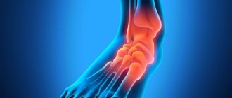
Hallux valgus is a disease in which the metatarsophalangeal joint of the big toe becomes bent, followed by the formation of a “bump” at its base. This condition is popularly known as “thumb bone.”
It is important to understand that a protruding bone is not just a cosmetic defect. This is a full-fledged disease that requires specialized treatment. In its absence, there is a risk of developing bursitis and curvature of other toes.
Causes of bunions in the foot
The following factors can provoke the development of hallux valgus:
- osteoporosis;
- genetic predisposition;
- flat feet;
- hormonal disorders;
- wearing tight shoes with narrow toes;
- diseases of cartilage tissue;
- excess body weight.
According to statistics, a protruding bunion bothers approximately 40% of women over 30 years of age. In men, the deformity is extremely rare. This is due to the greater elasticity of the ligaments in the fairer sex.
Indications for surgery
At the initial stage, conservative therapy helps to reduce the load on the affected joint and reduce pain. But you can completely get rid of the deformity only with the help of surgery. Among the main indications for resection:
- persistent calluses that develop into purulent wounds, ulcers that threaten sepsis, necrosis of the skin;
- loss of elasticity of the ligamentous apparatus, inability to bend or straighten the little finger;
- loss of support on the foot, which makes it difficult for a person to stand or walk;
- frequent relapses of purulent bursitis;
- loss of sensitivity due to damage to nerve endings.
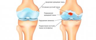
Taylor's deformity leads to decreased quality of life. Pain limits mobility and leads to disability at a young age. A persistent inflammatory process provokes the destruction of cartilage plates and accelerates the development of osteoarthritis.
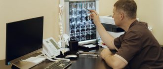
The main indication for surgical removal of a bone growth is the results of radiography. When receiving the image, the orthopedist measures the angle between the 4th and 5th fingers and the size of the head of the joint. The choice of tactics may be influenced by the severity of the deformity, the presence of flat feet or club feet in the patient, the development of bursitis and other inflammatory processes.
How to recognize the disease?
The following changes indicate the development of a bone in the leg:
- the thumb deviates outward, while forming a “ball” at its base;
- there is constant aching pain in the area of deformity;
- fast fatiguability;
- difficulties with choosing shoes.
In the absence of treatment and progression of the pathology, a hammer-shaped shape of the remaining toes is observed. The appearance of the slightest signs of disease should be a reason to contact an orthopedist.
Gout symptoms
The first stage of gout is described as hyperuricemia (increased uric acid in the blood).
Hyperuricemia is detected using a biochemical blood test. In most cases, no other symptoms of the disease are detected. Sometimes general weakness, sweating, itchy skin, and constipation may occur. Actually, it is worth talking about gout from the moment of the onset of acute gout attacks. An attack of gouty arthritis can be triggered by:
- drinking alcohol (single consumption of a significant dose);
- hypothermia;
- joint injury;
- common acute respiratory infections.
First of all, as a rule, the joint of the big toe (first metatarsophalangeal joint) is affected. Typically the joint is affected in only one leg. Quite often other small joints are affected - the wrist or phalanges of the fingers. In the future, other joints may suffer from gout attacks. In women, the disease can affect several joints at once at the very beginning.
Typically, an attack of gouty arthritis lasts no more than 5-7 days, after which complete remission occurs (all symptoms disappear) - until the next attack. This course of gout is called the intermittent (“interval”) stage. Then the disease can progress to the chronic stage.
Attack of gouty arthritis
An attack of gouty arthritis is manifested by acute pain in the joint. Quite quickly, the area of the affected joint swells and turns red. The color of the skin over the joint may take on a bluish tint. The patient feels chills and the temperature rises, possibly up to 38 °C or higher. Any touch to the joint increases the pain, the joint completely loses mobility. The pain can be very intense and cannot be relieved by analgesics.
In most cases, the attack occurs at night; by morning the pain usually subsides. However, in severe cases, severe pain can last up to 3 days, then its intensity slowly decreases.
Chronic gout
If attacks of gouty arthritis become frequent and are quite severe (periods of pain prevail over periods of remission), chronic gout is diagnosed. The chronic form of the disease is characterized by increased dysfunction of the affected joints, and as the patient stops using them, atrophy of the articular muscles develops.
A specific symptom of gout is the formation of tophi. Tophus is a subcutaneous or intradermal accumulation of urates (uric acid salts). Tophi look like nodules - dense, rounded formations. Their diameter can vary from 2 mm to 5 cm or more. Tophi usually appear approximately 5 years after the first attack of gout, slowly increasing in size. However, with an unfavorable course, their formation can occur more quickly. Typical places where they occur are fingers and toes, ears, knee and elbow joints, feet, and brow ridges. It doesn't hurt to touch these formations. The skin over tophi is usually dry and rough because its blood supply is impaired. Over time, a fistula may form in this place, through which a white pasty mass (urate itself) is released.
Tophi are not always formed (only in 50-60% of patients).
Chronic gout also often causes urolithiasis. Urates can form kidney stones, which can cause renal colic, block the outflow of urine, and contribute to the development of pyelonephritis.
Stages of hallux valgus deformity
The disease is divided into several stages depending on the severity of symptoms:
Stage 1 – the phalanx deviates by 20˚, there is aesthetic discomfort. Stage 2 – the finger deviates 30˚ from the normal position, pain appears with prolonged exertion. Stage 3 – deviation of the phalanx up to 50˚, there are problems with choosing shoes, the lump constantly hurts. Stage 4 – displacement of the big toe over 50˚, signs of bursitis are observed, severe discomfort is present even in the absence of any physical activity, curvature of the entire foot is noticeable.
If bunions begin to appear on your feet, do not put off visiting a specialist.
Peculiarities
When there is a painful bulge on the outside of the foot at the base of the little toe, podiatrists diagnose Taylor's deformity, or "tailor's disease." For the first time, a detailed description and diagnosis was carried out by the American specialist H. Davis in 1949, noting complete identity with the development of Hallux Valgus.
An inflamed area forms on the lateral surface of the head of the little finger bone, causing the finger to deviate inward. The pain can radiate to the lateral part of the foot and provoke inflammation of the ligaments, bone and cartilage tissue. Characteristics:
- the forefoot expands, the angle between the 4th and 5th toes increases;
- the little finger takes on the shape of an arc; with the varus subtype, it can overlap the ring finger in a cruciform manner;
- a “bone” is formed on the outside.
For a long time, the pathology develops asymptomatically. The foot gradually increases in volume due to a growth, an increase in the fourth intermetatarsal angle. Conservative treatment eliminates the disease only at the initial stage. In most cases, surgery is performed using different methods.
Treatment of a bunion on the big toe
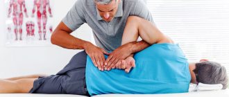
The development of a treatment program is carried out by an orthopedic surgeon after examining the patient and establishing the stage of disease progression. At the initial stages, deformation of the metatarsophalangeal joint can be eliminated using conservative methods.
The following methods for eliminating bunions are effective:
- abductor bandage – ensures fixation of the big toe in an anatomically correct position using a special device, used during sleep, since it is difficult to move with it;
- corrective pads and interdigital partitions – intended for use throughout the day, they provide correction of the position of the phalanx and prevent pressure on the pathological area;
- orthopedic insoles - used at any stage of hallux valgus, necessary for proper distribution of the load on the foot and spine during walking; ideally, insoles should be made individually, taking into account the characteristics of the anatomical structure of the patient’s foot.
The following are used as auxiliary methods for treating a protruding thumb bone:
- massage;
- physiotherapeutic treatment;
- exercise therapy;
- hardware traction.
In advanced stages of pathology, when there is severe deformation of the joints, effective treatment is only possible through surgery. In modern medicine, closed operations performed through several punctures are widely used. During the intervention, the bone is removed and the phalanx of the finger is fixed in the correct position.
After surgical treatment, the patient must always wear comfortable shoes with arch support and orthopedic insoles to avoid relapse of the pathology.
Methods for diagnosing gout
Diagnosis of gout includes physical, laboratory and instrumental research methods. The diagnosis of gout is made when several signs characteristic of this disease coincide
Physical diagnosis
Physical diagnosis involves visual examination of the patient and palpation. The examination results are compared with the patient’s complaints and medical history.
Laboratory diagnostics
Laboratory diagnostics make it possible to establish that the cause of pathological changes in the joint is a violation of uric acid metabolism. The following analyzes play a decisive role in this:
- biochemical blood test (the level of uric acid in the blood plasma is important);
- analysis of synovial (intraarticular) fluid for the presence of urates in it. To do this, a puncture of the joint is done.
- 24-hour urine collection can be performed to assess glomerular filtration rate.
If there is a need to confirm that the detected tumors in the joint are tophi, a biopsy is performed with further examination of their contents.
Instrumental diagnostics
X-rays can reveal pathological changes in the joints. They occur in late stages, so the onset of the disease cannot be determined using x-ray methods. To identify tophi (they do not always protrude above the surface of the skin), computed tomography or MRI are used.
In the early stages, ultrasound can be used. Using ultrasound, fairly small deposits of salts are detected, so tophi are visible on ultrasound even at the stage of their formation. Also a characteristic sign of gout is a “double contour” - the appearance of an echogenic strip running parallel to the line of the articular cartilage. It occurs because salt crystals do not penetrate the structure of the cartilage, but simply cover it on top, creating a duplicate contour that is clearly visible during examination.
Sign up for diagnostics To accurately diagnose the disease, make an appointment with specialists from the Family Doctor network.
Features during operations
The mechanism of development of Taylor's pathology or tailor's disease largely mirrors the pathogenesis of hallux valgus in Hallux Valgus. this allows surgeons to use identical treatment and surgical techniques.
The purpose of surgical intervention is to restore the biomechanical connections in the forefoot, improve the condition of the ligamentous apparatus, and eliminate the aesthetic defect. It is necessary to remove the valgus deformity of the fifth metatarsal bone and return the parabola of the little finger to its natural position.
Preparing for surgery
In most cases, manipulations are performed under spinal anesthesia. To exclude complications and develop a surgical plan, the patient is prescribed the following preparatory procedures:
- radiography of the foot in two projections;
- blood chemistry;
- coagulogram;
- tests for sexually transmitted diseases, HIV, hepatitis C;
- Ultrasound of the lower extremities;
- X-ray of the chest and respiratory organs.
For chronic diseases of the cardiovascular and endocrine systems, additional consultation and examination by specialized doctors is required.
Rehabilitation period after surgery
On the first day, the patient is recommended to remain in the clinic under the supervision of a surgeon. The patient is fitted with special orthoses - Baruk boots, which, when walking, relieve the load from the forefoot and toes on the heel. These shoes must be worn for at least 6 weeks. In a difficult situation, you can additionally rely on crutches and limit physical activity.
After surgery, for 2 weeks it is recommended:
- bed rest;
- place a pillow or cushion under your feet to stimulate the outflow of excess fluid;
- for severe pain, apply cold compresses for 30 minutes up to 4 times a day.
To reduce pain, the doctor selects medications individually and calculates the dosage for home use. Swelling begins to decrease on the 3rd–5th day after surgery. After 14 days, it is recommended to visit the surgeon again to remove the skin sutures.
After 6 weeks, the patient is sent for a repeat x-ray. With pronounced positive dynamics and the absence of contraindications, walking without corrective orthoses with a load on the toes is allowed.
In some older patients, swelling may persist for a long time, which is associated with decreased tone of the veins of the extremities. In such a situation, lymphotropic therapy and contrast foot baths are prescribed.
Advantages of minimally invasive techniques
With early treatment, in 80% of cases, surgeons prescribe low-traumatic foot surgeries. The work is carried out through small incisions or pinhole punctures. Advantages of the methods:
- allow you to correct complex deformities in one procedure;
- give predictable results;
- You can operate on two feet at the same time;
- with the percutaneous technique, the patient can stand on his feet and move freely from the first day;
- no plaster cast or crutches are required.
Minimally invasive operations are highly specialized and are performed by highly qualified orthopedic surgeons using a special technical base.
Surgeries for Taylor deformity take from 25 minutes to an hour and can be performed on both legs simultaneously. In most cases, there is no significant blood loss, which shortens the rehabilitation period.
Rehabilitation and recovery
The duration of the recovery period is individual and depends on the type of surgical intervention. The stronger the deformation, the longer the rehabilitation will take. On the first day, bed rest is indicated. During the following sessions, you can carefully stand on the operated leg.
Most modern methods of surgical treatment avoid the use of plaster splints and crutches, but for some time it will be necessary to bandage the lower leg and foot to prevent swelling. But after arthrodesis, you need to walk with the support of crutches, since the load on the damaged surface is not allowed.
Patients are allowed to walk in special Baruk shoes, which reduce the load on the forefoot when walking. Due to this, the healing of wounds, injuries, and the tightening of sutures occurs at an accelerated rate. Baruk's shoes have a non-slip surface, adjustable clasps, and an openable upper, so they can be worn even with bandages. On average, it is needed for a period of 4-6 weeks, then they switch to regular shoes with orthopedic insoles.
During the rehabilitation period, you must adhere to the following recommendations:
- when at rest, it is advisable to keep your legs elevated by placing bolsters;
- Before removing the sutures, try to protect the wounds from moisture;
- Do not step on your foot completely for at least a month - try to transfer the load to the heel;
- be sure to wear special shoes, use orthopedic insoles, then choose comfortable, wide shoes with flat soles;
- do foot exercises regularly;
- take medications prescribed by your doctor.
Full recovery ends in about six months. During this time, active sports should be excluded, especially those associated with heavy loads.
Contraindications for surgery
Absolute or temporary restrictions for performing an osteotomy on the foot are:
- osteoporosis and reduced bone tissue regeneration;
- exacerbation of rheumatoid arthritis;
- obesity 3-4 degrees;
- acute stage of infectious diseases.
Minimally invasive surgeries are allowed for varicose veins and joint bursitis.
Taylor syndrome leads to the formation of a painful growth at the base of the little finger, swelling, and calluses. New methods of surgical treatment can relieve the patient of complications and restore lightness to his gait.
In what cases is surgery necessary?
Surgical treatment is indicated in the following situations:
- severe pain that limits daily activities;
- inflammatory processes, chronic swelling in the joint of the first finger, which are not eliminated by taking medications;
- inability to wear regular shoes;
- restrictions on the movements of the first finger - the inability to bend or straighten it.
Most patients with hallux valgus note significant improvements from conservative treatment methods and selection of the right shoes. But if these methods do not produce an effect, especially in elderly patients, it is necessary to decide on surgical intervention.