Hemianopia is a partial loss of vision due to congenital or acquired damage to the visual system. Hemianopsia is not an independent disease, but accompanies other diseases. Therefore, hemianopsia is treated by doctors of different specializations - oncologists, neurologists, therapists, endocrinologists, etc. Let's consider the causes of the pathology, methods of treatment and prevention.
General information
With hemianopsia, a person cannot see on one side of the eye—right or left. The visual picture partially falls out of sight, as if visibility was closed or blocked. Sometimes the disease is accompanied by hallucinations. This condition occurs when the visual structures of the brain, which controls all functions and systems of the body, are damaged. Therefore, pathology is classified as a neurological disease and does not belong to ophthalmology.
Hemianopsia occurs:
- congenital;
- acquired.
The acquired form most often develops in women after 30 years of age. Symptomatic treatment against the background of the underlying disease is indicated.
The congenital form develops against the background of lesions of the central nervous system:
- underdevelopment of the brain and cranium;
- exit of the meninges outside the cranium;
- increased intracranial pressure due to the influence of cerebrospinal fluid;
- dropsy of the brain.
In multiple sclerosis, the nerve membranes are affected, including the visual apparatus.
From a clinical point of view, two forms of pathology are distinguished:
- homonymous (symmetrical);
- heteronymous.
Homonymous hemianopsia is characterized by loss of the left/right part of the visual field in both eyes. Heteronymous pathology has 2 forms:
- bitemporal;
- binasal.
Bitemporal hemianopsia is characterized by loss of the temporal halves of the visual field in both eyes. The names of the pathology with the prefix “bi” indicate that blind areas of the visual field appear on both sides.
There is also a binasal form of hemianopsia, which appears due to the “empty sella” syndrome or chiasmatic arachnoiditis. Binasal pathology is loss of visual fields on the side of the nose. Such a clinical picture is rare; it accompanies oncology, hydrocephalus, inflammatory foci in the arachnoid membrane, and diseases of the central nervous system.
There are cases of partial damage to the visual system in one eye and complete blindness in the other.
Depending on the volume of the affected area, hemianopia occurs:
- full;
- partial;
- square;
- scotoma.
In case of complete hemianopsia, half of the visual field is affected, in case of partial hemianopsia, part of half of the visual field is affected. With a square one, only a quarter of the field; with a scotoma, a round-shaped spot appears in the visual field.
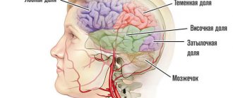
Mechanisms of visual impairment
The nerve impulse that, under the influence of light, appears on the retina - the nervous tissue of our eye - is transmitted along a chain of neurons as part of the optic nerve, the visual pathway, to specific areas of the brain to process the received information and transform it into a whole picture.
Since a person has two eyes, we should have two pictures, but thanks to the peculiarities of the anatomy and physiology of the nervous system, the two images merge into one.
Photo: https://pixabay.com/photos/eye-iris-look-focus-green-1132531/
One of the reasons for this transformation is that the nerve fibers from one eye at a certain moment are divided into two groups, one of which continues to transmit information to the brain on its side, and the second goes to the opposite side, connecting with the first group of the other eye. This crossing of fibers is called chiasma.
When the nerve pathway is compressed or its blood circulation is disrupted, the affected area ceases to function, and the transmission of nerve impulses from the eye to the brain is interrupted, and the eye seems to go blind in patches. When examining visual fields, the doctor can make an assumption about where the dysfunctional area is located and can suggest where to look for the pathological process.
In the future, to determine the cause of the development of visual field impairment, an MRI or CT scan of the brain is done, sometimes with contrast.
- If the pathological process is located before the chiasm, blindness or partial loss of vision occurs in only one eye.
- If the chiasm is compressed, then the temporal halves of vision of both eyes fall out - the person perfectly sees what is happening in front of him, but does not notice what is on the side of him until he turns his head in the right direction.
- If the lesion is localized after the chiasm, then loss of the temporal half of vision occurs on one side and the nasal half on the other, or vice versa.
The size of the lost field depends on the force of compression of a particular part of the visual pathway or on the degree of disruption of its blood circulation.
Causes
The acquired form of pathology develops when:
- occipital cortex;
- central parts of the visual pathways;
- visual tracts.
Damage to brain structures is caused by:
- acute cerebrovascular accidents;
- inflammatory processes of the meninges;
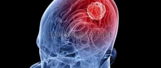
- volumetric intracranial formations - tumors;
- angiomatosis;
- traumatic brain injuries;
- iatrogenic factor;
- congenital pathology of the central nervous system.
- multiple sclerosis.
An acute disorder of cerebral blood supply is a stroke that can occur against the background of thrombosis of the vessels supplying the brain, arterial hypertension, atherosclerosis and other vascular pathologies. When blood circulation is impaired, the brain suffers from a lack of oxygen and glucose.
Inflammatory foci in the meninges are formed due to infection. This is possible with purulent otitis media, head trauma, as well as with inflammatory diseases of internal organs - the infection enters the brain through the bloodstream. For example, during an inflammatory process in the nasal sinuses, the chiasm located at the base of the brain may be affected. Inflammatory processes in the cerebral cortex can be influenced by purulent foci in the tissues of the lungs or bronchi.
Angiomatosis is a series of hereditary pathologies that affect the nervous system and skin. The disease is characterized by active proliferation of blood vessels and the formation of neoplasms - fibroids, plaques, etc.
An iatrogenic factor is unintentional damage to the visual apparatus during surgery on the central nervous system.
Symptoms
Hemianopsia immediately begins with an acute form and accompanies a traumatic brain injury or stroke. Patients complain that entire areas of the visual image disappear from the field of vision or that large spots appear before the eyes - they prevent clear visualization.
Often the acute form of hemianopsia is accompanied by the following symptoms:
- dizziness;
- nausea, vomiting;
- violation of spatial orientation;
- increased blood pressure;
- violation of facial expressions;
- numbness of the limbs;
- speech disorder.
But sometimes the disease occurs without pronounced symptoms: the patient complains of constant headaches, which are accompanied by partial loss of visual fields; unmotivated weakness and general malaise appear. If such symptoms appear, you should immediately see an ophthalmologist or therapist.
Patients with hemianopsia find it difficult to cope with everyday responsibilities and perform normal work. Walking around the city is dangerous: they do not always see approaching vehicles, do not notice many objects on the road surface, and are poorly oriented in space.
When peripheral vision is lost, it is difficult for a person to navigate space, take care of himself, and perform daily work.
A healthy visual system provides a person with central and peripheral vision. With hemianopia, peripheral vision is absent, which leads to disorientation in space. The field of view is the totality of objects that a person sees simultaneously when fixing his gaze on one thing. When part of the visual field is lost, orientation in space is difficult or impossible.
General symptoms
Based on clinical manifestations, it is customary to divide homonymous and heteronymous hemianopsia. In the first form of the disease, patients will say that they are unable to see in the right or left half of the eye.
When the left and right optic tracts are affected, the patient's right and left eyes go blind, respectively.
As the disease progresses, the patient may experience visual hallucinations. This manifests itself in failure to recognize familiar objects, the appearance of flashes, etc. Patients often develop prosopagnosia, that is, the inability to recognize familiar people.
If patients remain oriented, they most often deny their disease. This condition indicates positive Anton-Babinsky syndrome. In this case, patients can fix their gaze on only one object, and surrounding objects will not enter their field of vision.
Most often, the first symptoms indicating hemianopsia appear after a person has suffered a stroke. Patients who begin to develop the disease mainly complain that their performance decreases because it becomes much more difficult to perform any actions.
People face the following problems: inability to find the right object, loss of spatial orientation, difficulty eating, etc.
Clinical manifestations
The clinical course is complicated by agnosia or hallucinations. A patient with agnosia cannot distinguish between familiar objects: he simply does not recognize them. With prosopagnosia, the patient does not recognize familiar faces. With “mental gaze paralysis,” patients see only one object: surrounding objects completely fall out of sight.
Hallucinations occur in patients who have had a stroke. They can be either formalized (the patient sees familiar images) or unformed (various lines, geometric shapes). Hallucinations always appear unexpectedly.
The lesion can affect different parts of the visual apparatus:
- optic nerve;
- optical chiasmus;
- cortical visual analyzer;
- cerebral cortex.
Optic nerve
Complete damage to the optic nerve leads to absolute blindness. With partial damage, individual “pieces” fall out of the field of vision - the person sees a “cropped” picture or blind sectors appear in the field of vision.
If the optic chiasm is affected, the person may develop complete blindness. But in practice, partial damage most often occurs, which manifests itself in the absence of peripheral vision. Patients feel like a “horse in blinders,” that is, they cannot see anything on the sides. They have central vision, which can be sharp, but lateral vision is completely absent. This pathology is called bitemporal hemianopia.
Sometimes visibility disappears not on the sides, but in the central part. In this case, patients cannot see their own nose and complain that a black spot has appeared in front of their eyes on the side of the nose. This pathology is called binasal hemianopia.
Cortical visual analyzer
Homonymous (of the same name) hemianopsia is the most common clinical picture when the visual analyzer is damaged. Same name means on the same side. For example, right hemianopsia is loss of the left half of the visual field in both eyes. Less common is lower or upper hemianopsia.
If the cortical projection area is partially damaged, then a partial loss of the visual field is observed: either the squares appear in the upper left or lower right.
Cortex
If the cerebral cortex is damaged, hallucinations may occur. For example, the patient sees shiny lines or dots in front of him. The figures move with the movement of the eyeball. This phenomenon is called photopsia.
When the cerebral cortex is damaged, agnosia (inability to recognize familiar objects) or prosopagnosia (non-discrimination, failure to recognize familiar faces) develops.
Complications
Pathological damage to the optic tract leads to optic nerve atrophy. If the underlying disease that led to hemianopsia is incurable, then the patient’s quality of vision will gradually decrease until complete blindness.
Some patients develop persistent depression due to ophthalmological pathologies; they prefer to spend time alone and withdraw from society. Ridicule of ridiculous movements and sloppiness causes them psychological discomfort and a desire to avoid it by any means. If left untreated, depression can lead to more serious mental health problems. Therefore, patients should be consulted by a psychologist or psychotherapist.
Complications of hemianopsia
Hemianopia negatively affects a person's quality of life. People with this diagnosis are unable to independently carry out their usual daily activities – eating, reading, driving. They often get lost in space and have difficulty communicating with other people.
As hemianopsia progresses, there is a risk of complications of the disease, including complete blindness. The outcome of the pathology depends on its type, age category, individual characteristics of the human body and many other factors.
With timely initiation of treatment, the prognosis is favorable. In this case, vision can be restored completely. Otherwise, the person goes blind and becomes disabled. If hemianopia occurs due to a stroke, the disease has a favorable prognosis. As a rule, visual functions are restored within six months.
Diagnostics
Diagnosis of any visual pathology begins with an ophthalmological examination - viziometry, autoreflectometry, examination of the pupil and measurement of intraocular pressure.
To establish hemianopia, you need to perform a topical diagnosis:
- perimetry;
- campimetry.
- Ultrasound Dopplerography;
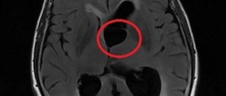
- MRI;
- computed tomography;
- CT angiography.
Using perimetry, “blind” areas of the visual field are identified. This is the key diagnosis in this case. With homonymous pathology, symmetrical areas are formed: at the temporal part on one side and at the bridge of the nose on the other. That is, the right lobes or the left.
If perimetry cannot be performed due to the patient’s age or other reasons, other diagnostics are used. For example, the doctor holds a stick or towel in his hands and asks the patient to find the middle. With hemianopsia, the patient will divide only part of the stick in half, since he does not see the entire object. Also, the patient does not see the movement of the doctor’s fingers on the affected side of the eye.
With bitemporal pathology, there is a violation of the temporal halves of the visual field for both eyes. With binasal pathology, the central halves (near the nose) fall out.
Using computer campimetry, disturbances in color perception and color perception in a specific area of the visual field are determined.
MRI and CT can determine the etiology of the disease, which will help select the desired treatment algorithm. Using computer diagnostics, it is possible to determine the localization of tumors, areas of brain damage during a stroke, and the extent of traumatic injuries.
CT angiography visualizes the vascular network, using this diagnosis to determine the degree of damage to blood vessels.
Doppler ultrasound is a non-invasive examination method to determine the speed of blood flow.
Hemianopsia – simple things to say. Features of medical terminology
The medical term "hemianopsia" comes from the Latin "Hemianopsia", where "Hemi" is a prefix meaning half of something, and "anopsia" is the absence or defect of vision, which literally means "half blindness".
Hemianopsia is a violation of the perception of a certain area of the visual field or the complete loss of half of it in one or two eyes. Unlike small, barely noticeable areas of prolapse (scotomas), hemianopsias are usually noticed by patients on their own, or are detected randomly during routine examinations.
The presence of hemianopsia indicates anatomical changes in the visual pathway and certain areas of the brain in the form of their compression or circulatory disorders and is an important diagnostic criterion for the most accurate determination of the affected area even before specific examination methods are carried out.
Hemianopsia can be detected using a special device - a perimeter; as a rule, neurologists, neurosurgeons, and ophthalmologists are sent for this study if they suspect anatomical changes in the brain.
Treatment
Without eliminating the underlying disease that caused the pathology of the visual analyzer, it is impossible to get rid of hemianopsia. Healing from a somatic disease will lead to full or partial restoration of visual fields. Treatment is carried out either by a neurosurgeon or a neurologist.
Treatment of encephalitis, stroke, and traumatic brain injury is carried out in a hospital. Some acute clinical conditions require emergency surgery. The development of a tumor process in the brain requires treatment at an oncology center: the patient is prescribed radiation therapy or chemotherapy.
If a genetic form of pathology is identified, consultation with a geneticist is required. It is impossible to cure a genetic pathology, so patients are prescribed symptomatic treatment. You will have to take medications and treatment for the rest of your life.
Congenital pathology can be eliminated with surgery or drug treatment. The result will depend on the degree of tissue damage, anatomical abnormalities, and concomitant diseases.
What does the ophthalmologist do in this case? He prescribes symptomatic treatment that relieves pain and partially restores visual function. For example, a doctor may prescribe medications that improve blood microcirculation in the tissues of the visual organ. To strengthen the immune system, immunomodulators and vitamin complexes are prescribed. Physiotherapy stimulates the functions of the optic nerve.
Patients are asked to wear special glasses that complement the missing visual picture.
If the underlying condition is not treated, the optic nerve may become damaged. Optic nerve atrophy leads to vision loss and complete blindness. It is important to consult a doctor in a timely manner and undergo medical examination if necessary.
Prevention
It is impossible to prevent the development of pathology. However, the patient can improve the quality of vision if he follows the recommendations of doctors:
- when reading books, you should not turn your head to the sides;
- when reading books, keep your head straight, and try to cover as much of the text as possible with your eyes;
- you need to read the text not horizontally, but vertically;
- When reading, the book should be held at an angle of 90 degrees.
To expand the visual range, special ophthalmological programs developed by leading doctors are used.
In order not to provoke the development of pathology, you need to protect your head from blows and injuries. It is unacceptable to overstrain the visual organs so as not to injure the optic nerve and blood vessels. Avoid smoking tobacco and staying in environmentally unclean areas. Sometimes intoxication of the body leads to eye pathology, so follow safety precautions when working with chemicals.
Preventative examinations once a year are important for every person after 30 years of age. Left-sided or right-sided hemianopia most often develops in women aged 30-50 years. Pathology at the initial stage can only be noticed at a doctor’s appointment, so do not neglect regular professional examinations. This disease may not show itself in any way, but subsequently vision will begin to decline sharply. Along with decreased vision, the quality of life will deteriorate, even to the point of disability.
Useful video
Irina Sergeevna Mironova’s story about the causes and features of the development of the pathological condition:
Author's rating
Author of the article
Alexandrova O.M.
Articles written
2100
about the author
Was the article helpful?
Rate the material on a five-point scale!
( 1 ratings, average: 2.00 out of 5)
If you have any questions or want to share your opinion or experience, write a comment below.
Bottom line
Hemianopsia is a neurological disorder that causes blind spots in the visual field of both eyes. This pathology is not an independent disease; it accompanies diseases of a neurological and other nature. Hemianopsia can be congenital, acquired, or genetic. Acquired and congenital pathology can be cured; genetic pathology cannot be corrected. To ensure that the level of vision in a genetic disease does not decrease, symptomatic treatment is carried out. Sometimes you have to be treated for the rest of your life.
Sources used:
- Kvasova M. D. Vision and heredity. - Moscow / St. Petersburg: Dilya, 2002.
- Skoromets A. A. Topical diagnosis of diseases of the nervous system: A guide for doctors. — 1st ed. - L.: Medicine, 1989
- D. Hubel. Eye, brain, vision. — ed. A. L. Byzova. - M.: Mir, 1990.
- Wikipedia article