Magnetic resonance imaging has high tissue and fluid contrast. The procedure allows visualization of loose structures of the body without significant invasive manipulations. MRI is actively used in neurology and neurosurgery to diagnose diseases of the nervous system. Magnetic resonance scanning is considered the most informative method of studying the brain. According to the results of tomography, inflammatory, degenerative, dystrophic changes, and tumors are detected.
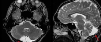
Colloid cyst on MRI
A brain cyst is a benign neoplasm inside the skull. It is a closed pathological formation with predominantly liquid contents. Congenital cysts appear when intrauterine development is disrupted; acquired ones can be the result of head trauma, infectious disease, cerebrovascular accidents and other conditions. The formations are not prone to malignant degeneration, but when they reach an impressive size they can compress surrounding structures. It is important to diagnose a cyst as early as possible and determine treatment tactics. MRI scanning allows you to identify and differentiate formations and track the dynamics of the process.
MRI of the brain showed a cyst, what does it mean?
Colloid cyst in the third ventricle of the brain (circled with a dotted line)
As a result of magnetic resonance imaging, multiple layer-by-layer images of the anatomical area are obtained. Sections are made in three planes, which allows for a detailed study of each area of the skull. The fact that the MRI showed a cyst in the brain is reported when the results are issued. In conclusion, the doctor specifies the location and type of formation, size, and relationship of the cavity with surrounding structures. If tissue metaplasia is suspected (the appearance of cells in the study area that are not typical for this area), it is recommended to perform an MRI with contrast enhancement. The distribution of the drug can suggest the nature of the tumor.
Detecting a brain cyst on an MRI means that there is a round-shaped cavity filled with fluid in the skull. Based on specific signs, the radiologist determines the type of the latter and describes in detail the changes in the surrounding tissues. Detecting a pathology is not a reason to panic. Most cysts are congenital, grow slowly, and do not become malignant. With the results of the study, you need to contact your doctor (who initiated the tomography), neurologist or neurosurgeon. The specialist will determine how dangerous the deviations are, make a prognosis for the development of the pathology, and prescribe additional examination.
Etiology and pathogenesis
The causes of a congenital cyst are unfavorable factors that occur during the development of the fetus. These are:
- fetal hypoxia during delivery;
- fetoplacental insufficiency;
- taking a specific group of medications by a pregnant woman;
- Rh conflict between mother and child;
- intrauterine infections.
Factors that provoke the development of a congenital cavity with fluid are drug, alcohol or nicotine addiction of the mother. In this case, the child’s development takes place under conditions of intrauterine intoxication, which negatively affects the brain structures. The causes of the cavity can also be chronic decompensated diseases of the expectant mother.
An acquired cavity with fluid in the head develops as a result of:
- inflammatory diseases (encephalitis, brain abscess, arachnoiditis, meningitis);
- traumatic brain injuries;
- injuries of newborns received during childbirth;
- cerebrovascular accidents (subarachnoid hemorrhage, ischemic stroke, hemorrhagic stroke).
Depending on the etiology, the following types of liquid cavities are distinguished:
- A cyst of iatrogenic origin forms as a complication after brain surgery.
- Parasitic - develops with paragonimiasis, cerebral form of taeniasis and echinococcosis.
A cavity with fluid can also replace cerebral tissue during degenerative and dystrophic processes in the head.
If a cyst is present, there are a number of factors that can trigger its growth. These include obstruction of venous outflow from the skull, strokes and other vascular disorders, as well as head injuries, hydrocephalus, and neuroinfections.
What does a cyst look like on an MRI of the brain?
Cystic structure of brain metastases on MRI
The brain is studied using high-field equipment with a power of 1.5 Tesla, which allows creating detailed and clear images. With magnetic resonance imaging, monochrome sections of the area under study are obtained in increments of 1-2 mm. On MRI images, brain cysts appear as round formations of varying diameters with clear contours. The deviation can be determined by the change in the signal. The intensity of the latter will be different compared to healthy areas. The difference is clearly visible in the photographs. Special scanning modes allow you to obtain more detailed information about the cyst and determine its type:
- Epidermoid. It is usually located in the area of the cerebellopontine angle, prepontine cistern, quadrigeminal tract, ventricles, and less commonly in the hemispheres. Growth is slow, with the risk of compression of the brain stem, entrapment of nerves and blood vessels. There are signs of inflammation around the epidermoid cyst. The intensity of the signal from the formation is usually heterogeneous, but corresponds to the cerebrospinal fluid (if there is no fat inside). On DWI and FLAIR images, the cyst appears lighter than the brain fluid. Epidermoids with fat inside T1-weighted images give a hyperintense signal, on T2 - hypointense (compared to the cerebrospinal fluid).
- Dermoid. Usually the formation is located in the midline of the skull. Due to the high lipid content, T1 imaging gives a hyperintense signal. Unlike the epidermoid, it always has a heterogeneous structure.
- Lipoma. Fatty cysts are most often located in the area of the corpus callosum, interhemispheric fissure, pituitary infundibulum and hypothalamus. MRI scans reveal clear contours of the tumor. Mass effect and swelling of surrounding tissues are not observed. On T1 WI the cyst is sharply hyperintense. On T2 VI it is the opposite (compared to cerebrospinal fluid). Calcifications and vessels look like hypointense areas in the structure of the formation.
- Ependymal. A rare type of cyst that forms under the membrane lining the brain cavity. On MR scans it looks like a formation with clear contours, the signal intensity corresponds to the cerebrospinal fluid.
- Arachnoid. It is formed as a result of the accumulation of cerebrospinal fluid between the sheets of the arachnoid membrane of the brain, most often in the middle cranial fossa, interhemispheric fissure, cerebellopontine angle, at the level of the quadrigeminal. On T1, the VI looks somewhat lighter than the cerebrospinal fluid. In FLAIR mode it produces a hypointense signal.
Arachnoid cyst (indicated by arrow) on an MRI scan
- Neuroglial. Found in the cerebral parenchyma, in the area of the choroid plexus of the ventricles. In the photographs it looks like a round formation with liquor contents, often combined with anomalies of brain development.
- Colloidal. Usually located in the anterior superior part of the third ventricle, between the foramina of Monroe. It has a round shape, a fibrous capsule, and clear contours. In the presence of protein in the contents, it gives a hyperintense signal on T1 VI and a hypointense signal on T2 VI. Contrast does not accumulate.
- Rathke's pouch cyst. Rarely seen. Located in the area of the sella turcica. The signal intensity depends on the nature of the content. When serous, it gives a typical fluid response. Mucoid contents are characterized by a hyperintense signal on T1-weighted images. When contrasting, accumulation of the substance is observed in the capsule area.
Arachnoid cyst (circled in red) on MRI
An experienced radiologist knows exactly what a cyst looks like in an MRI photo. An ordinary person will not be able to independently identify and differentiate education. In doubtful cases, it makes sense to show the research results to several specialists.
Treatment methods for liquor cysts
The most common indications for surgical treatment of a cyst: it grows too quickly, causes seizures, causes neurological deficits, the cyst interferes with the child’s development.
The main, safest and most effective method for treating cerebrospinal fluid cysts today is fenestration. That is, dissection of the walls of the cyst for communication between the cyst and other cerebrospinal fluid spaces, where this accumulated fluid is normally located.
It is possible to install a stent (catheter) connecting the cavity of the cyst and the ventricles. It happens that the operation turns out to be ineffective and forces you to use another method of treatment - bypass surgery.
Shunt surgery (today practically not used as a primary method of treatment), although it allows you to quickly reduce the size of the cyst, but creates a lifelong dependence on the shunt system. If this method can be avoided, neurosurgeons try to avoid it.
However, when treating young children (up to two years old), as well as when treating cysts that were complicated by hemorrhage, shunt operations are inevitable.
In general, surgeries to treat cysts have a low risk of complications in the hands of experienced doctors. This operation involves hospitalization for only three to four days and several subsequent consultations with a surgeon. Deviations from this rule occur only in 10% of cases.
Most often, in such a situation, endoscopic surgery is used, but in some cases, an open microsurgical approach is more correct. An uninitiated person will not see the difference after the operation; the size of access to the brain cavity will be the same. In this case, it is not so important for the patient which instrument is used to perform this operation. It is important to make the patient feel better.
Forecasts
In most cases, a frozen cavity with liquid of insignificant size does not bother the patient and does not cause any symptoms. In other cases, with adequate and timely treatment, the outcome is favorable.
In rare cases, patients after surgical removal of a cyst experience a residual moderate-to-severe liquor-hypertensive symptom. If a focal neurological deficit develops, it persists after treatment.
With the removal of the cyst, epileptic paroxysms disappear, but often appear later. This is justified by changes in the operated area of the head, in particular the formed adhesions. Secondary epilepsy is practically uncontrollable by anticonvulsant therapy.
Preparation rules
During a routine examination, doctors recommend following a few simple recommendations:
- If it is necessary to administer contrast, you must come to the clinic on an empty stomach, that is, do not eat food for 7-8 hours.
- If you are afraid of research or confined spaces, be sure to consult with a specialist. Your doctor may prescribe you sedatives to make the examination process more comfortable.
- Do not bring any metal products inside the tomograph. For this reason, doctors recommend removing metal accessories in advance.
- Prepare a change of clothes that do not have metal or metal-containing accessories.
- The duration of the diagnostic test is about 40 minutes. Empty your bladder in advance to avoid discomfort during the procedure.
Diagnostics
After an oral conversation and a physical examination (coordination, mental state, work of analyzers and other external signs are studied), the patient is examined using:
- tomography (CT, MRI, PET);
- ultrasound diagnostic methods (echoencephalography);
- EEG (electroencephalogram);
- angiography of cerebral vessels;
- ophthalmological examination;
- scintigraphy and others.
The most reliable method is biopsy (tissue collection is performed when cancer is removed or stereotactic biopsy). Histological analysis of tissue samples gives a complete picture of the type of neoplasm and all its features.