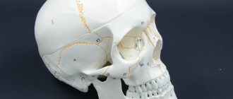
A fracture of the base of the skull is one of the most severe traumatic brain injuries. This condition is accompanied by damage to one or even several bones that form the base of the skull. Damaged in this case may be:
- occipital bone;
- temporal bone;
- ethmoid bone;
- sphenoid bone.
A fracture of the base of the skull can occur from a variety of serious physical injuries: car accidents;
- when a person falls from a great height;
- when hitting the face in the area of the lower jaw or base of the nose.
In most patients, the base fracture occurs from the arch. Statistics say that the number of such patients reaches 59%.
Causes of skull fracture
All causes of skull fracture are inherently mechanical injuries. In such cases, injury to the bone tissue of the head may occur:
- Falling from a great height or high speed;
- Hit your head hard with a heavy object;
- As a result of a traffic accident (car accident).
All these situations can arise as a result of non-compliance with safety rules on the road and at work, as a result of street fights and aggressive sports in sports clubs, or extreme sports.
When assessing age and social categories, groups at increased risk of skull fracture include children, active teenagers and middle-aged people, as well as people dependent on drugs and alcohol.
Skull fracture: classification of types
Classification of cranial bone injuries is made according to several criteria. Thus, depending on the appearance and physical condition of the victim’s head, injuries can be divided into open and closed fractures. An open fracture is a condition in which the scalp is damaged. A closed fracture is an injury in which the soft tissue remains intact.
The basic classification of injuries is based on the location of the injury:
- Fracture of bone tissue at the base of the skull;
- Broken bones at the base of the skull.
It is also possible to fracture the vault and base of the skull at the same time.
Depending on the nature of the resulting injuries, damage to the bone tissue of the head can be:
- Interferes. This type of injury is extremely dangerous because with a deep fracture, a cavity is formed in the skull from fragments of broken bone. If ingested, sharp fragments of the skull can damage the meninges, the brain, its arteries and cause a leak of cerebrospinal fluid. More often, such injury is accompanied by intense bleeding in the brain.
- Perforated. The main cause of injury was a gunshot wound to the head. The result of such an injury is almost always the immediate death of the victim.
- Shards. It is most often caused by being hit in the head with a heavy, sharp object or falling onto a protruding piece or hard object. In this case, the bones of the skull split, forming sharp fragments. Skull fragments damage the meninges and blood vessels. In most cases, such injuries end in death even before first aid is provided to the victim.
- Linear. With this type of injury, there is no critical displacement of bone tissue and fragments. The main injury of a broken bone is a shallow fracture. In the case of a linear skull fracture, the damaged portion of the bone may heal on its own over time.
Skull fracture.
Of all types of traumatic brain injury, a linear fracture is the least dangerous. This type of injury is most often diagnosed in children.
Fractures of the base of the skull
This type of injury is extremely dangerous because the integrity of the major components of the skull that protect the brain is compromised. In this case, the main parts of the brain, nerve bundles and the brain stem are damaged. A fracture of the base of the skull can cause an acute inflammatory process in the brain.
Bone fractures are accompanied by a rupture of the protective membrane of the brain and create a high risk of infection. Fractures due to trauma spread from the base of the skull to the bones of the orbit and nose. If the skull is damaged in the middle fossa, the fracture will extend to the ear.
Cranial vault fractures
The cranial vault is the line connecting the bones of the head. In different places the intersection may be as follows: the skull is not only jagged or folded, but also flat. On the sides of the cranial vault there are temporal protrusions that smoothly turn into depressions. In front there is a pronounced protrusion, which represents the frontal bone. On the back there are two parietal tubers and an occipital bulb. Between these areas of the cranial vault is a temple.
When the cranial arch is fractured, the internal bone plate is destroyed. Damages the dura mater when pressed. If a fracture occurs when the damaged bone reaches the vessels of the dura mater, these vessels rupture and bleed. With a closed fracture, hematomas do not have clear boundaries.
Types of traumatic brain injuries
Doctors divide all traumatic brain injuries into 2 large groups:
- open injuries are the most serious type of injury, in which not only the skull is damaged, but also the soft structures of the brain;
- closed injuries - a brain contusion or a fracture of the skull bones is likely, but the integrity of the soft tissues is not compromised.
Open head injuries include:
- fracture of the base and vault of the skull with soft tissue injuries;
- fracture of the base of the skull with injury to blood vessels.
1 MRI of the brain
2 Treatment of traumatic brain injuries
3 Treatment of traumatic brain injuries
Closed view of TBI
This type of injury is diagnosed in 70% of cases and includes:
- brain concussion;
- brain contusion without compression;
- brain contusion with compression.
Types of traumatic brain injuries by severity:
- mild degree (concussions and some types of brain contusions in which consciousness remains clear);
- moderate degree (brain contusions, characterized by short-term loss of consciousness and the appearance of some neurological abnormalities);
- severe degree (concussion with numerous injuries and compression of the brain, leading to prolonged loss of consciousness, disruption of vital systems and even death).
The consequences of traumatic brain injuries depend on the severity of the specific injury. A mild head injury may not cause any complications, while a moderate head injury may cause neurological impairment, and a severe head injury may result in death.
In all cases of head injury, it is necessary to urgently transport the victim to the nearest hospital or emergency room, where he will receive the necessary diagnostics and qualified treatment for a traumatic brain injury!
Treatment of traumatic brain injuries
Therapy for such injuries is carried out in a hospital setting with mandatory bed rest (from several days to a month).
Treatment of traumatic brain injuries is mainly conservative and symptomatic. Drugs are used to relieve cerebral edema, suppress the gag reflex, painkillers, as well as antihistamines and cardiovascular drugs.
Treatment of traumatic brain injuries when a hematoma and signs of compression of the brain are detected allows for surgical intervention.
1 MRI of the brain
2 MRI of the brain
3 MRI of the brain
Skull fracture: clinical manifestations
The symptoms of the pathology depend on the nature of the existing injuries. However, there are also common symptoms that indicate a skull fracture. These include:
- Sudden, short-term, partial or complete loss of sensation in certain areas of the head or body;
- Loss of consciousness;
- Coma;
- Paresis;
- Arrhythmia;
- A state of increased excitement or complete immobility;
- Paralysis;
- Involuntary urination;
- Prolonged tension headache; Nausea; Vomit;
- Nausea and vomiting;
- Rhythm disturbance and respiratory arrest;
- Students react sluggishly to external stimuli;
- The circulatory system is disrupted.
Loss of consciousness is one of the possible symptoms of a skull fracture.
Often after a skull fracture, there is periodic loss of balance, as well as confusion in space. The more clearly the symptoms of the pathology appear, the more severe the injury and the more extensive the brain damage.
Symptoms of a basal skull fracture
Depending on the location of the injury, the victim may experience different symptoms of a basal skull fracture. Trauma to the anterior fossa of the skull often causes:
- Intense and prolonged nosebleeds;
- Sometimes there is a leak of cerebrospinal fluid through the nostrils;
- After two or more days, bruises and bruises appear around the eyes and on the whites of the eyes.
- With parallel damage to the hyoid bone, emphysema develops at the site of injury.
With a fracture of the base of the skull in the area of the medial fossa, pathological processes are often observed, such as:
- Bleeding from the ear;
- Disturbances in the functioning of the facial nerves;
- The appearance of hematomas around or behind the ears;
- Intensive leakage of cerebrospinal fluid through the auricle;
- Loss of sensation of individual taste buds;
- Occasional difficulty maintaining balance.
Moreover, these types of injuries are often accompanied by partial or complete hearing loss.
Damage to the base of the skull from the posterior lobe is accompanied by the following unpleasant conditions:
- The appearance of hematomas around the auricle of one or both ears;
- Disturbances in the functioning of nerve endings.
Often such damage leads to the development of paralysis of the stamens. Accompanied by impaired articulation, speech pronunciation and problems with swallowing. In this case, the victim's voice becomes hoarse and nasal, and speech becomes slurred.
Signs of a calvarial fracture
The following symptoms are typical for a skull fracture:
- Bleeding from the patient's nose, ear, or mouth;
- Intermittent exudation of cerebrospinal fluid. The volume of leaked liquid can exceed 200 ml per day. In this case, intracranial pressure is significantly reduced. The pathological process lasts up to 6 days;
- The appearance of late bruises around the eyes and on the whites of the eyes. This symptom appears several days after the injury;
- Hearing impaired.
The occurrence of nosebleeds is one of the possible symptoms of a skull fracture.
Paresis and paralysis of the facial nerve can occur when the bony canal is damaged, ptosis and one or both eyes looking outward or downward can occur due to rupture of the oculomotor nerve.
A significant increase in bleeding occurs when the victim tries to turn his head. Therefore, turning your head with this type of fracture is strictly prohibited.
Symptoms of a basal skull fracture
A fracture of the base of the skull is an open traumatic brain injury. It is worth noting that injuries accompanied by the release of blood or cerebrospinal fluid from the ear canals or nose are classified as open penetrating head injuries.
Based on location, injuries are divided into fractures:
- middle cranial fossa;
- anterior cranial fossa;
- posterior cranial fossa;
The most common fractures encountered in medical practice are middle cranial fossa fractures. Such fractures can be longitudinal, transverse or oblique. Most often, doctors fix longitudinal fissures in the temporal lobes. In this case, fractures can spread through various holes, crevices and thinning of the skull bones.
Among the main manifestations of fractures:
- bleeding from the ear canal;
- decreased hearing acuity;
- release of cerebrospinal fluid;
- the appearance of bruises in the area of the temporal muscle.
All of the above damage occurs when there is a blow to the occipital region.
It is worth noting that transverse fractures are clinically characterized by the patient being completely deaf. The patient may also exhibit disturbances in vestibular function, loss of taste in the two anterior thirds of the tongue, and may also experience peripheral facial palsy.
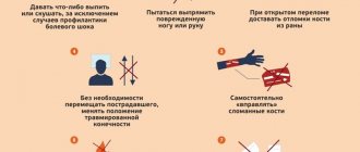
First aid
If you notice signs of a skull fracture, seek medical attention immediately. Before arriving, doctors do not want to give the victim anesthesia, since some medications can significantly increase bleeding or breathing problems, as well as provoke the patient into a coma.
Before the ambulance arrives, it is important to provide first aid:
- The patient should be placed on his back on a hard surface, with his head buried in the ground. The pillow should not be placed under the head. If the patient is unconscious, he should also be placed on his back, but in a half-turn position with a roll of clothing on one side. You need to tilt your head slightly so as not to choke when vomiting.
- Treat the head wound with an antiseptic and apply a sterile bandage.
- Remove the victim's dentures, jewelry, watches, and glasses.
- Remove any clothing that may be tight fitting, obstructing blood flow and breathing.
- Bring a cold object wrapped in a clean cloth to your head.
If the victim is not breathing, empty the vomit from the mouth and perform mouth-to-mouth resuscitation using chest compressions. To avoid direct contact with the victim's mucous membranes, use a piece of clean, permeable cloth.
If there are no breathing problems, it is permissible to give the injured person Analgin with diphenhydramine. When transporting a patient to a medical facility, the following activities are performed:
- Glucose, diuretics and cardiac drugs are administered intravenously. However, in case of severe bleeding, diuretics should not be used. Instead, Polyglucin or Getinol is administered. Lasix is most often used as a diuretic, and cordeamine and sulfacamphocaine are the main medications used to support heart muscle function.
- If breathing problems occur, the patient is inhaled through an oxygen mask.
- Convulsions and increased motor activity are suppressed by Suprastin.
Polyglucinum is prescribed for severe bleeding.
The use of narcotic anesthetics is unacceptable. Such drugs can cause respiratory arrest.
Skull fracture: diagnostic methods
Diagnostics is carried out comprehensively and includes the following activities:
- Medical examination and collection of information about the patient’s ailments. During the examination, the doctor assesses the general condition of the victim, the reaction of the pupils, and measures the pulse and blood pressure. The position of the tongue and the symmetry of the patient's jaw are also assessed. Neurological reactions are studied;
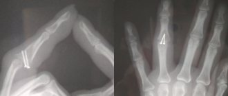
- X-rays of the skull are taken in two projections;
- A computed tomography (CT) or magnetic resonance imaging (MRI) scan is performed.
In extremely severe cases, when it is impossible to make a complete diagnosis, treatment of the patient is prescribed based on the external symptoms of the pathology.
Treatment options
Patient treatment methods are selected depending on the type of injury. Therapy can be carried out either surgically or conservatively. For linear skull fractures, conservative methods are mainly used. They are also acceptable for use in mild to moderate injuries where the flow of cerebrospinal fluid can be stopped without surgery.
Conservative treatment includes:
- Strict adherence to the rules of bed rest.
- Perform lumbar punctures 2-3 times a day every day. At the same time, oxygen is introduced into the subarachnoid space of the spinal cord.
- Taking diuretics.
- Daily disinfection of the oral cavity, middle ear and nasopharynx to prevent the development of purulent inflammation.
Surgical intervention is necessary in the following cases:
- Shock fracture of the skull;
- Linear fracture of the skull bones with the formation of a large number of fragments;
- Bone damage that puts pressure on the brain and causes blood vessels and nerve endings to rupture;
- Recurrent purulent inflammation.
In case of a deep skull fracture, intervention is required. surgical
Treatment of linear fractures, as well as other types of fractures during surgery, is carried out by craniotomy. After removing all fragments and purulent formations, the skull bed, previously removed with bone or a special titanium plate, is closed. The most common type is a plate prosthesis.
Brain contusion
Compared to a concussion, a brain contusion is a more serious injury that requires urgent treatment. With this type of TBI, small vessels of the brain are affected, hemorrhage occurs, and cerebrospinal fluid (cerebrospinal fluid) may leak.
The injury may be accompanied by compression of soft tissues or occur without it. The method of treating a brain contusion depends on this.
The main symptoms of a brain contusion without compression :
- loss of consciousness (or coma);
- frequent vomiting;
- tachycardia and arrhythmia;
- decreased muscle function in the arms and legs (paresis) and loss of coordination;
- high blood pressure;
- the appearance of blood in the cerebrospinal fluid;
- prolonged severe headaches;
- vestibular dysfunction;
- loss of memory for immediate events.
Compression of the brain may occur during the head injury itself or some time later as a result of the occurrence of an intracranial hematoma.
Hematomas can be acute, subacute and chronic. If in the first case the patient’s condition begins to rapidly deteriorate, then with subacute and chronic hematoma the inflammatory process can last for months.
When the brain is compressed by a hematoma, the following clinical signs :
- convulsions;
- increased intracranial pressure;
- the appearance of delusions and hallucinations;
- weakening of abdominal and tendon reflexes;
- paresis or paralysis of the limbs;
- damage to the optic nerves.
Skull fracture: complications
There are always complications after a skull fracture. With a linear skull fracture, the negative consequences are minimal and may only include occasional nausea and headaches. This type of skull fracture is most common in infants and older children.
Complications after such a fracture in newborns most often appear in adulthood, but already in infancy the pathology can be accompanied by the accumulation of blood at the site of injury.
In general, a fracture of the vault and base of the skull can cause the following pathologies:
- Rapid deterioration of vision and hearing;
- Regular migraines in the head area;
- Frequent loss of consciousness;
- Breathing disorders;
- Jumps in blood pressure, up to hypertensive crisis;
- Loss of spatial orientation;
- Hyperactivity and nervousness;
- Epilepsy;
- Bleeding from the ear and nose.
Survival for fractures of the base of the skull and roof depends on the correctness and timeliness of medical care. For injuries of the cranial vault, in which there are no complications in the form of purulent formations and multiple fragments, the survival rate is more than 65%.
In victims with basal skull fractures, the survival rate is about 50% without significant complications. In contrast, in cases of trauma to the base of the skull with concomitant epileptic seizures and periodic nosebleeds, the chances of survival are 24-50%. However, the final result largely depends on the timeliness and adequacy of treatment.
Brain concussion
A concussion is considered a fairly mild form of traumatic brain injury with reversible consequences.
Symptoms of a concussion can appear within the first day after a head injury.
It is important to pay attention to the following possible signs of a concussion:
- dizziness and headache;
- nausea, vomiting (may occur repeatedly);
- fainting (depending on the degree of damage, loss of consciousness can be short-term or long-lasting);
- insomnia;
- violation of orientation in space and time;
- noise in ears;
- flushes of blood to the face;
- increased sweating.
In older people and children, a concussion is possible without loss of consciousness, but older patients may experience disturbances in orientation in space and time.
Diagnosis and treatment of concussion
For the diagnosis of all types of traumatic brain injuries, the most informative are x-rays and magnetic resonance imaging. This allows you to exclude a fracture of the skull bones.
When a concussion is treated, the prognosis is usually favorable.
Drug treatment is aimed at improving the condition of the brain, relieving pain, eliminating dizziness, insomnia and anxiety. Therefore, when treating a concussion, the patient is given a course of metabolic and vascular therapy.
Rehabilitation
For any skull fracture in an adult or child, in addition to treatment, a long rehabilitation period is required. While the injury is healing and for at least 6 months afterwards, the patient should not undertake any physical activity.
During the recovery period, the patient is advised to periodically use the Chantz collar. It is also possible to participate in sessions of magnetic and acupuncture therapy, massage and electrophoresis. The patient is recommended to attend classes with a psychologist and psychiatrist, and in some cases, classes with a speech therapist are necessary.