The term “ischemic attack” is the modern name for transient cerebrovascular accidents in accordance with the International Statistical Classification ICD-10. The paroxysmal manifestations or “attacks” that a person experiences are temporary (transient) in nature and often go away on their own.
Without fail, against the background of another ischemic attack, there is a reduction in the blood supply to a certain area of the brain. The critical period for neurological symptoms is 24 hours. If cerebral insufficiency lasts longer, the condition is regarded as a stroke.
Therefore, doctors consider types of transient ischemic attacks (TIAs) as a very likely harbinger of acute ischemic stroke. The name microstroke has become popular among people. It is practically important to begin intensive therapy during this period. Rational treatment allows you to avoid serious consequences.
International classification
Due to the inconsistent nature of complaints, not all patients come to the clinic. Therefore, it is impossible to provide reliable data on the frequency and prevalence of this brain pathology. The presence of previous transient cerebral ischemia within five years before stroke has been established in 30–50% of patients.
ICD-10 identifies a subgroup of transient cerebral ischemic attacks and related syndromes with code G45.
Their options reflect the most common location of the occurrence of short-term mechanical obstruction in the arteries supplying the brain:
- G45.0 — level of the vertebrobasilar arterial system;
- G45.1 - impaired blood supply to the cerebral hemisphere due to temporary occlusion of the carotid artery;
- G45.2 - multiple nature of vascular lesions on both sides;
- G45.3 - the symptom of transient blindness predominates in the clinic;
- G45.4 - leading manifestation - temporary amnesia (memory loss);
- G45.8 - transient ischemic attack associated with other causes;
- G45.9 - the code is used in the diagnosis if signs of TIA are present, but the causes are not specified.
Causes
The main causes of transient ischemic attack are arterial hypertension, thromboembolism and atherosclerotic lesions of large vessels that bring blood to the brain, for example, the vertebral and carotid arteries. In addition, disturbances in the nervous regulation of the state of blood vessels and metabolic processes in the brain, the presence of morphological changes such as stenosis, as well as changes in the physicochemical and biochemical properties of the blood play a significant role in the pathogenesis. These include an increase in viscosity, adhesion and aggregation of blood cells.
People most at risk for developing TIA are:
- senile and elderly people;
- suffering from arterial hypertension or heart disease, including atrial fibrillation, left ventricular aneurysm, cardiomyopathy, bacterial endocarditis , previous myocardial infarction or the presence of an artificial heart valve;
- patients with diabetes mellitus ;
- when the vertebral artery is compressed by osteophytes formed as a result of cervical osteochondrosis or ankylosing spondylitis (Bechterew's disease) ;
- exposed to stress and occupying responsible positions;
- excessively heavy loads and difficult working conditions;
- with a long history of smoking;
- with reactive changes in myocardial and endothelial tissue caused by Fabry disease ;
- having deposits of globosides in the tissues and organs of the cardiovascular system.
What happens in the blood vessels and cells of the brain?
During an ischemic attack, the arteries that carry oxygen and nutrients to different parts of the brain undergo a short-term spasm. This is caused by a disturbed vascular reaction, a failure of the “control” function of the cortical nuclei.
Perhaps their negative role is played by:
- vascular inferiority due to genetic predisposition;
- impaired coagulating properties of blood (hyperprothrombinemia increases thrombus formation);
- the process of autoallergy - the formation of antibody complexes on the inner walls of blood vessels;
- inflammatory reactions in vasculitis.
Even a short-term interruption in the supply of brain cells (neurons) disrupts the internal energy production process, causes oxygen deficiency (hypoxia), and suspends all types of metabolism.
Clinical symptoms depend on the extent of the lesion and its location. They differ from the manifestations of a stroke by returning to normal within 24 hours.
Causes and factors contributing to temporary ischemia
The causes of cerebral TIA coincide with the main provoking factors of ischemic stroke:
- Men over 50 years of age are most susceptible to attacks;
- atherosclerotic changes in blood vessels;
- hypertension;
- systemic vascular diseases of inflammatory and autoimmune nature (lupus erythematosus, vasculitis);
- excess weight (obesity) and endocrine pathology;
- diabetes;
- changes in the bony processes of the spine in the cervical region;
- heart disease, arrhythmias;
- nicotine poisoning from smoking;
- the effect of alcohol.
All of these factors disrupt the correct response of brain vessels in response to an increase in the need for nutrients, mental work, and physical activity. Instead of increased blood supply, a spasm occurs, which leads to a more or less pronounced disproportion between the “request” of neurons and the supply.
In the presence of severe cardiac and endocrine diseases, transient ischemia is possible in childhood and adolescence.
Pathogenesis
At the initial stage, the blood vessels of the brain dilate and blood flow increases. This compensates for the local slowdown in blood flow. If compensatory mechanisms fail, cerebral blood flow gradually decreases, and tissues experience oxygen starvation. This stage is called ischemic penumbra. The process leads to dysfunction of neurons. There can be two outcomes: in the first case, the deficiency is compensated by collateral circulation, in the second, irreversible changes occur - a stroke.
Clinical manifestations
Symptoms of TIA are determined by the location of the lesion. In diagnosis, they indicate a dysfunctional area of the blood supply to the brain. In neurology there are:
- general cerebral symptoms - dizziness, headache, nausea, weakness, short-term loss of consciousness;
- local manifestations are more specific, typical for certain areas of the lesion.
For any dizziness or staggering when walking, you need to find out the cause
It is by focal manifestations that one form of TIA can be distinguished from another.
Vertebrobasilar attacks are the most common manifestation of temporary ischemia (up to 70% of all cases). They have very diverse clinical symptoms. Occur when turning the head or spontaneously.
“Cervical” migraine syndrome is associated with damage to the vertebral arteries during spondylosis deformans and osteochondrosis of the cervical vertebrae. Manifests:
- acute pain in the back of the head and neck with irradiation along the surface of the head in the form of a “helmet” to the eyebrows;
- dizziness and fainting;
- nausea;
- tinnitus.
Vestibular disorders - a feeling of “rotation of objects”, loss of balance, nystagmus of the eyeballs.
Atonic and adynamic changes - transient weakness, loss of muscle tone.
Convulsive syndrome - characterized by convulsions in the arms and legs without loss of consciousness, extension and stretching of the limbs occurs.
Vascular visual disturbances - the patient describes a sudden blurred vision, spots and dots before the eyes, optical figures, changes in color perception.
Transient speech disorders.
Paroxysmal contractions of the diaphragm cause coughing attacks, hypertension, palpitations, lacrimation and salivation, and constriction of the pupils.
When studying the patency of the carotid arteries, it is possible to identify pathology
Carotid transient ischemic attacks are associated with impaired blood circulation at the level of the carotid arteries. Characteristic symptoms:
- headache;
- short-term disturbance of consciousness or orientation;
- temporary acute weakness and loss of sensitivity in the arms and legs (muscle hypotonia and paresthesia);
- mild speech disturbances are possible.
Paroxysmal systemic dizziness
Paroxysmal systemic dizziness is the most common symptom of focal circulatory disorders, both in the brain stem and in the structures of the inner ear.
A reliable criterion for brainstem vertigo, according to Sarklinik, is the combination of dizziness with cerebral symptoms proper, for example oculomotor, visual, pyramidal, and sensory symptoms. A reliable criterion for peripheral dizziness is the combination of dizziness with cochlear disorders.
The manifestation of paroxysmal dizziness in the form of a monosyndrome in most cases indicates its peripheral origin.
In the differential diagnosis of peripheral or central dizziness, the phenomenon of accelerated growth of vestibular excitability - vestibular recruitment - is of great importance. With central vertigo it is negative, with peripheral vertigo it is positive.
Signs of aortic-cerebral attacks
When blood circulation is disrupted in the area of the aorta to the outgoing carotid and vertebral arteries, the attacks are of a more severe carotid-vertebral nature. Patients develop:
- short-term darkening of the eyes;
- dizziness and noise in the head;
- spatial orientation is impaired;
- sudden weakness in the limbs;
- speech disorders.
Pathology can occur with coarctation of the aorta. In this case, against the background of high blood pressure, the following occurs:
- severe headaches;
- feeling of heaviness in the back of the head;
- a feeling of objects swaying or spinning around;
- decreased muscle tone;
- staggering when walking;
- nausea and vomiting.
Manifestations intensify when changing the position of the head.
The tinnitus is painful
Diagnostics
Diagnosis during an attack is complicated by its transience. But the causes of ischemic attack remain, so it is necessary to determine them with the greatest accuracy. The following must be taken into account:
- similar symptoms occur with organic pathology of the brain (tumors, migraines, meningitis), so all available diagnostic methods should be used;
- the patient has an increased risk of stroke;
- Specialized neurological hospitals have the most complete technical base; it is better to undergo the examination in a hospital setting.
The examination plan should include:
- peripheral blood analysis;
- biochemical tests indicating the functioning of the liver and kidneys, the presence of tissue necrosis;
- lipidogram with determination of the ratio of high and low density lipoproteins, triglycerides;
- detailed coagulogram to study coagulation processes;
- urine analysis to confirm liver and kidney function, identify elements of inflammation, impaired permeability of the vascular wall;
- Dopplerography of the arteries of the neck and brain will allow you to determine changes in blood flow speed, the initial stage of atherosclerosis, narrowed zones, space-occupying formations from brain tissue and vascular origin (tumors, aneurysms);
- angiography of the vascular system of the cerebral arteries is used to identify the degree of circulatory disorders, thrombosis, and the development of the network of auxiliary vessels;
- an electroencephalogram allows you to distinguish signs of vascular pathology from other organic brain lesions;
- An electrocardiogram helps identify arrhythmias, myocardial diseases and impaired contractility of the heart.
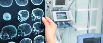
Magnetic resonance imaging (MRI) and computed tomography are performed to exclude a connection between symptoms and tumors, the presence of intrathecal hematoma
An ophthalmoscopic examination of the fundus of the eye, which is carried out by an eye doctor, is used as a “mirror” of the cerebral vessels.
To make a correct diagnosis and prescribe treatment, the participation of several specialists is necessary, including a therapist, neurologist, ophthalmologist, and cardiologist.
EPIDEMIOLOGY
Numerous cohort studies in different countries have demonstrated mixed incidence of TIA, which ranged from 0.37 to 1.1 per 1000 people per year. Like strokes, the risk of TIA increases significantly with age. In one British study, the incidence of TIA among people over 85 years of age reached 6.41 per 1000. According to the Cardiovascular Health Study (1993), the prevalence of TIA was 2.7% among men 65–69 years of age and 1.6% among women of the same age. age, and in persons 75–79 years old it increased to 3.6% and 4.1% in men and women, respectively. At the same time, in people who have had a stroke, a history of TIA is observed in a fairly significant number of cases - from 7 to 40%. This wide variation is believed to be explained by a number of factors, including different criteria for diagnosing TIA in different studies. Many TIAs remain undiagnosed.
ETIOLOGY AND PATHOGENESIS
Various risk factors can lead to the development of TIA as one of the variants of acute cerebrovascular accident.
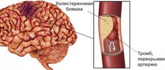
The leading risk factor is atherosclerosis of the main arteries of the head with the formation of hemodynamically significant stenoses and embologenic plaques. In almost 90% of cases, the development of TIA is caused by atherothrombosis of the carotid arteries, most often at the site of their bifurcation into external and internal.
The second most common factor is the presence of blood clots in the cavities of the heart and on its valves in atrial fibrillation (AF), dilated cardiomyopathy, myocardial infarction, artificial heart valves, bacterial and non-bacterial endocarditis.
Much less often, TIAs develop as a result of:
- coagulopathies;
- angiopathy caused by arterial hypertension, diabetes mellitus or other reasons;
- developmental anomalies (kink, duplication, hypo- or aplasia) of the carotid or vertebral arteries, coarctation of the aorta;
- extravasal compression of the vertebral arteries by pathologically altered cervical vertebrae;
- dissection of the carotid or vertebral arteries.
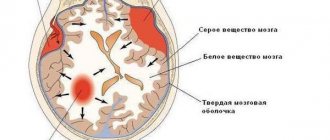
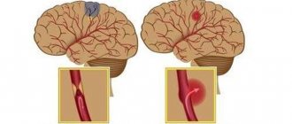
The main mechanism for the development of TIA is the entry of microemboli from the main arteries of the head or heart into the small vessels of the head. This is accompanied by an acute, but reversible (without the formation of a heart attack) critical decrease in blood supply to a region of the brain in a certain arterial basin. The key point in the pathogenesis of TIA is precisely reversible local cerebral ischemia, which develops when cerebral perfusion decreases below 18–22 ml per 100 g/min. (at a norm of 50–60 ml per 100 g/min), which is the functional threshold for ischemia.
A transient drop in blood flow in the area distal to the site of arterial occlusion leads to the appearance of focal symptoms. When blood flow is restored as a result of autoregulation mechanisms and the opening of collaterals, regression of focal symptoms and completion of the vascular episode are observed. In the event of a further drop in perfusion below the threshold of reversible changes (8–10 ml per 100 g/min), a cerebral infarction is formed.
CLASSIFICATION
There is no generally accepted classification of TIA.
In clinical practice, a classification corresponding to the classification of ischemic stroke (TOAST classification) is often used, according to which the following types of TIA are distinguished:
- atherothrombotic - due to atherosclerosis of large arteries, leading to their stenosis or occlusion. When an atherosclerotic plaque or thrombus fragments, an arterio-arterial embolism develops, which is also included in this type of stroke;
- cardioembolic – the most common causes are atrial fibrillation, valvular heart disease, myocardial infarction, especially less than 3 months old;
- lacunar - due to occlusion of small-caliber arteries, the damage of which is associated with arterial hypertension or diabetes mellitus;
- TIA associated with other rarer causes - non-atherosclerotic vasculopathies, hypercoagulation, hematological diseases, hemodynamic mechanism of development of focal cerebral ischemia, dissection of the arterial wall;
- TIA of unknown origin, which includes strokes with an unknown cause or with two or more possible causes, when it is impossible to make a definitive diagnosis.
DIAGNOSIS
The main objectives of diagnosing TIA are:
- exclusion of cerebral infarction in the first hours from the development of TIA;
- exclusion of other diseases with a clinical picture similar to TIA;
- determination of the etiology of TIA with the aim of timely initiation of targeted prevention of cerebral infarction.
The diagnostic algorithm for TIA is the same as for ischemic stroke.
Considering the high risk of developing ischemic stroke in a patient with TIA, examination and treatment should be carried out immediately in a hospital setting.
Clinical signs and symptoms
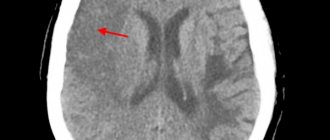
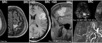
TIA is a cerebrovascular disease with transient neurological symptoms. Symptoms of TIA are mostly the same as those of patients with ischemic stroke and include unilateral limb weakness, difficulty speaking, sensory disturbances, and difficulty walking. Since these symptoms are temporary, they are often assessed on the basis of the patient’s story; the diagnosis of TIA is made retrospectively, since at the time the patient is examined by a specialist, focal neurological symptoms are no longer present (Fig. 1).
TIAs in the carotid artery system are manifested by transient mono- or hemiparesis, sensory disturbances (mono- or hemianesthesia), and speech disorders.
TIAs in the vertebrobasilar system are characterized by short-term vestibular and cerebellar disorders (ataxia, systemic dizziness, dysarthria). Sometimes hemianopsia or transient visual impairment in both eyes occurs.
Transient blindness. Transient monocular blindness (TMB), or amaurosis fugax, is a condition of often unilateral, sudden short-term (usually within a few seconds) loss of vision due to transient ischemia in the area of the blood supply of the ophthalmic, posterior ciliary arteries or retinal arteries. Visual impairment is often described by patients as a “curtain” or “damper” that has moved from top to bottom or bottom to top. Sometimes vision loss is limited to the upper or lower half of the visual field. TMS usually occurs spontaneously, without provoking factors, but can sometimes be triggered by bright light, changes in body position, exercise, or a hot bath. Painful sensations are not typical for TMS.
Transient global amnesia (TGA) is a unique syndrome in which a patient, usually of middle age, suddenly loses short-term memory with relative preservation of memory for distant events. The patient's consciousness and orientation in his own personality are not impaired, but there is incomplete orientation in space and the environment. There is a stereotypical repetition of the same questions and confusion. The etiology of this dramatic syndrome remains unknown; they are trying to explain it by ischemia of the hippocampal-fornical system. Some authors consider this pathology an epileptic phenomenon or a variant of migraine. Episodes of TGA are often triggered by certain factors, such as emotional distress, pain, and sexual intercourse. Attacks usually last from several tens of minutes to several hours. Memory recovery after the attack is complete. As a rule, the patient fully or partially remembers the events that occurred during the amnesia episode. There is no neurological deficit. The tendency to recur is small, the frequency is rare - once every few years.
Differential diagnosis
In order to carry out a differential diagnosis of TIA and diseases that mimic it, a thorough history taking, clinical examination, palpation and auscultation of the carotid arteries are necessary (in the presence of a systolic murmur at the site of bifurcation of the common carotid artery into the external and internal - in the area of the angle of the lower jaw, contralateral neurological symptoms, it is highly likely to assume the development of a TIA), as well as appropriate imaging methods (Fig. 2).
The difficulty of diagnosing TIA is due to a wide nonspecific palette of symptoms, which determines the need for differential diagnosis. Differential diagnosis is carried out with various pathological processes that can manifest as transient neurological disorders:
- ischemic stroke with rapid regression of focal symptoms;
- hemorrhagic stroke with rapid regression of focal symptoms;
- a brain tumor;
- traumatic brain injury;
- partial epileptic seizure;
- associated migraine;
- demyelinating disease;
- hyperventilation syndrome;
- conversion disorder;
- autoimmune processes;
- hypoglycemic state and other dysmetabolic processes.
- Symptoms not typical for TIA:
- disturbance of consciousness;
- isolated non-systemic dizziness;
- general weakness;
- fainting;
- flickering (sparkling) scotoma;
- isolated tinnitus;
- urinary and/or fecal incontinence;
- gradual progression of symptoms (especially sensory ones) involving several parts of the body;
- acute behavioral disorders.
Laboratory and instrumental studies
Laboratory research
Basic methods:
- general clinical blood test with determination of hematocrit and platelet count;
- general urine analysis;
- international normalized ratio (INR), activated partial thromboplastin time (APTT), prothrombin time, fibrinogen;
- blood glucose;
- total cholesterol, high and low density lipoproteins, triglycerides;
- blood electrolytes (potassium, sodium, calcium, chlorides);
- transaminase activity, total direct bilirubin;
- urea, creatinine;
- creatine phosphokinase, C-reactive protein;
- total protein.
Additional (according to indications):
- determination of antiphospholipid antibodies;
- identification of various hypercoagulopathies;
- determination of homocysteine;
- protein fractions;
- blood test for HIV, syphilis, hepatitis B, C;
- molecular genetic tests for suspected hereditary syndromes (MELAS, CADASIL Fabry, etc.).
Instrumental studies
Basic methods
The main methods for diagnosing TIA are imaging studies (brain, blood vessels, heart), which allow:
- exclude ischemic and hemorrhagic stroke (cerebral infarction, hematoma, subarachnoid hemorrhage);
- carry out differential diagnosis with non-vascular diseases of the brain;
- determine the mechanism of development of TIA (atherothrombosis of a large vessel, cardiac source of embolism) and choose rational stroke prevention.
Computed and magnetic resonance imaging
Magnetic resonance imaging (MRI) or computed tomography (CT) of the head is indicated in all patients in whom a TIA is suspected based on the clinical picture.
The method of choice for neuroimaging for suspected TIA is MRI using diffusion-weighted images (DWI), which provides the most accurate early assessment of ischemic brain damage.
If MRI is contraindicated (patients have pacemakers, metal staples on an aneurysm, metal heart valves) or is unavailable, an X-ray CT scan is performed. This method allows for differential diagnosis with spontaneous and traumatic intracranial hemorrhages.
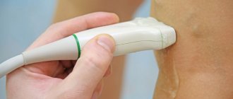
Urgent ultrasound examination of the vessels of the head and neck
A mandatory component of diagnosing a patient with TIA is non-invasive ultrasound visualization of the vessels of the neck and preferably intracranial vessels. This method makes it possible to identify steno-occlusive lesions of carotid vessels and determine the need for surgical restoration of cerebral blood flow.
Duplex scanning (DS) of the main (extracranial) arteries of the head (MAG) is used to diagnose stenoses, potentially embologenic atherosclerotic plaques in the MA, violation of the integrity of the vascular wall with the formation of intramural (intramural) hematoma and the presence of anatomical abnormalities. Based on the results of DS, additional information is obtained about blood flow, hemodynamic disorders and the degree of their severity.
Transcranial Dopplerography (TCDG) of cerebral arteries with microembolodetection makes it possible to assess the condition of intracranial arteries and detect the circulation of emboli in them.
Magnetic resonance angiography (MRA) or CT angiography is indicated in cases where DS does not provide a reliable result. As a rule, this occurs with the development of ischemia in the vertebrobasilar system.
Additional instrumental methods (aimed at clarifying risk factors for TIA):
EchoCG is indicated for suspected cardioembolic mechanism of TIA in cases where the history and/or physical examination indicate the possibility of cardiac pathology, when the patient’s age exceeds 45 years, and also when the results of examination of the vessels of the neck, brain and blood tests do not reveal the cause TIA. Transesophageal echocardiography is more informative than transthoracic echocardiography in determining the pathology of the interatrial septum (septal aneurysm, patent foramen ovale, etc.), atrial thrombi and valve diseases.
Carotid angiography is a standard diagnostic procedure before performing carotid endarterectomy. It is also indicated in patients with TIA if DS and MRI (CT angiography) give conflicting results or if they are not possible.
EEG is indicated for patients who need to make a differential diagnosis of TIA and epileptic seizure.
Holter 24-hour ECG monitoring according to indications.
Daily blood pressure monitoring according to indications.
Fundus examination, perimetry as indicated.
Formation of diagnosis
The clinical diagnosis of TIA reflects the vascular system or basin in which transient cerebral ischemia occurred (carotid artery syndrome or vertebrobasilar arterial system syndrome), as well as the background process that led to the acute vascular complication of TIA, similar to how it is formulated for stroke. Two types of TIA have their own codes in the international classification of diseases - transient blindness (amaurosis fugax), which occurs as a result of embolism of the ophthalmic artery, and transient global amnesia, which develops with bilateral ischemia of the mediobasal parts of the temporal lobes.
TREATMENT
According to international recommendations for the management of patients with ischemic stroke and transient ischemic attacks, if TIA is suspected, patients should be immediately hospitalized in the department for the treatment of acute cerebrovascular accidents (Fig. 3). This approach is due to their high risk of developing a stroke in the coming hours, as well as the need for differential diagnosis with other diseases and conditions resembling TIA.
Treatment and secondary prevention of stroke in patients with TIA are inseparable and begin from the moment of diagnosis.
Non-drug treatment
Surgery:
- is the leading direction in the treatment of patients who have suffered a TIA, as well as the prevention of stroke in the presence of hemodynamically significant stenosis (≥70%) or acute occlusion of the arterial artery;
- Angiosurgical restoration of blood flow is recommended within 24 hours after TIA;
- if there are individual contraindications to surgery in the acute period, the decision on surgical intervention is made as planned over the next 6 months;
- Carotid thromboendarterectomy is more often performed. Vascular stenting is carried out in special cases (difficult direct access to the vessel, high risk of perioperative complications, restenosis after carotid endarterectomy, etc.);
- Antiplatelet therapy is carried out before and after surgical treatment.
Drug treatment
Emergency pathogenetic therapy for in-hospital TIA in a neurovascular department should be carried out in the amount of AI therapy, especially on the first day of the disease, when in most cases both clinical and instrumental (CT, MRI) diagnostics do not yet provide convincing evidence in favor of one or another option .
As with AI, therapeutic measures consist of basic and specific therapy.
Basic therapy (includes control over the basic functions of the body and maintaining homeostasis):
- correction of blood pressure - it is advisable to maintain the level of mild arterial hypertension 160–180/90–100 mm Hg, and avoid a significant decrease in blood pressure, especially in patients with bilateral carotid artery stenosis;
- control of blood glucose levels, for hyperglycemia above 11 mmol/l - consultation with an endocrinologist regarding insulin therapy, for hypoglycemia below 3.3 mmol/l - administration of a glucose solution;
- control of water and electrolyte balance to avoid an increase in hematocrit and a decrease in the rheological properties of blood;
- electrocardiography - if arrhythmia is detected - daily monitoring and consultation with a cardiologist.
If it is necessary to lower blood pressure in severe arterial hypertension (>220/120 mm Hg), preference is given to the following drugs:
- the ultra-short-acting cardioselective beta-blocker esmolol is administered intravenously at a dose of 500 mcg/kg for 1 minute (loading dose), then 50 mcg/kg per minute over the next 4 minutes; maintenance dose – 25 mcg/kg per minute (or less);
- the α1-adrenergic receptor blocker urapidil is prescribed in a dose of 12.5–25 mg (1 ampoule contains 5 ml of a 0.5% solution, i.e. 25 mg). This amount of the drug is administered intravenously slowly over 5 minutes. The drug begins to act within 3–5 minutes. Duration of action is 4–6 hours. If the effect is insufficient, the administration of the same dose is repeated (with an interval of at least 15 minutes). The effective dose can vary from 25 to 100 mg. If the hypotensive effect is not persistent enough, a slow drip infusion of the drug is prescribed;
- ACE inhibitor captopril (used sublingually at a dose of 25 mg), enalapril orally at an initial dose of 5 mg, enalaprilat (administered orally intravenously slowly or drip over 5 minutes at a dose of 1.25 mg every 6 hours).
- in cases resistant to this therapy, it is possible to use an intravenous infusion of the peripheral vasodilator sodium nitrop-russide (start at a rate of 0.5 mg per 1 kg of body weight per minute under constant blood pressure monitoring).
Specific antithrombotic therapy
Antithrombotic therapy for patients with TIA begins immediately, performing both therapeutic and preventive functions, given the high risk of stroke in the first days after the development of TIA. The choice of drug is determined by the type of TIA - cardioembolic and non-cardioembolic.
For non-cardioembolic TIA, one of the platelet antiplatelet agents is prescribed:
- acetylsalicylic acid (Cardiomagnyl, Thrombo-ass, Cardio ASA, Aspirin Cardio, etc.) at a dose of 325 mg/day for 48 hours with a transition to 50–100 mg/day (at the rate of 1 mg per kg of patient weight);
- clopidogrel (Plavix, Egitromb) 75 mg;
- dipyridamole (75 mg 3 times a day);
- small doses (25 mg) of aspirin + 200 mg of slow-release dipyridamole in capsules 2 times a day (Agrenox).
For the cardioembolic mechanism of TIA development (most often as a result of AF), the treatment of choice is oral anticoagulants:
- warfarin is used with mandatory monitoring of the INR, maintaining it at a level of 2–3. The initial dose of warfarin is 2.5 mg, if necessary, the dose is increased until the target INR level is 2-3. Monitoring the INR and increasing the dose by 1.25 mg is carried out once a week. After dose titration, INR monitoring is reduced to once a month. Warfarin is used to prevent recurrent TIA or stroke in various cardiac sources of thrombi, both valvular (especially with artificial heart valves) and non-valvular;
- the so-called new oral anticoagulants are recommended for use only for non-valvular AF: dabigatran 150 or 110 mg (persons over 80 years old) 2 times a day, rivaroxaban 20 or 15 mg (for moderate renal impairment) 1 time a day, apixaban 5 or 2.5 mg (persons over 80 years of age with body weight <60 kg or serum creatinine ≥133 μmol/L) 2 times a day. In cases of severe renal impairment, all anticoagulants are contraindicated.
If there are contraindications to oral anticoagulants, ASA is prescribed at a dose of 325 mg/day for 48 hours, with a transition to 50–100 mg/day.
Evaluation of treatment effectiveness
In a patient who has suffered a TIA, the criteria for the effectiveness of treatment are:
- complete regression of neurological deficit;
- stabilization of vital functions and homeostasis;
- normalization of laboratory parameters;
- achieving target blood pressure values;
- restoration of blood flow, confirmed by the results of angiographic studies and ultrasound methods.
Complications and side effects of treatment
The most common complication of antithrombotic therapy is bleeding. When prescribing this therapy, it is necessary to find out the presence of potential sources of bleeding in the patient.
Risk factors for bleeding include:
- state of the blood coagulation system;
- age over 65 years;
- arterial hypertension;
- impaired renal and liver function;
- history of bleeding;
- history of peptic ulcer;
- alcohol or drug use;
- concomitant therapy (NSAIDs, corticosteroids);
- combined antithrombotic therapy.
But even with the risk of bleeding, in most cases the risk of developing a cerebral or myocardial infarction after a TIA is higher than the risk of bleeding.
Errors and unreasonable assignments
Gross errors are:
- refusal to hospitalize patients with TIA;
- prescribing antithrombotic therapy at the prehospital stage;
- administration of diuretics at the prehospital stage;
- lack of monitoring of vital signs and limited ability to provide adequate care to patients with TIA;
- the desire to lower blood pressure with moderate arterial hypertension.
FORECAST
With timely emergency hospitalization of a patient with TIA and adequate emergency treatment, the symptoms of TIA undergo a reverse development.
More than half of patients who have suffered a TIA remain at risk of developing cardiovascular complications, such as stroke, myocardial infarction, and vascular death.
The risk of developing a stroke within 3 months after a TIA ranges from 10 to 20%. In half of them, cerebral infarction occurs in the first 2 days after transient neurological symptoms.
To predict the risk of stroke after a TIA, the AVSD2 scale was developed (Tables 1, 2).
Almost 60% of TIA patients die within 5 years from other cardiovascular diseases.
After a TIA, ongoing therapy is recommended to prevent stroke and other cardiovascular diseases.
Management of patients who have suffered a TIA at the outpatient stage of treatment
Prevention of recurrent TIAs and strokes
Antithrombotic therapy continues almost lifelong, especially for patients who have a high risk of stroke according to the ABCD2 scale.
In patients with arterial hypertension, continuous antihypertensive therapy is indicated to prevent second TIAs and strokes. The target blood pressure level is determined individually, but it should not be lower than 130/80 mm. Hg with surgically unresolved unilateral stenosis of the carotid artery more than 70% of the vessel diameter and below 150 mm. Hg – with hemodynamically significant stenosis of the carotid arteries on both sides. The choice of antihypertensive drug is determined by the presence of concomitant and background diseases.
In case of atherothrombotic processes, especially in the presence of loose embologenic plaques in the carotid arteries, as well as against the background of lipidemia, the prescription of statins is indicated.
Lifestyle modification (giving up bad habits, dieting, rational physical activity).
To activate the processes of restoration of ischemic neurons, maintain endogenous protective mechanisms of nervous tissue, and stimulate neuroplasticity of the brain, it is advisable to prescribe neurotrophic drugs. One of the proven drugs in this group is Cerebrolysin, which contains fragments of neurotrophic factors, biologically active peptides and amino acids. The ECOMPASS study, using modern neuroimaging techniques (tractography/fMRI), demonstrated earlier and more complete disclosure of the potential of neuroplasticity in the combination therapy group (Cerebrolysin + rehabilitation measures). Several domestic randomized studies (Gusev E.I., Chukanova E.I.) have shown that the addition of Cerebrolysin to the standard treatment of patients with CCI (10 ml IV for 10 days every six months) or those who have had a stroke (20 ml IV /c for 10 days with a repeat course after six months) reduces the number of new/recurrent strokes and TIAs.
Prevention of cognitive impairment after TIA
Cognitive impairment (CI) is detected in almost 40% of patients who have suffered TIAs or “minor strokes”. CIs are more often detected in elderly and senile people, in whom the vascular process can stimulate the progression of subclinical neurodegeneration.
Currently, from the standpoint of evidence-based medicine, the role of regular physical activity in the prevention of CI in patients who have suffered a stroke or TIA is shown.
It is important to conduct cognitive training, diverse sensory stimulation, and maintain social contacts.
It is also advisable to include drug support for cognitive functions with neurometabolic drugs. It is desirable to have additional properties aimed at the pathogenetic mechanisms of cerebrovascular diseases and reducing the pathological impact of underlying diseases. From this standpoint, such drugs as deproteinized hemoderivate (Actovegin) and citicoline (Cerakson®) have proven themselves well. Both drugs provide modest but consistent improvements in memory and behavior in patients with CI.
The ARTEMIDA study showed a significant improvement in cognitive function in patients with ischemic stroke who took Actovegin for 6 months at a dose of 2000 mg intravenously for 3 weeks, followed by oral administration of 1200 mg/day for 21 weeks. The persistent therapeutic effect persisted for another 6 months after discontinuation of therapy.
Numerous clinical trials have proven the positive role of citicoline for patients in the acute period of stroke and during the rehabilitation stage. The IDEALE study noted the effectiveness and safety of oral citicoline (1 g/day) in elderly people with moderate vascular CI.
Treatment
The main goal of treatment is to prevent stroke. Therefore, it is necessary to start therapy early, without waiting for severe ischemic attacks and their recurrence.
Hospitalization is necessary for frequent attacks that impair the ability to work. If the examination is carried out and ischemic attacks occur rarely, treatment can be carried out at home under the supervision of the attending physician at the clinic.
Directions of therapy:
- for blood thinning, such popular drugs as Aspirin, ThromboAss, Cardiomagnyl are recommended; if due to concomitant stomach diseases they are poorly tolerated, Ticlopedin is prescribed;
- in stationary conditions, Reopoliglucin is administered intravenously;
- statin drugs are widely used to lower blood cholesterol levels and delay the development of atherosclerosis, these include Atorvastatin, Simvastatin, Pravastatin;
- To relieve vascular spasm, coronary agents are used: Papaverine, nicotinic acid, Nikoverine;
- drugs that restore microcirculation of cerebral vessels include Cavinton, Vinpocetine;
- Nootropics (Piracetam, Nootropil, Cerebrolysin) help preserve neurons and provide them with additional energy for recovery.
The drug, coming from Germany, requires caution in use in patients with kidney disease, liver disease, and diabetes mellitus
Recently, there has been a debate about the advisability of using statins, since the craze and hopes have turned into complications in the form of a disturbed psyche. Women are especially prone to them. Therefore, the prescription is considered justified if a two-month strict diet does not lead to normalization of blood cholesterol levels. And if the lipoprotein content is normal, there is no need to use them.
It is necessary to monitor blood pressure levels and take medications for hypertension, diuretics if indicated. Patients with diabetes cannot get rid of transient attacks without maintaining normal blood glucose levels with the help of hypoglycemic drugs.
If the initial phenomena of thrombosis are detected in a hospital setting, fibrinolytic therapy is carried out in an attempt to dissolve and remove the thrombosis.
Auxiliary therapies:
- if there is a connection between ischemic attacks and cervical osteochondrosis, electrophoresis with drugs that relieve muscle spasms, careful massage of the collar area, Darsonval currents on the head are prescribed;
- Oxygen, pine, and radon baths have a good relaxing effect; they are best carried out in courses in sanatorium conditions;
- physical therapy helps restore impaired blood circulation and develop a network of auxiliary vessels.
From folk remedies, any reasonable recipes for the prevention of atherosclerosis and strengthening the immune system are suitable. These include lemon-garlic tincture, hawthorn, rosehip infusion, clover, and fish oil supplements. You should not try to replace medications with them. It is impossible to get rid of atherosclerotic plaques using folk remedies.
Blood pressure control and lipid-lowering therapy
Aggressive therapy for TIA, in addition to DAPT, involves prescribing lipid-lowering and antihypertensive therapy with achieving target blood pressure values (130/80 mm Hg) as soon as possible. This treatment strategy differs significantly from the procedure adopted in the current recommendations for providing care to patients with stroke, which do not mention statins, and arterial hypertension up to 180 mm Hg. Art. is considered acceptable in the first hours of the disease [12]. Arterial hypertension is the most important risk factor for IS and a frequent accompaniment of the acute period of vascular accident. The causes of arterial hypertension in the acute period of IS are associated with activation of neuroendocrine and neurogenic mechanisms of blood pressure regulation, ischemia of brain stem structures, hospital stress, and disruption of autoregulation of cerebral circulation [20]. The optimal values of systolic and diastolic blood pressure in the acute phase of IS have not been established, and clinicians do not have a consensus on this matter. Large studies that should resolve this issue have not yet been completed [20–22]. The only argument justifying a high level of blood pressure in the acute period of IS is the supposed possibility of the best perfusion of brain tissue in the area of the ischemic focus at high systemic pressure in conditions of failure of autoregulation [23, 24]. However, the results of recent studies do not support this idea [24]. The assumption about the beneficial effect of high systolic blood pressure on the damaged brain also seems unconvincing with regard to perivascular edema, since in patients with high blood pressure values and developing cerebral infarction, edema in the damaged area is more pronounced [24, 25]. The preservation of “therapeutic” arterial hypertension in patients with TIA seems even less justified. Transient focal cerebral ischemia is very often associated with hypertensive crisis, cerebral vasospasm and thromboembolism. In all cases of TIA, conditions for restoring cerebral blood flow will be better when blood pressure levels are close to normal. Lipid-lowering therapy with statins is not mentioned in guidelines for the management of patients in the acute period of stroke, but is actively used in patients with TIA [2, 3, 25]. The pleiotropic effects of statins, which reduce edema, stabilize atherosclerotic plaques and restore microcirculation, may be useful not only for preventing a recurrent vascular event. Studies in which biomarkers of inflammation and tissue destruction (high-sensitivity C-reactive protein and matrix metalloprotease 9) were used to verify the results of intensive lipid-lowering therapy in patients with atherothrombotic IS showed the effectiveness of atorvastatin at a daily dose of 60 mg [25].
Is it possible to predict the likelihood of a stroke with TIA?
Clinical studies have confirmed a reduction in the incidence of ischemic stroke in the group of patients receiving treatment by 30–45%. This is with reliable data on stroke detection:
- in the coming years in ¼ of patients with ischemic attacks;
- during the first week - in 43% of cases.
Therefore, the demands of doctors to go to the hospital, even if the symptoms have disappeared, clearly provide the patient with the prevention of serious disorders.
The left column of the table contains links to the authors of the studies; the reliability is beyond doubt and is confirmed by the number of participants
There is no need to rely only on medications without following personal protection rules. The consequences depend on a person’s lifestyle more than on medications. Recommended:
- eliminate smoking and alcohol;
- adhere to an anti-atherosclerotic diet for the rest of your life (sharply limit animal fats, high-calorie foods, switch to vegetable oils, fish, dairy products with reduced fat content, be sure to eat vegetables and fruits at any time of the year);
- physical activity is limited only to heavy sports, walking, swimming, fitness, cycling are recommended;
- Monitoring blood pressure will help prevent TIA in a timely manner and get rid of brain problems.
Transient ischemic attacks should be treated as a warning signal about an increasing risk of stroke. By listening to the “signals” of your body, you can avoid severe pathology, prolong your active life and not be a burden to your family and friends.
Neuroimaging and antiplatelet therapy
Neuroimaging results can have a significant impact on the choice of treatment options. Aggressive management of patients with DAPT increases the risk of intracranial hemorrhage (ICH). And this significantly limits the treatment of patients with TIA in contrast to patients with ACS. And although current clinical guidelines allow prescribing DAPT to all patients with TIA and minor IS, it is safer to start treatment with a loading dose of acetylsalicylic acid (300–325 mg), and include clopidogrel (as the second component of DAPT) in a loading dose of 300 mg in the treatment regimen after performing MRI (CT) [3, 4, 11]. It should be noted that the 2021 Russian clinical guidelines demonstrate a more cautious approach to DAPT, prescribing the prescription of two antiplatelet drugs only to patients who, along with the picture of acute cerebral ischemia, also have cardiac indications for more aggressive therapy [12]. If, according to the results of neuroimaging, biomarkers of a high risk of ICH are detected in a patient, the doctor may therefore limit the aggressiveness of treatment (Table 1).
If MRI (CT) does not detect biomarkers of high risk of ICH, then DAPT for 2–3 weeks. significantly reduces the likelihood of all vascular events and IS. Loading doses (300–325 mg for acetylsalicylic acid and up to 300 mg for clopidogrel) have been used in clinical trials, which showed a 32% reduction in the likelihood of stroke within 90 days after TIA [13–16]. Studies [14, 17–19] have established that patients with TIA and minor IS without signs of amyloid cerebral angiopathy receive benefits from DAPT. The 2021 AHA/ASA clinical guidelines allow the use of DAPT for TIA and IS, allowing for the possibility of extending such therapy up to 3 months. [eleven]. Recent European guidelines demonstrate a more cautious approach to aggressive antithrombotic therapy, limiting the duration of DAPT to 10–21 days [17]. Thus, active antiplatelet therapy for patients with TIA is not only possible, but also highly advisable [10, 17, 18]. The trend in recent years towards an increase in the activity of antithrombotic therapy for TIA seems obvious. Antiplatelet drugs are prescribed as early as possible in combinations and doses that best prevent recurrent vascular events. The relatively new concept of “early secondary prevention” involves active or even aggressive management of patients in the first hours and days after the onset of symptoms of circulatory decompensation in the cerebral vascular system. Antiplatelet agents are prescribed regardless of the suspected stroke subtype. However, several days after cardiac examination, patients with atrial fibrillation should receive anticoagulants instead of antiplatelet agents. The principle approach to the active management of patients with TIA is not limited to antithrombotic therapy. Antihypertensive and lipid-lowering therapy for patients with signs of TIA differs from the strategy used for large-focal strokes.