- Types of dysplasia
- Degrees of dysplasia
- Diagnosis of the disease
- Treatment of dysplasia
- Recovery period
Dysplasia is a violation of the structure of body tissues, with a simplification of their structure, deformation of cells and their components. Congenital dysplasias caused by genetic causes, as a rule, have diverse manifestations, often multiple and in different systems.
Dysplasia of the mucous membranes has a local connection to a specific organ, such as the stomach, intestines, cervix. Unlike all other dysplasias, this type of pathology is not inherited and is not associated with a global genetic failure, but is caused by the vital activity of pathological microflora.
Types of dysplasia
Dysplasias are very diverse and heterogeneous, congenital and acquired dysplasias are essentially very different processes, some involve several systems, others only areas of the mucous membrane.
Congenital dysplasias are manifested by developmental anomalies - deviations in the structure that do not interfere with normal functioning, and developmental defects - these anomalies already disrupt the functioning of the anatomical region.
Mucosal dysplasia does not form defects or developmental anomalies, but can lead to malignant processes. Absolutely different manifestations of dysplasia do not allow creating a unified classification; each researcher of the problem offers his own systematization of heterogeneous pathology, not free from shortcomings.
Congenital dysplasias are formed in the prenatal period; clinical manifestations affect the integumentary tissues and the musculoskeletal system; their division is very conditional.
Dysplasia of the ectoderm is distinguished - the superficial tissue of the embryo from which the outer integument is formed: skin with nails and hair and the oral mucosa with teeth. Pathology can manifest itself at any age, in any set of symptoms and with any severity. This can only be dry skin with small spots of atrophy and an almost complete absence of teeth, or baldness with no nails and moon-shaped defects in the tooth enamel with a full set of teeth. It is also possible that there is simply a decrease in the number of sweat glands in the skin, which leads to rapid overheating already in infancy and the danger of sudden death.
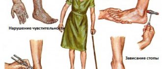
The vast majority of those suffering from genetic connective tissue dysplasia may experience disorders of all body systems from minor to life-threatening and with any set of symptoms: neurological disorders, heart valve defects, skeletal changes, vascular aneurysms, softening of the tracheal rings, prolapse of the kidneys, and also leading to sudden death due to combined damage to the cardiovascular and respiratory systems. The most active surge in manifestations is observed in adolescence, when, for example, scoliosis and flat feet progress sharply, cardiac arrhythmia and myopia appear. Several syndromes with characteristic external manifestations have been formed, again with variable severity - from scanty individual manifestations to extremely pronounced ones with early death from complications, such as Marfan syndrome and osteogenesis imperfecta.
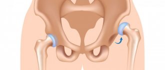
Joint dysplasia is manifested by congenital malformations of large - hip and knee, as well as small joints of the hand and foot, and in essence can be considered the same connective tissue dysplasia, but in a separate articular system. As a rule, the pathology is detected in infants, but in a minimal form it can go unnoticed, which is discovered during an examination for an accidental leg injury.
Fibrous dysplasia is indicated by single and multiple cysts in the bones. Some people live healthy lives into old age and learn about genetic pathology in the form of a small brush by chance during an X-ray for another reason. Others suffer from chronic pain and limb deformity, often with shortening, due to multiple cystic cavities in the bone tissue.
Dysplasia of the mucous membranes of internal organs is “earned” during life. In most cases, the process does not manifest symptoms or has uncharacteristic and unstable minimal signs, which are found by microscopy of a piece of tissue. Dysplasia can progress from mild, through moderate and severe, up to stage zero cancer - and this is its main danger. Severe dysplasia is difficult, and sometimes impossible, to reliably distinguish from non-invasive carcinoma in situ.
Gastric dysplasia can be clinically manifested by gastric pathology; the patient is often infected with Helicobacter. The pathology lies in the simplification of the cellular structure - decreased differentiation, changes in cell nuclei and other intracellular structures. Mild dysplasia not only turns into moderate, but also regresses; moderate dysplasia proceeds similarly, while severe dysplasia is considered a precancerous process and is subject to serious treatment and observation. Gastric dysplasia in frequency of occurrence is significantly inferior to metaplasia - the formation in the mucosa of areas of cells very similar to the cells of the large or small intestine. During microscopy, areas of intestinal metaplasia are always found around any dysplasia, and this is also a precancerous process.
Primary urothelial dysplasia is an uncommon pathology, manifested by symptoms of an irritable bladder with frequent and painful urge to urinate. This variant of dysplasia is not usually divided into degrees; the probability of developing cancer on a dysplastic background is slightly more than 15% and the transition to stage zero cancer will take from 4 to 8 years.
Cervical or dysplasia of the cervical mucosa is a very common pathology, since its leading cause is infection with the human papillomavirus. The virus itself cannot be killed - it is resistant to any drugs, but it is fatal and in the vast majority of women it will heal on its own in the next two to three years.
Connective tissue dysplasia in the practice of a pediatrician
Translated from Greek as “deviation in formation”
“Medical Bulletin” has touched upon the topic of DST more than once.
But so far only isolated manifestations of this congenital pathology have been described. Thus, in “MV” No. 21 for 2010, Professor L.M. Makarov spoke about primary channelopathies, often leading to sudden cardiac death in children and caused by mutations of genes expressed in the myocardium. In “MV” No. 4 for 2011, Professor I.V. Viktorova described joint hypermobility syndrome, and in No. 17-18 for 2011, Professor E.A. Gallyamov spoke about the most important pathogenetic factor of gastroesophageal reflux disease - congenital hiatal hernias, formed due to the increased elasticity of the tissues limiting this hole.
These publications already show how widespread and significant diseases caused by CTD are. However, many years of research by Z.V. Nesterenko and the world science data that she analyzes indicate that DST affects not three, but much more body systems, and this problem is incomparably wider and more acute than it was thought about in 1989, when the Scottish doctor R. Beighton first proposed to designate the congenital pathology of TS, manifested by a decrease in its strength, by the term dysplasia, which translated from Greek means “deviation in formation.”
If proline changes to arginine
The frequency of detection of CTD (according to various authors) is quite high - from 26 to 80% for all ages and from 74 to 85% among children.
In the development of such dysplasias, mutations of genes encoding the synthesis and spatial structure of collagen and responsible for the formation of matrix components are of key importance. Accordingly, according to one of the first classifications of DST, they were divided into diseases caused by impaired synthesis or catabolism of the fibrous components of DST or its main substance.
But in the 1990s, a classification that is more common today was adopted. The first group of dysplasias includes fairly rare differentiated dysplasias (DDSDs), which have a monogenic and specific type of inheritance, more often autosomal dominant, and clearly defined clinical symptoms. These are Marfan syndrome, Ehlers-Danlos syndrome, osteogenesis imperfecta and several others.
To get acquainted with DDST, let's turn to the most common (frequency of occurrence among newborns is 5:100,000) of their representative - Marfan syndrome (MS). All proven cases of this syndrome are a consequence of a mutation in the fibrillin gene. It is localized in the long arm of chromosome 15, field 21 (15ql5-q21.3). The essence of the mutation is the replacement of the amino acid proline with arginine in the fibrillin protein. As a result, the synthesis of collagen type 3 increases and the content of collagen type 1 decreases. If normally the ratio of collagen-1:collagen-3 is 6:4, then with SMA it drops to 3:7.
The clinical picture in a typical case of SMA is manifested by a characteristic triad of signs related to the musculoskeletal and cardiovascular systems, as well as the visual organs.
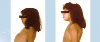
Changes in the musculoskeletal system include: tall stature, asthenic physique (the length of the limbs is disproportionate to the length of the body), arachnodactyly (long thin fingers), chest deformity, high arched palate, kyphoscoliosis, ligamentous weakness. Lesions of the cardiovascular system include: dilatation of the aortic root, aortic regurgitation, dissecting aortic aneurysm, mitral valve prolapse, blood regurgitation due to insufficiency of this valve. A characteristic pathology of the visual organs is iridodenesis (tremor of the lens due to weakness of the ligament of Zinn), subluxation of the lens, a high risk of retinal detachment and high myopia.
Before the widespread use of surgical correction of cardiovascular pathology, almost all patients with SMA died before the age of 30-35 years. Moreover, the main group is still in childhood. However, in the 21st century, with adequate therapy, the life expectancy of most patients with SMA is only slightly inferior to this indicator in the general population.
Tall, freckled, stooped
The second group of DST, which makes up the disorders most often encountered by practical pediatricians, includes disorders united by the term undifferentiated dysplasia (UDTD). Unlike differentiated dysplasias, UCTD are genetically heterogeneous pathologies. The main characteristic of such dysplasias is a wide range of clinical manifestations without clear symptoms. That is, UCTD is not a nosological entity, and there is no place for it in ICD-10 yet. In turn, UCTDs are divided into 2 groups: diseases with an established and unidentified (and this is the vast majority of UCTDs) gene defect.
UCTD in a child can be diagnosed already at the stage of physical examination with a comprehensive assessment of the so-called phenotypic markers. External and visceral markers are distinguished, the predominance of which depends on what type of TS lesion occurs - dense or loose. The critical number of external markers that allows one to make a conclusion about the presence of UCTD is now considered to be 3-6. Based on the diagnostic significance of individual signs of UCTD, diagnostic tables are proposed with a score of external and visceral markers, as well as biochemical parameters.
For example, external changes in the skin in children with UCTD usually indicate damage to loose connective tissue and are characterized by the presence of hyperelasticity, increased extensibility, stretch marks, keloid scars, a pronounced subcutaneous venous network, characteristic pigment spots of the “café au lait” color, or depigmentation, a large number nevi.
But lesions of dense TS are manifested by changes in the skeleton: poor posture in the form of kyphosis and scoliosis, stooping, chest deformities, and flat feet. Such children, as a rule, are tall and asthenic in build.
All children with UCTD exhibit so-called minor developmental anomalies, or dysmorphia. The most common dysmorphias include: fair skin, fused eyebrows, wide bridge of the nose, hyper- and hypotelorism, blue sclera, epicanthus, abnormal tooth growth, deformed auricle, fused lobe, curved little fingers, incomplete syndactyly of the fingers, light or red hair color. Diagnostically significant for identifying UCTD is the presence of 6 or more dysmorphias in a child.
Markers external and internal
Let us now turn to the internal markers of UCTD, and we emphasize that there is a direct correlation between the number of external and internal markers.
If we talk about the integral indicators of internal UCTD markers detected by biochemical methods, then the most informative is determining the level of molecules formed during the breakdown of collagen. These are hydroxyproline and glycosaminoglycans in daily urine, and in blood serum - lysine and proline. A change in the ratio of collagens of different types in UCTD allows the use of the indirect immunofluorescence method according to Sternberg LA (1982) in diagnosis.
On the membranes of leukocytes, an increased representation of HLA histocompatibility antigens is usually detected - A28, B35, Cw5, Cw52, and on the other hand, a reduced number of antigens such as A2, B12, Cw3. The most promising, of course, are molecular genetic diagnostic methods that identify specific gene mutations. However, in cases of UCTD, these assays are still at an early stage of development.
One of the most often involved in the pathological process in UCTD in children is the cardiorespiratory system.
This leads to serious pediatric errors
With UCTD in children, the formation of the elastic framework of the lungs is disrupted at the beginning of life, which causes the valve mechanism of bronchial obstruction and the formation of emphysematous bullae due to rupture of morphologically incompetent interalveolar septa. The consequence of subpleurally located bullae can be spontaneous pneumothorax. A congenital defect in the cartilaginous and connective tissue framework of the trachea and bronchi leads to their increased mobility, the occurrence of bronchiectasis, and pneumosclerosis. Dyskinesia of the trachea and bronchi leads to the development of broncho-obstructive syndrome (BOS).
There is a high correlation between the severity of bronchial asthma in children and the manifestations of dysplasia. And the more common and severe the latter are, the earlier the child develops pulmonary hypertension and pulmonary fibrosis in addition to bronchial asthma.
Anomalies in the structure of the bronchopulmonary system in UCTD lead to a deterioration in the elimination of pathogenic agents in conditions of altered immune reactivity and contribute to the long-term persistence of bacteria and the formation of a recurrent course of pneumonia. It is also noteworthy that the level of atypical childhood pneumonia is growing, caused by intracellular pathogens - chlamydia and mycoplasma, with a predominant lesion of the interstitium of the lungs, with a recurrent course of which progresses, as in cases of asthma, pneumofibrosis, pulmonary hypertension.
With UCTD, congenital defects are often diagnosed: tracheobronchomegaly, tracheobronchomalacia, cystic hypoplasia of the lungs. Against the background of dysplasia, the listed diseases are especially often accompanied by the development of severe complications in the form of pneumofibrosis, pulmonary hypertension, bronchiectasis, and spontaneous pneumothorax.
Unfortunately, in contrast to the above-mentioned pronounced manifestations of UCTD in children, subclinical variants are usually not diagnosed. This often leads to incorrect interpretation of the pathological process and serious pediatric errors.
Truly small heart
Since 1987, the classification of diseases of the cardiovascular system of the New York Heart Association has identified the syndrome of connective tissue dysplasia of the heart (CDS), which accompanies both differentiated and undifferentiated dysplasia. In turn, cardiac dysplasia includes several syndromes.
Valvular syndrome combines isolated and combined heart valve prolapses and myxomatous valve degeneration. In approximately 70% of cases, the syndrome is represented by prolapse of the mitral valve, less often - tricuspid or aortic, enlargement of the aortic root and pulmonary trunk; ectopically located chordae, aneurysms of the sinuses of Valsalva. In some cases, such changes are accompanied by regurgitation phenomena, which is reflected in the indicators of myocardial contractility and volumetric parameters of the heart.
With thoradiaphragmatic syndrome, we are talking about the asthenic shape of the chest or its deformation, spinal deformities (scoliosis, kyphoscoliosis, hyperkyphosis, hyperlordosis), changes in standing and excursion of the diaphragm. Among children with CTD, the most common deformity of the chest is pectus excavatum, and the second most common is keeled. Deformations of the sternum, ribs, spine and the associated high position of the diaphragm reduce the chest cavity, increase intrathoracic pressure, disrupt the inflow and outflow of blood, and contribute to the occurrence of arrhythmias.
Vascular syndrome involves damage to elastic arteries, in which their walls expand and saccular aneurysms appear. Arteries of both muscular and mixed types are affected. As a result, bifurcation-hemodynamic aneurysms appear, as well as pathological tortuosity of vessels up to looping.
Thoradiaphragmatic heart syndrome (THDS) is formed in parallel with the progression of deformation of the chest and spine, against the background of valvular and vascular syndromes. In children with a typical asthenic constitution, the asthenic variant of STDS is more often manifested. In this case, the size of the heart chambers decreases, but while maintaining their normal thickness. In a word, a “true small heart” is being formed, functioning without serious deviations.
Unfortunately, in some children with chest deformation due to displacement of the heart, when it “moves away” from the mechanical influences of the chest bone, rotating and accompanied by “torsion” of the main vascular trunks, the so-called false stenotic variant of TDS is formed, which is especially severe.
Metabolic cardiomyopathy syndrome combines cardialgia, arrhythmias, and disorders of repolarization processes. The development of metabolic cardiomyopathy is determined by the influence of cardiac factors (valvular syndrome, variants of CVD) and extracardiac conditions (autonomic dysfunction, deficiency of micro- and macroelements, etc.). Cardiomyopathies with DST usually do not have specific symptoms.
Arrhythmic syndrome includes ventricular and atrial extrasystoles, paroxysmal tachyarrhythmias, pacemaker migrations, atrioventricular and intraventricular blocks, anomalies of impulse conduction along additional pathways, ventricular preexcitation syndrome, and QT interval prolongation syndrome.
A practical pediatrician should remember that in children suffering from CTD, cardiomyopathies and arrhythmic syndrome are very common (in 60-64% of patients). And it is they who determine the increased risk of sudden cardiac death in such children.
A universal remedy has not yet been found
A universal remedy that restores connective tissue for all forms of dysplasia has not yet been found. An individual treatment program is selected for each child with CTD. Its three main tasks are: improving the metabolic processes of connective tissue, eliminating existing complications and preventing new ones.
In any case, we are talking about the actually coordinated work of a team of pediatricians of several specialties. And about a very complex, complex treatment that affects the entire childhood period of life (in the Russian Federation - up to 18 years). We will have time to give only one example of such therapy, related to Marfan syndrome.
Non-drug therapy includes strict adherence to the child’s diet, including nutrition, where the emphasis is on the consumption of complete proteins and foods containing polyunsaturated fatty acids; and physical activity, certain types of which are prohibited for such patients. There is a list of professions for which schoolchildren suffering from CTD cannot be trained.
Drug treatment includes symptomatic administration of b-blockers. In the case of aortic dilatation, and especially in the presence of regurgitation, they reduce the ejection into the aorta and, accordingly, the load on its walls, correcting concomitant hypertension. It is believed that these cardiotropic drugs reduce the risk of sudden death in children with cardiac damage due to SMA and any other CTD.
Since it has been proven that the progression of skeletal pathologies in Marfan syndrome slows down when the deficiency of microelements (calcium, magnesium, zinc, copper) necessary for the formation of TS is eliminated, nutritional supplements containing the above substances, as well as hyaluronic acid, synthetic analogues of vitamins K and D3. In the blood of patients with SMA, there is often an increased level of somatotropic hormone. Therefore, to reduce its secretion, high-fat Omega-3 enzymes are prescribed in the diet from early childhood.
Let us now turn to therapy affecting connective tissue. It includes ascorbic acid in the form of special children's milkshakes, as well as correctors for impaired synthesis and catabolism of glycosaminoglycans - chondroitin sulfate and succinic acid. Another drug recommended as a corrector of bioenergetic processes is carnitine chloride.
What surgeons can do
For aortic aneurysm, dissecting aneurysm, aortic valve defect with symptoms of heart failure, and children with Marfan syndrome, only surgical treatment can help. Clear indications for prosthetics have been developed. For example, an aneurysmally dilated aorta must be replaced with an endo- or exoprosthesis. In case of mitral valve prolapse accompanied by “stable” regurgitation, valve replacement is not performed. However, with the rapid progression of regurgitation, up to the addition of left ventricular failure, valve replacement becomes necessary.
Surgical treatment of deformities of the chest and spine is an extremely traumatic procedure, carried out in several stages, often complicated by pleurisy, pericarditis, and pneumonia. The question of its feasibility was discussed many times at symposiums dedicated to DST. Specialists from various countries adopted a common position denying the advisability of such operations for any DST.
Degrees of dysplasia
For every three women with mild dysplasia of the cervical mucosa, there is one patient with moderate and severe dysplasia. In nine out of ten women, the site of pathological changes in the mucous membrane is located at the transition of the squamous epithelium to the glandular epithelium, which is normally located at the beginning of the cervical canal, but due to childbirth and abortion it can move deeper into the canal closer to the uterine cavity.
Dysplasia is characterized by the appearance of irregular cells - atypical, but the intercellular structures do not suffer at all. Cells change their shape, their nuclei also change size, shape and color. The nuclei are larger, uneven with a thick outer shell, the intracellular fluid becomes lighter and increases in volume. Instead of absolutely identical epithelial cells, a complete cellular re-sorting appears, intensively dividing into the same defective specimens. The probability of developing cancer in this group of different-sized cells reaches 50%, although this will take many years.
The degrees of dysplasia or, according to the modern classification, emphasizing the possibility of progression to a malignant process, cervical intraepithelial neoplasia or CIN are graded as follows:
- Mild cervical dysplasia or CIN I - pathology is located in one third of the epithelial layer. Studies have shown that in two thirds of women, within three years from the moment of infection with human papillomavirus types 16 and 18 (HPV), the process will spontaneously resolve due to the natural death of the cell affected by the virus, which the virus does not have time to leave and is exfoliated along with its cellular “house”. In three out of ten infected women, HPV will continue to live, and in one it will develop into moderate dysplasia; however, mild dysplasia is not considered precancer.
- Moderate degree of cervical dysplasia or CIN II - the lesion covers more than half the thickness of the epithelium - up to two thirds. In three out of a hundred, such dysplasia progresses to severe over time.
- In severe cervical dysplasia or CIN III, only the surface of the epithelium remains free of atypical cells; visually, even in an electron microscope, this pathological condition is very similar to cancer in situ. It is assumed that every seventh woman diagnosed with CIN III already has malignant cells, however, in every third patient, severe dysplasia can become moderate or mild and even disappear. The problem is one thing - it is impossible to say who will have cervical cancer in the future and who will not get it.
The risk of cancer is real in a woman with long-standing cervical neoplasia, so even mild dysplasia that has not disappeared after 36 months of observation is operated on. By the way, HPV infection does not necessarily promise dysplasia; in every fourth infected woman, the virus does not penetrate the cells - it “sweeps” past to the exit of the genital tract.
DST syndromes
Each patient with dysplasia is a carrier of a unique defect, which makes it difficult for doctors to create a general classification of pathologies. One thing is known: disorders in connective tissue during DST appear, as a rule, in several organs and systems of the patient’s body at once and can take the following forms:
- myopia, astigmatism, myopia;
- scoliosis, flat feet, osteoporosis, early osteochondrosis, diaphragm deformation;
- disturbances in the functioning of the gastrointestinal tract, kidneys;
- arrhythmia, changes in blood pressure, disorders of the heart valve apparatus and vascular function;
- hypermobility of joints, for example, hands, feet, shoulders, shoulder blades;
- diseases of the bronchi and lungs;
- asthenia: decreased performance, increased fatigue;
- Immune system disorders: immunodeficiency, allergies;
- neurological disorders: panic attacks, vegetative-vascular dystonia;
- asymmetrical facial features, incorrect placement and growth of teeth, curvature of the neck, etc.;
- thinness, transparency and extensibility of the skin;
- mental disorders: depression, eating disorders, hypochondria, etc.
Diagnosis of the disease
Mucosal dysplasia does not hurt, does not interfere with life - it has no symptoms.
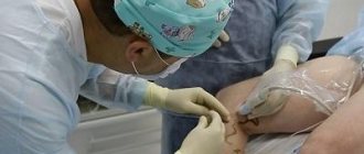
The simplest way to detect pathology of the cervical mucosa was invented by Papanicolaou in the 1940s and consisted of taking scrapings of surface cells . Today, modified instruments are used to collect more material. Examination of cells under a microscope - cytology allows us to determine the next diagnostic stage - colposcopy.
Extended colposcopy - examination of tissues under high magnification from five to 30 times, with additional enhancement of the “picture” with special solutions, which helps in choosing the optimal place for taking a piece of tissue - a biopsy of the area of dysplasia. Pieces of mucous membrane measuring at least 3 millimeters are sent for microscopy - histology. A biopsy is excluded in case of inflammation and infections, but only temporarily.
Further, with morphological confirmation of dysplasia the mucous membrane of the cervical canal is scraped to identify its changes; in a woman, dysplasia can be localized in the glandular crypts - the pits of the mucosa and the epithelial transition zone can move higher. Curettage visualizes the pathological substrate hidden from the eye.
Treatment of dysplasia
For congenital dysplasia, there is no radical treatment; for some life-threatening malformations, operations are performed, but all therapy is aimed only at reducing symptoms with increasing motor activity and is palliative in nature.
For joint dysplasia, effective but painful methods of multi-month fixation have been developed to create peace and provide time for development that did not happen in the prenatal period.
In case of gastric dysplasia, Helicobacter is eliminated, the diet is adjusted, the diet and lifestyle are modified towards normal, and medications are used to protect the mucous membrane from damage.
Treatment of cervical dysplasia depends on its severity: with mild dysplasia, conservative measures are resorted to. The human papillomavirus is resistant to drug effects, but its vital activity is supported by disorders of the mucous membrane during chronic inflammation, and contributes to a decrease in immunity due to hormonal disorders and systemic diseases.
It is easier for a healthy woman to get rid of HPV, but if she has diseases, it is necessary to help the body - to cure acute inflammation, transfer a chronic process into long-term remission, achieve hormonal balance, normalize sugar and other blood elements.
For long-term CIN I, which has been observed for almost 3 years without a tendency for the tissue to normalize its structure, surgery is also performed - the cervical sector is removed. The sector is similar to a geometric figure - a cone, hence the name of the operation - conization.
Moderate and severe dysplasia is treated only with surgery - conization of the cervix is performed using radio wave or laser methods. The operation is highly effective, leaves almost no scars and does not interfere with future pregnancy and childbearing.