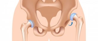
Anatomy
The synovium is located around joints and ligaments, providing them with protection. This occurs due to the release of a small amount of fluid (lubricating secretion), which reduces friction and provides shock absorption.
The exact cause of hip synovitis is unknown, but some theories include a history of trauma to the femur or a recent viral illness such as an upper respiratory tract infection, bronchitis, or otitis media.
One of the reasons for the development of inflammation of the hip joint is infection in the synovial fluid. This occurs against the background of any other infectious disease. In this case, fever, a significant increase in body temperature and general poor health are added to the classic symptoms. In this case, it is necessary to first deal with the source of infection.
Main reasons:
- sports injury;
- stretching;
- blood diseases;
- metabolic problems;
- endocrine pathologies;
- dystrophic joint pathology;
- overweight;
- allergy;
- bruises.
Among the infectious pathogens that most often cause synovitis are pneumococci, streptococci, staphylococci, Koch's bacillus (tuberculosis) and STIs (sexually transmitted infections).
Signs
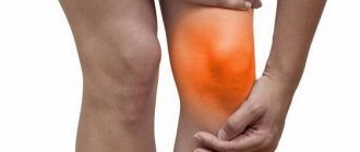
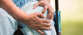
The disease can occur in acute or chronic form. In the acute form, pain, swelling, and redness of the skin occur in the joint area. Movements are limited due to pain. In the chronic form, the same symptoms are present, but they are less pronounced. They then subside, then a new exacerbation develops. Inflammation of the articular membrane when an infection is attached can turn into a purulent form. The joint swells even more, severe pain occurs, and pus accumulates under the skin. The general condition of the patient is disturbed, body temperature rises.
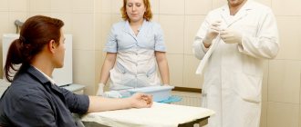
At CELT you can get a consultation with a traumatologist-orthopedic specialist.
- Initial consultation – 3,000
- Repeated consultation – 2,000
Make an appointment
Typology
In medicine, the following classification of synovitis is proposed:
- infectious – there is infection with a pathogen;
- traumatic – due to mechanical injury;
- reactive – develops due to prolonged mechanical trauma, for example, at work (car mechanic, builder, athlete, etc.);
- aseptic – for endocrine diseases (for example, diabetes), blood diseases (hemophilia) and metabolic disorders (gout);
- allergic - allergens affect the synovial membrane when it has increased sensitivity.
To make a correct diagnosis, it is imperative to tell the doctor about all possible signs and sensations during the inflammatory process.
Treatment of synovitis of the knee joint
The pathology is treated by a wide group of specialists at various levels. In acute and purulent types, the patient must be hospitalized.
The first stage is the removal of fluid accumulated inside the joint, and only then complex therapy is prescribed using medications and physiotherapeutic methods.
The main goal of therapeutic treatment is to eliminate the factors that provoke an increase in the volume of joint fluid.
Drug treatment
The use of medications falls into the category of conservative methods and involves puncture with evacuation of accumulated fluid and the use of a number of drugs that eliminate symptoms and help combat the causes.
The following groups of drugs are prescribed as drug therapy:
- glucorticosteroids;
- chondroprotectors;
- painkillers;
- antibiotics.
Particular attention should be paid to chondroprotectors, the use of which can accelerate the process of tissue regeneration. One of the most effective among them is considered to be the drug “Artracam”.
Physiotherapy
Physiotherapeutic techniques help restore the functionality of the affected joint. Already 3-4 days after the start of treatment, patients are referred to activities such as:
- phonophoresis using corticosteroid drugs;
- UHF therapy;
- therapy using magnetic and electric fields.
Surgical intervention
Radical methods are used in situations where the patient’s treatment is not amenable to conservative methods. Thus, surgical intervention is indicated for recurrent chronic synovitis, the progression of which is accompanied by changes in the tissue of the joint capsule.
Surgery involves partial or complete removal of the inflamed joint membrane.
The postoperative period involves immobilization and the prescription of drug therapy with the use of antibiotics, painkillers and, of course, chondroprotectors, which provide the opportunity to accelerate regenerative processes within the joint tissues.
Therapeutic physical education (PT) as a method of complex treatment of synovitis
The use of exercise therapy complexes is an integral part of complex treatment and allows you to speed up the recovery process, helping to improve metabolic processes within the affected tissues.
A set of exercises is selected individually, in strict accordance with the recommendations of the attending physician.
Preventive actions
There is no way to insure against all diseases, but there are measures to prevent their occurrence, in particular, such an unpleasant disease as synovitis.
Among the most effective methods of prevention are:
- wearing comfortable orthopedic shoes;
- reduction of strong physical activity;
- lesson with a physical therapy instructor;
- maintaining balance in the daily diet;
- giving up bad habits;
- preventive examinations with the attending physician.
Pay special attention to health. At the first signs of pathology, seek medical help and follow all the instructions of your doctor in order to live a full life.
Symptoms
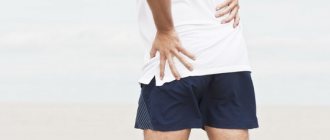
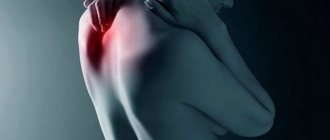
The general clinical picture of the pathology consists of severe pain, weakness/poor health and local hyperthermia. The patient's body temperature may also rise to low-grade levels (37-37.5 °C). Often inflammation is accompanied by swelling and redness of the skin over the joint. The pain syndrome intensifies when leaning on the leg, moving it to the side and during palpation.
With an infectious etiology, the disease begins acutely, with an increase in body temperature and general malaise. Then bursting pain appears in the joints (at the site of suspected synovitis). Within a few hours, severe swelling, hyperthermia and local hyperemia develop. The patient feels a headache, an increase in general body temperature, and sometimes there are other signs of intoxication.
With non-infectious etiology, the pathology does not develop so quickly, but this will depend on the cause. The primary symptom complex is represented by pain when moving. After a couple of days, swelling, hyperthermia, hyperemia, joint deformation and increasing pain are added.
Joint damage in childhood
Hemorrhagic vasculitis (Henoch-Schönlein disease)
Joint syndrome: arthralgia or arthritis, polymorphic, predominantly hemorrhagic rash on the lower extremities, large joints, buttocks. Joint syndrome is unstable.
Features: combined with abdominal and renal syndrome.
Chronic ulcerative colitis and Crohn's disease
Articular syndrome: peripheral asymmetric arthritis, with predominant damage to the joints of the lower extremities.
Features: spondylitis, sacroiliitis, associated with the activity of the underlying disease. High detection rate of HLA B27.
Tuberculosis
Articular syndrome: severe arthralgia, spinal damage, unilateral drive, coxitis. Diffuse osteoporosis, marginal bone defects develop, and rarely, a limited bone cavity with the presence of sequestration; destruction of the articular ends of bones, their displacement and subluxation. There is also reactive polyarthritis that develops against the background of visceral tuberculosis. Characteristic damage to small joints.
Features: combined with positive tuberculin tests.
Lyme disease (systemic tick-borne borreliosis)
Articular syndrome: mono-, oligo-, symmetrical polyarthritis. Erosion of cartilage and bones may develop.
Features: combined with tick-borne erythema, damage to the nervous system, heart. Detection of antibodies in blood serum to Borrelia burgdorferi of the IgM and IgG class by indirect immunofluorescence.
Viral arthritis
Arthritis may be associated with viral infections.
Joint syndrome is short-term, completely reversible.
Features: found in acute viral hepatitis, rubella, mumps, chickenpox, arbovirus infection, infectious mononucleosis, parvo- and adenovirus infection, etc. Determination of antibodies to viruses is rarely indicated, since rapid spontaneous recovery usually occurs.
Hypertrophic osteoarthropathy (Marie-Bamberger syndrome)
Articular syndrome: defiguration of fingers in the form of “drumsticks”, hypertrophic periostitis of long tubular bones, arthralgia or arthritis with effusion into the joint cavity. Symmetrical damage to the distal joints of the upper and lower extremities (wrist, tarsus, knee joints).
Features: found in tuberculosis, fibrosing alveolitis, lung cancer, sarcoidosis.
Hemophilia
Joint syndrome: accompanied by bleeding in the joints followed by an inflammatory reaction and effusion. The knee joints are affected, and less commonly, the elbow and ankle, wrist, shoulder and hip joints. Relatively rare - joints of the hands, feet and intervertebral joints.
Features: begins in early childhood.
Leukemia
Joint syndrome: ossalgia, flying arthralgia, asymmetrical arthritis with sharp constant pain in the joints, exudative component and painful contractures.
Features: must be excluded in case of systemic variants of juvenile rheumatoid arthritis (JRA).
Neoplastic processes. Neuroblastoma, sarcoma, osteoid osteoma, metastases in leukemia
Articular syndrome: may be accompanied by myalgia, ossalgia, arthralgia, monoarthritis. Characterized by severe pain in the periarticular areas, a severe general condition that does not correlate with the activity of arthritis.
Features: combined with typical hematological and radiological changes.
Osteoid osteoma
Osteoid osteoma is a benign bone tumor.
Usually the pain is moderate and constant. Typically, the pain occurs in the middle of the night and is severe enough to wake the child.
In these cases, it is necessary to conduct a thorough examination of the child to exclude malignant neoplasms of bone tissue, which have similar symptoms and cause chronic bone pain that can wake the child at night. With osteoid osteoma, taking paracetamol or ibuprofen is usually sufficient to relieve pain. In all cases where a malignant bone tumor is suspected in a child, as well as in cases where the pain is not relieved by paracetamol or ibuprofen, it is necessary to consult the child with an experienced orthopedist familiar with this pathology.
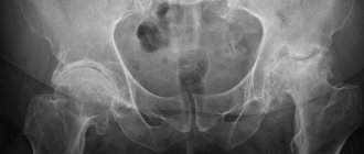
Osteoid osteoma is more common in boys than in girls. In most cases, clinical manifestations appear in adolescence, however, in some cases, the onset of pain and associated anxiety occurs at an earlier age. The usual location of osteoid osteoma is in the hip joint (see below), but sometimes the tumor is located in the knee area. When localized in the hip joint, the diagnosis of osteoid osteoma is not difficult and is easily detected on a regular x-ray. In most cases, once osteoid osteoma is definitively diagnosed, no treatment is required. However, in some cases the pain syndrome can be quite pronounced. In the most severe cases, surgical removal of the area of bone damaged by osteoid osteoma is possible.
Hypothyroidism
Articular syndrome: arthralgia with slight swelling of soft tissues and non-inflammatory effusion into the joint cavity. The knee, ankle and hand joints are affected, and carpal tunnel symptoms may develop.
Features: impaired skeletal formation, slower growth of long tubular bones and ossification, osteoporosis. Muscle weakness and myalgia are evident.
Congenital dislocation of the femur
Congenital dislocation of the femur is detected in newborns during a thorough examination of the hip joints in the maternity hospital and during the first subsequent examination. Immediately after diagnosing the dislocation, an abduction splint should be applied. Congenital dislocation of the femur occurs in approximately 0.7% of newborns. Family cases are common. The hip joints should be examined at the first regular medical examination after birth. If there is a dislocation, the femur returns to its normal position with an audible click. A provocative test is performed by dislocating the femoral head while pressing on the medial sides of the thighs. Different lengths of the lower limbs may indicate a dislocation. It is best detected by bending the legs at the hip and knee joints at an angle of 90°. The presence of asymmetrical inguinal folds suggests congenital dislocation of the femur, however, such a sign is detected 10 times more often than the dislocation itself. The calcaneal-gluteal test for dislocation of the femur is often positive: with the child lying on his stomach, the legs are bent at the knee joint and the hips are extended so that the heels touch the buttocks. The heel on the side of the dislocation crosses the midline and touches the buttock of the opposite side. Congenital dislocation can be bilateral. In such cases, asymmetry tests do not detect dislocation.
Structural abnormalities of the limbs
Clubfoot (talipes equinovarus) can always be diagnosed immediately after birth. With congenital calcaneovalgus clubfoot (talipes calcaneovalgus congenitus), the newborn’s foot is turned into a valgus position and is in dorsiflexion. For this condition, treatment is rarely prescribed; parents are advised to develop the foot. The adductor clubfoot of the metatarsus (talipes metatarsus adductus) is characterized by internal rotation of the forefoot, visible as an inverted curvature of its medial part. Sometimes a plaster cast, adhesive tape, or surgery is indicated.
Flat feet are considered benign if the forefoot can be moved up and down freely and the valgus angle disappears when the child stands on tiptoes.
Walking with internal or external rotation of the foot is a normal condition that does not require treatment.
O-shaped curvature of the legs (genu varum). Consultation with a specialist is indicated for unilateral damage or worsening of the deformity after the second year of life.
X-shaped curvature of the legs (genu valgum). Consultation with a specialist is indicated for unilateral lesions (Blunt's disease), as well as if there is a minimum distance of 10 cm between the ankles at the start of school.
Toes that overlap each other rarely require surgery.
In case of syndactyly of the fingers, the condition is assessed by a pediatric surgeon immediately after birth, if the first and second fingers are involved in the process. In other cases, surgical correction must be performed at the age of 4–5 years. In all cases of syndactyly, consultation with a pediatric surgeon is necessary. For toe syndactyly, treatment is usually not indicated.
Aseptic synovitis of the hip joint
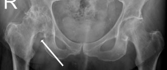
Aseptic synovitis of the hip joint is diagnosed based on typical symptoms, and unnecessary tests should be avoided. The most common cause of acute lameness in children under 10 years of age. In addition to lameness, the child complains of pain in the hip or knee joints and fixes the hip in a position of flexion and external rotation. A typical clinical sign is limited range of motion and pain during internal rotation of the hip. A slight increase in ESR is possible (not higher than 35 mm/h). Effusion into the cavity of the hip joint is easily detected by ultrasound. If possible, ultrasound should always be performed to confirm the diagnosis. The disease is rarely bilateral. Simultaneous damage to other joints excludes aseptic synovitis of the hip joint.
The child is prescribed to rest with his legs bent at the hip joints. Aspiration of the contents of the joint cavity in a hospital setting is necessary only if septic arthritis is suspected or with severe pain. The prognosis is favorable.
Epiphysiolysis of the femoral head
Early diagnosis and surgical treatment of the disease are necessary. If left untreated, medial and posterior displacement of the femoral head progresses and treatment outcome worsens. With moderate and severe diseases, the likelihood of early onset of secondary osteoarthritis increases. The epiphysis of the femoral head slips to the side relative to the femoral neck. The condition is considered stable if the epiphysis is fixed in a new position. An unstable condition means that the pineal gland is not fixed and pain often occurs. The disease occurs between the ages of 10 and 16 years (a little earlier in girls), often in combination with obesity. The disease occurs 2.5 times more often in boys than in girls. Bilateral lesions are noted in 20–30% of cases.
Symptoms: lameness and pain in the knee, hip and groin area occur when supporting the affected limb. Typically, the patient holds the lower limb in a slightly externally rotated position.
Diagnosis: X-ray reveals posterior displacement of the epiphysis. If a disease is suspected (symptoms of hip damage appear at a typical age), it is necessary to evaluate the condition of both limbs in the anteroposterior projection and Lauenstein projection. Indentation caused by recent displacement can be seen on ultrasound. If an effusion is detected on ultrasound, then epiphysiolysis is unstable.
Treatment: Treatment is always surgical. The epiphysis is usually stabilized using a single screw. The smaller the volume of surgical intervention, the better the outcome. It is necessary to consider the possibility of prophylactic fixation of the opposite unaffected hip if the patient is young or if there is an underlying endocrine disease or metabolic disorder (in 7% of cases).
Perthes disease (Legg–Calvé–Perthes disease, pseudocoxalgia)
This is aseptic necrosis of the femoral head, leading after 4–8 months to a clinically manifested subchondral fracture due to physical activity. Fracture and delayed ossification of the cartilage of the femoral head lead to softening of the epiphysis. If left untreated, the femoral head may flatten. It is necessary to diagnose the disease early and treat the tendency to subluxation. The prognosis is favorable if the round shape of the femoral head is preserved. The disease occurs mainly in boys aged 2 to 12 years. The incidence ratio among boys and girls is 5:1. In 10% of cases, the disease is bilateral, but both femoral heads are usually not affected at the same time. The main manifestation is lameness. The disease resembles aseptic synovitis of the hip joint, but begins gradually and becomes protracted or recurrent. The pain is localized in the area between the groin fold and the knee joint. ESR, C-reactive protein level and the number of leukocytes in the blood were not changed. Bone age lags behind passport age by 2 years. Diagnosis is based on radiography: the first sign is a subchondral disorder of the bone structure, subsequently accompanied by the formation of cysts and flattening of the epiphysis. The prognosis for young children (up to 6 years) is favorable.
Osguth–Schlatter disease
Osgut-Schlatter disease is repeated damage to the apophysis of the tibial tuberosity during physical activity. The onset of manifestations coincides with the period of rapid growth in adolescence. Spontaneous healing should be promoted by avoiding excessive stress. Pain occurs in the upper third of the lower leg or knee when running or jumping. While running, the pain is more severe when the heel touches the supporting surface; when the heel is lifted from the surface, the pain is less pronounced (biomechanics of the knee extensor apparatus!). The tibial tuberosity is prominent and sensitive to palpation. Radiography reveals fragmentation of the tuberosity. In typical cases, there is no need for x-rays.
Spondylolisthesis
The most common cause of long-term or recurrent back pain in adolescents. Lower back pain that occurs after exercise and often radiates to the hips may be a sign of spondylolisthesis. Clinical signs:
- excessively pronounced lumbar lordosis;
- the displacement of the vertebra is palpated (palpable “threshold”);
- prominent spinous process.
The diagnosis is based on identifying the displacement of the vertebra on a lateral x-ray (an anteroposterior x-ray is not necessary). The course of spondylolisthesis is monitored by performing radiographs 2-3 times a year (consultation with a pediatric surgeon is also necessary) in children with characteristic manifestations. Excessive stretching (lifting weights, gymnastic exercises) should be avoided.
Scheuermann's disease
Scheuermann's disease is an osteochondritis affecting the anterior part of the vertebral cartilage, occurring in late puberty (ages 13–15 in girls and 15–17 years in boys). The disease occurs 4 times more often in girls than in boys. The most common manifestations are pain in the thoracic spine and back stiffness, and stiffness of the hamstrings.
Diagnosis is based on radiographic findings.
- Wedge-shaped deformity of the vertebral bodies with a flattened anterior part.
- Deformed marginal vertebral plate; Schmorl's hernia (a depression towards the vertebral body) develops in the later stages.
Scoliosis
Scoliosis is idiopathic, progressive in 85% of cases and functional in 15%. Scoliosis is a disease, not bad posture; exercise therapy is not effective for this disease. The disease develops in a phase of rapid growth (at the age of 10–12 years in girls, in boys several years later). For screening, examination of the spine is used in the position of bending the torso forward with straightened legs. Scoliosis is confirmed if one shoulder blade is higher than the other. In mild or controversial cases, children should be monitored at 6-month intervals. The severity of scoliosis is assessed using radiographs, determining the maximum angle of deformity. X-ray examination is indicated if obvious scoliosis is clinically detected.
Calve's disease (vertebra plana)
A rare disease in children aged 2–10 years, manifested by complete flattening of the vertebral body. The clinical picture includes local pain, increased ESR, and sometimes leukocytosis. When carrying out differential diagnosis, it is necessary to exclude tuberculosis. A patient with suspected Calve's disease should be sent to the hospital for further research.
Discosis
Usually aseptic, but can also be caused by bacteria. Difficulty walking and sitting are typical clinical manifestations in preschool children. The diagnosis is based on local tenderness and pain when moving the spine. The diagnosis can be confirmed by radioisotope bone scanning using technetium. The child must be hospitalized for further examination. In many cases, the condition is benign and resolves spontaneously.
Growing pains (clinically insignificant leg pain)
- Growing pains never appear during the daytime.
- Regardless of the severity of the pain attack at night, if it is growing pains, then in the morning the child always feels good/great.
- Any child who experiences pain in the morning or during the day requires a full medical examination.
Clinically insignificant leg pain (so-called “growing pains”) is diagnosed based on clinical manifestations after ruling out arthritis. In children under 4 years of age, another disease should be considered. It is necessary to evaluate the psychosocial background, especially in the presence of other manifestations (headache, abdominal pain). Recurrent pain in the limbs without organic causes occurs in children during growth. 50% of children experience leg pain at some point during childhood. Leg pain usually occurs around 4 years of age. The condition is most common between the ages of 6 and 11 years. Often the condition is hereditary. Most often the muscles of the legs and thighs and the area of the knee joints hurt. The upper extremities are rarely affected. The child may also have a headache or stomach pain.
The child unexpectedly wakes up at night during deep sleep and complains of pain in his leg. Parents immediately understand that the child has a health problem because he wakes up and cries in bed. Most often, such episodes occur a few hours after the child has fallen asleep, but sometimes the child may wake up late at night. Typical symptoms include pain in the knee, either in the front or back, or pain in the muscles directly above the knee. Usually the pain disappears after ten or fifteen minutes with a light massage and does not remind you of itself in the morning. Most cases of growing pains affect large joints, such as the knee, rather than the fingers or toes. Sometimes a child may wake up in pain for several nights in a row, but more often these episodes occur sporadically over a period of several weeks or several months. Growing pains often appear after days when the child has been especially physically active. Growing pains may disappear for several months or years and then reappear during periods of particularly rapid growth.
The main point when assessing growing pains is the fact that if these are growing pains, then in the morning after waking up the child feels absolutely healthy.
The best explanation for the mechanism of growing pains is the tension of the muscles and tendons as the leg bones rapidly increase in length.
During the day, the child is active, and the information that his brain receives from tense muscles and tendons is lost in the flow of other information from many other events. At night, when the child falls asleep and there are no distractions, the pain impulses that come from the tense muscles and tendons reach the higher nerve centers and the child wakes up in pain. As a result, it can be summarized that growing pains are observed during the period of the child’s most intensive growth and after days of high physical activity.
Growing pains can be a worrying experience for both parents and baby, especially when painful episodes occur over several nights in a row. In most cases, a light massage and conversation is enough to get the child to sleep. Children with more severe pain usually stop complaining after taking the usual dose of paracetamol or ibuprofen, exactly the same as for a headache. If the child wakes up for several nights in a row, then pain medication can be given before the child goes to bed. This will reduce pain sensitivity so your child can sleep peacefully through the night. After two or three nights without pain, you should stop giving the child the medicine.
It is necessary to assume another cause of pain if the child has the following manifestations:
- prolonged pain in one limb;
- combined symptoms (for example, lameness or change in general condition);
- Symptoms occur in the morning or during the day.
If atypical symptoms appear, it is necessary to conduct at least a general blood test and determine the ESR.
Damage to joints and ligaments in children
Incomplete dislocation of the radial head
Typical injury in children aged 1 to 5 years.
Injury may occur if the child is pulled or lifted by the arm. A recent injury can be easily reduced by first rotating the arm into a supinated position and then pressing on the head of the radius, pushing it through the annular ligament. At this moment, you can hear a click and the severity of symptoms immediately decreases. In old lesions, reduction is often unsuccessful. In such cases, treatment includes the application of a sling bandage. Symptoms disappear spontaneously.
Foot subluxation
A torn anterior tibiotalar ligament is the most common tendon injury. If the traction test is positive (when you pull the foot forward, there is a feeling of separation of the foot from the lower leg), the application of a support bandage, sometimes a plaster, as well as surgical treatment (rarely) may be indicated. Specialist consultation is required.
Patella dislocation
The most common dislocation in teenagers is patellar dislocation.
If the patella is displaced to the lateral side, it is necessary to carry out reposition as soon as possible.
Literature
- Brewer EJ Jr., Bass J., Baum J. Current proposed revision of JRA criteria // Arthritis Rheum. 1977, 20 (Suppl): 195.
- Cassidy J., Petty R. eds. Textbook of pediatric rheumatology, 5 nd ed. // New York: Churchill Livingstone. 2005.
- Ilona S. Szer, Yukiko Kimura, Peter N. Malleson, Taunton R. Southwood. Arthritis in children and Adolescents. Juvenile Idiopathic Arthritis // Oxford University Press. 2006. 456 p.
- Alekseeva E. I., Litvitsky P. F. Juvenile rheumatoid arthritis. Etiology, pathogenesis, clinical picture, diagnostic and treatment algorithms. M.: Publishing house "VEDI". 2007. 360 p.
- Neeck G., Michels H. Endocrine aspects of pediatric rheumatic diseases // Bailliere's Clinical Rheumatology. 1996. No. 2. P. 349–363.
- Alekseeva E. I., Zholobova E. S. Reactive arthritis in children // Issues of modern pediatrics. 2003, vol. 2, no. 1, p. 51–56.
- Nasonov E. L., Nasonova V. A. Rheumatology. National leadership. Geotar-Media, 2008. 720 p.
- Alekseeva E. I., Bzarova T. M. Rheumatology. Clinical recommendations. Ed. E. N. Nasonova. M.: Publishing group "GEOTAR-Media". 2005, 2006, p. 120–140.
- Alekseeva E. I., Zholobova E. S., Chistyakova E. G., Valieva S. I. Reactive arthritis. Educational and methodological manual. M.: Publishing house MMA im. I. M. Sechenova, 2004. 134 p.
E. I. Alekseeva , Doctor of Medical Sciences, Professor T. M. Bzarova
Scientific Center for Children's Health, Russian Academy of Medical Sciences, Moscow
Contact information for authors for correspondence
Diagnosis and treatment
Before making a diagnosis, a traumatologist must conduct a physical examination, interview, prescribe tests and diagnostics. Differential diagnosis may require X-ray examination and/or ultrasound. If an infectious etiology is suspected, it is necessary to undergo a biochemical and general blood test, urine test, and an antibiogram to determine sensitivity to antibiotics.
Treatment for hip synovitis includes simple home remedies such as rest, heat, and massage of the painful area. Avoid putting weight on the affected side until the pain goes away.
If the inflammatory process is mild, it is possible to carry out conservative treatment in the form of analgesics, NSAIDs, vitamin complexes and the application of warming ointments. For more severe development, injections with glucocorticosteroids are prescribed. A mandatory condition for treatment is bed rest. For rehabilitation, exercise therapy, massage and electrophoresis are prescribed (after the end of the acute period). In case of irreversible degenerative disorders, they resort to synovectomy - removal of part of the affected tissue.
When there is a large accumulation of fluid, arthrocentesis is used for diagnosis and treatment - puncture of the hip joint. During this procedure, an anesthetic is administered. With its help, the quantity, color, consistency and presence of impurities in the form of pus and blood are assessed. The liquid is sent for analysis to identify the infectious agent. The joint is washed and an anti-inflammatory drug is injected. After determining the cause of infection, the question of injecting an antibiotic or hormone into the joint is decided.
The success of treatment directly depends on how quickly the patient consulted a doctor, the severity of the lesion and the duration of therapy. With the correct response, complete recovery and restoration of joint function occurs. On average, this takes up to 10 days, taking into account at least 2 weeks of rehabilitation period.
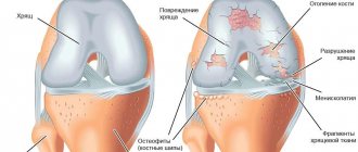
What is synovitis of the knee joint?
Synovitis of the knee joint is a common pathological inflammation, the development of which results in the accumulation of fluid that can become purulent. Synovitis belongs to the category of serious pathologies that require timely diagnosis and an integrated approach to treatment.
The pathological process occurs mainly as a result of trauma or as a concomitant inflammation with arthrosis.
It is important to note that synovitis is not an independent disease, but only becomes the result of other general or local pathologies.
Classification and etiology
For ease of diagnosis, synovitis of the hip joint, depending on the etiology and nature of the course, is classified as follows:
- infectious. Inflammation occurs due to the penetration of pathogenic microorganisms into the joint cavity by lymphogenous or hematogenous routes. It can be specific, caused by gonococci, Treponema pallidum, and Mycobacterium tuberculosis. Nonspecific synovitis manifests itself after infection of the synovial membrane with staphylococci, streptococci, and viruses;
- reactive. Pathology develops as a result of a decrease in the functional activity of the immune system. It recognizes the body's own cells as foreign proteins and begins to produce antibodies to destroy them. Reactive synovitis is often diagnosed together with rheumatoid or psoriatic arthritis;
- traumatic. After injury to the hip joint, the integrity of the synovial membrane is disrupted, which leads to excessive production of intra-articular fluid. The development of inflammation is caused by bruises, dislocations, subluxations, tendon ruptures, fractures, deep cuts, wounds;
- spicy. It occurs immediately after injury or infection and is characterized by a rapid course and severity of symptoms. With timely medical care, it responds well to treatment and does not cause any complications;
- chronic. A sluggish inflammatory process with alternating acute relapses and stages of remission. Develops as a result of untreated acute synovitis. The danger of the disease lies in the gradual irreversible destruction of the hip joint.
Rehabilitation and exercise therapy
To speed up the restoration of limb mobility to the previous extent after conservative or surgical treatment of synovitis, physical therapy exercises are recommended. The time frame for starting them is determined by the attending physician.
For classes, a special set of exercises that do not cause pain is selected. The goal is to eliminate high stress on muscles and joints. Exercises are carried out from static positions lying or sitting, affecting relaxation or stretching of muscles. Regular exercise increases physical endurance and makes tendons elastic.
The positive effect of gymnastics is to reduce pain, stabilize the production of synovial fluid, expand the range of organ mobility, and create a positive attitude toward recovery.
Positive progress in well-being can be consolidated by walking, as well as exercising on an exercise bike or elliptical machine.
Taking medications
Only medications prescribed by the attending physician are indicated for use:
- NSAIDs. Relieves pain, reduces the severity of the inflammatory process;
- Antiviral drugs. In the event that the tests performed reveal the viral nature of the disease;
- Antibiotics. Cures lesions caused by bacterial infection;
- Antihistamines. Relieves allergic manifestations.
- Immunostimulants, vitamin-mineral complexes, dietary supplements. Restores the immune system and strengthens the body.
Depending on the stage of the disease, we choose one or more treatment methods in Moscow:
Therapeutic massage, osteopathy, manual therapy in Moscow
Helps bones and joints take the correct physiological position, relieves pain and spasms, relaxes muscles.
Acupuncture in Moscow
Work on biologically active points. It affects the affected area and the body as a whole. Eliminates the cause of the disease and removes the symptoms.
In addition, according to indications, the following are used: taping, pharmacopuncture, FormTotix insoles, exercise therapy with an instructor and other methods. The choice of procedures depends on the current condition; taken together, they act faster and give a more lasting result.
private chiropractor
Physiotherapeutic procedures
NSAIDs and glucocorticosteroids quickly eliminate pain and inflammation, but also have many side effects. Rheumatologists reduce their dosages, and to maintain clinical effectiveness, prescribe physiotherapeutic measures to patients. After 5-10 sessions, blood circulation is restored in the affected tissues, and exudate stops accumulating in the joint capsule. By stopping the inflammatory process, the intensity of pain and swelling decreases, and joint mobility increases. In the treatment of left-sided and right-sided synovitis of the hip joint, the following physiotherapy procedures are used:
- electrophoresis with analgesics, chondroprotectors, NSAIDs. A sterile cotton-gauze swab soaked in drug solutions is applied to the joint area. A metal plate is placed on top of it, through which weak discharges of electric current are passed. Under their influence, drug molecules penetrate the inner lining of the synovial bursa;
- sinusoidally modulated currents. They irritate basic receptors, providing a distracting effect, reducing the severity of pain. Blood begins to flow to the damaged tissues, saturating them with molecular oxygen and nutrients. The use of sinusoidally modulated currents promotes the removal of products of the inflammatory process from the joint capsule;
- UHF therapy. The area of pain and inflammation is affected by ultra-high frequency electromagnetic fields. Heat penetrates the hip joint, accelerating regenerative processes. Swelling decreases, range of motion in the joint increases. UHF therapy, unlike other physiotherapy procedures, can be carried out even during an acute inflammatory process.
The use of magnetic therapy in the treatment of large joints, especially the hip, has been proven to be highly effective. The joint is exposed to a magnetic field - high or low frequency, alternating or constant. As a result, lymphatic vessels contract, promoting tissue detoxification. The removal of excess fluid accelerates, the condition of blood vessels, nerve and muscle fibers improves.
Surgery
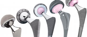
If conservative methods of treating synovitis are ineffective, the patient is prescribed a surgical operation - synovectomy. It can be arthroscopic or performed in the traditional open way. The disadvantages of the latter technique include a high likelihood of developing complications: the formation of postoperative hematomas, scars and cicatrices, painful sensations that do not subside for a long time and limited range of movements. As a result, the duration of hospitalization and rehabilitation period increases. Therefore, open synovectomy is usually performed only for complicated synovitis of the hip joint.
Prognosis for the course of the disease
For a favorable prognosis, factors such as the condition of the patient’s body, as well as timely effective treatment, are important. Complete recovery with preservation of the same motor activity is possible with the mildest forms of the lesion, as well as with the serous or allergic variant of the disease.
The severe course of the purulent form poses a threat to life, since a septic lesion develops. With the most favorable developments in this case, there is a high probability of stiffness or complete immobility of the affected organ, as well as dangerous complications on other organs and systems of the body.
Folk remedies that help with synovitis
To treat diagnosed synovitis of the knee joint of a non-dangerous form, you can use some traditional medicine:
- lotions based on herbal decoctions;
- rubbing with medicinal tinctures;
- taking herbal teas or herbal decoctions;
- ointments made from animal fats.
Any means are used only as an addition to the main treatment prescribed by a medical specialist. A combination of drug therapy, physical therapy and home treatment can provide an accelerated positive effect.