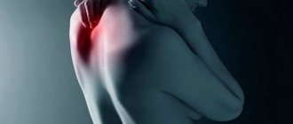
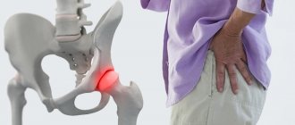
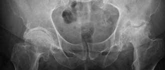
The hip joint is the largest and most powerful joint in the human body, responsible for functions such as moving the hips, lifting and lowering, bending and extending the legs, and also takes part in bending the body. Dislocation of the femur in the hip joint is an injury in which the head of the femur is displaced beyond the acetabulum. These dislocations account for no more than 5% of all dislocations. Dislocations can be divided into 2 types: anterior and posterior dislocations. Anterior dislocations are extremely rare and represent a downward displacement of the femoral head with rupture of the joint capsule. Posterior dislocation occurs more often, as a result of which the head of the hip joint pops back.
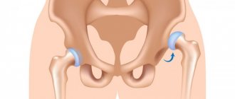
In cases where the hip joint loses its functions (the smooth cartilage that allows the femoral head to glide easily in the acetabulum is worn away) and any movements cause unbearable pain, patients undergo endoprosthetic surgery. An endoprosthesis is an artificial substitute for a human organ (performing its function), which is located inside the human body. Hip replacement is an operation to replace the affected components of the joint with an endoprosthesis that has the anatomical shape of a healthy joint and allows full range of motion. Dislocations during such an operation occur very rarely, but they do occur, and these are mainly dislocations of the head of the endoprosthesis.
Signs of dislocation.
The symptoms of dislocation of a healthy joint and an endoprosthesis are the same.
- Acute pain in the thigh area
- Standing on an injured leg is unbearable
- Inability to move the hip joint.
- Obvious deformation of the hip joint (depending on the type of dislocation)
- Forced position of the leg (depending on the type of dislocation)
- Increased pain when trying to make any movements with the injured leg
- Swelling and bleeding in the buttock, groin and thigh area
Subluxation of the hip joint in adults: treatment in Moscow
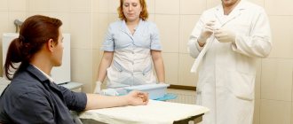
Treatment of subluxation and dislocation in Moscow can be done in orthopedic and traumatology departments of clinics and hospitals, as well as in the Yusupov Hospital and other medical institutions. At the Yusupov Hospital, patients undergo rehabilitation after endoprosthetics. The hospital is equipped with high-tech equipment that is used in the treatment of the musculoskeletal system. The hospital includes a 24-hour hospital and a rehabilitation department.
Orthopedic rehabilitation helps correct deformities that preceded surgery, helps restore the functionality of the affected hip joint after surgery, and prevents the development of complications and relapse of the disease. In the hospital you can undergo diagnostic examination using MRI, CT. You can make an appointment with a doctor by calling the Yusupov Hospital.
Treatment.
Treatment should be carried out in the traumatology department by qualified specialists.
- A dislocated femur can be reduced without any complications under general anesthesia in a closed manner (using various reduction techniques).
- Surgical treatment is indicated in cases where fractures of the neck or femur are added to the dislocation. (If, when the hip is dislocated, the cartilage is damaged (which can lead to coxarthrosis in the long term), there is a need for hip replacement).
- Applying a plaster splint or skeletal traction for 3-4 weeks.
- After this, the patient is recommended to walk on crutches for 10 weeks and is prescribed rehabilitation procedures.
Mechanism of occurrence
This injury mainly occurs to drivers and passengers of cars and motorcycles who do not use seat belts. A typical mechanism of injury is a strong impact of the knee of a bent leg on the dashboard of a car when colliding with an obstacle. In this regard, concomitant injuries occur due to the impact of the knee on the dashboard: bruise of the distal femur, fracture of the patella, with an extended knee - fractures in the ankle joint
The second, less common cause, falling from a height, is observed in elderly patients. Of more than a hundred patients observed by the author, several people received dislocations when falling from a height, one - when being run over by a car. Also, one received this injury in a fall from a horse at the hippodrome, whereas in the historical literature this was previously the main mechanism of injury. All the rest were drivers and passengers sitting in the front seats of cars.
The type of dislocation depends on the degree of hip flexion, the presence of adduction or abduction, external or internal rotation at the time of injury.
Thus, posterior dislocation usually occurs with at least 45° of flexion, adduction, and internal rotation of the hip. With each type of dislocation, the femoral head and the entire lower limb occupy a certain position. With all types of dislocations, hip flexion is detected, however, for each type its degree is different. In addition, posterior dislocations are characterized by adduction and internal rotation. With iliac dislocations, adduction and internal rotation are less pronounced than with sciatic dislocations. With posterior dislocations, a greater roundness of the gluteal region is determined.
For anterior dislocation to occur, flexion is also necessary, but in combination with abduction and external rotation.
Isolated dislocations occur only in 10-20% of all dislocations. But even among them, as a result of a study of isolated hip dislocations using computed tomography, it was found that in fact in 13% of cases they are accompanied by a fracture of the anterior part of the femoral head, combined with a fracture of the posterior edge of the acetabulum.
To diagnose hip dislocations and acetabular fractures, it is optimal to use computed tomography and three-dimensional computer reconstruction of the damaged joint. A CT scan provides a lot of additional information regarding, for example, impacted fractures of the acetabular wall, the presence of bone fragments in the joint, the degree of their fragmentation, unrecognized dislocation, and pathology of the sacroiliac region. The main advantages of CT include the ability to recreate the annular shape of the pelvis, allowing visualization of the complete picture.
Marginal fractures account for 40% of all acetabular fractures. Despite the fact that many authors classify fractures of the posterior wall of the acetabulum as simple fractures, unrecognized or discovered late, they cause rapid progression of anatomical, vascular, and trophic disorders in all structures and tissues of the hip joint, which often leads to severe disability.
How to avoid dislocations: a list of prevention rules
An artificial analogue of hip joints can serve efficiently for 15-30 years, but only under the condition of conscientious compliance with lifelong requirements. It is necessary to clearly understand that dislocations often occur due to the fault of the patient himself, who violated the principles of proper lifestyle and physical activity. So, to prevent postoperative consequences, including its relapse, it is necessary:
- regularly take control photographs of the operated area (for the first time they are taken after 3 months, then 6 and 12 months after the operation, then once annually);
- do exercise therapy daily – 1-2 times a day;
- avoid jumping, crossing legs, squatting, any sudden body maneuvers, overloading the section, twisting in the pelvic girdle area;
- sit on chairs of normal height with level back support, keep your spine straight while sitting;
- take all precautions to prevent traumatic situations;
- wear comfortable shoes with orthopedic soles, avoid high-heeled shoes and high-platform shoes;
- eat a balanced diet, monitor your weight (if your body weight is higher than normal, consult a nutritionist for help in losing weight);
- do not lift heavy objects and do not allow work associated with heavy physical labor;
- If you experience supporting and motor discomfort, pain, or swelling in the area of the endoprosthesis, immediately consult a doctor;
- if the problem persists, fully recover after the reduction of the head of the joint is performed in a medical facility.
Attention! To prevent dislocations, doctors recommend a special set of exercises aimed at effectively working out and increasing the endurance of the hip and gluteal muscles. It is extremely important to strengthen these muscle groups, because they are the main regulators of movement and stabilizers of the implant joint.
Features of the postoperative period
Prosthetics, technically performed at “5+”, are not yet a 100% guarantee of the successful functioning of the implant in the future. After correct installation of the implant, excellent rehabilitation must be carried out; this is the only way to minimize the risks and consequences. From the first day they begin systematic physical rehabilitation aimed at:
- increasing muscle tone with the help of therapeutic exercises;
- early transfer of the patient from a lying position to a standing position;
- practicing proper walking on crutches, and later without support;
- training in the technique of sitting down, taking a sitting position;
- correction of adaptive stereotypes developed in the preoperative period - vicious posture, incorrect manner of walking, sitting, etc.;
- accelerating the regeneration of the surgical wound and stimulating the integration of the prosthesis with the bone through physiotherapy procedures;
- providing the patient with full information about the mandatory need to limit certain elements of physical activity in order to prevent complications.
The transition from crutches to a cane, increasing the load on the operated leg and other important points are carried out according to the dynamics of recovery, well-being, age and weight criteria of the patient. Crutches are used for approximately 2.5-3 months, then they walk with the support of a cane. In general, walking without support is usually allowed after 4-6 months after surgery. No self-appointments! A person must strictly adhere to the step-by-step recovery plan recommended by the physical instructor and surgeon.
Content
- 1 Signs and symptoms 1.1 Posterior dislocation
- 1.2 Anterior dislocation
- 4.1 Classification 4.1.1 Posterior dislocation
- 5.1 Easy
- 6.1 Exercises
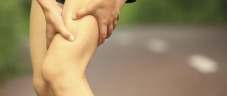
Sprain of the hip joint
Excessive physical impact on the connective tissues of the hip joint is the main cause of sprains. A large load on the ligaments of the hip joint occurs during sudden jumps, flexion or extension. People who lead a sedentary lifestyle are most often susceptible to such injuries, because the ligaments lose their elasticity.
At risk are children, elderly patients, as well as people suffering from obesity, diabetes and hypertension. Deformed posture, as well as bad habits, can play a role.
The causes of hip sprains can also be considered:
- sports or domestic injuries;
- long walking on uneven surfaces;
- conducting training without preparation;
- improperly performed physical exercises;
- deformation of the thigh and lower leg;
- incorrect placement of the foot when moving.
It is much easier to prevent ligament damage than to treat its consequences later, so you need to beware of situations that can lead to injury. But, if you fail to protect yourself, then you need to act very quickly to prevent complications.
Main causes, symptoms and diagnosis of hip joint diseases
Pathological conditions in the hip most often develop against the background of:
- mechanical damage to joint elements;
- a tendency to diseases of the articular tissue of the hip area at the genetic level;
- functional impairment due to infection;
- systemic diseases in connective tissue;
- damage to the integrity of the joint due to degenerative-dystrophic transformations.
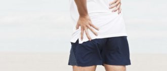
Any disease of the hip joint is characterized by the appearance of common symptoms:
- pain on the inner or outer thigh surface and in the groin area is of an acute, aching or pulling nature;
- motor activity is impaired, movements become limited;
- the skin becomes inflamed, the local temperature rises to subfebrile levels.
To correctly determine hip disease, a high-quality biochemical study is required. In addition, to accurately establish the diagnosis, instrumental diagnostic methods are shown:
- CT and MRI;
- Radiography;
- Ultrasound;
- Scintigraphy.
Let's consider the most common clinical diseases of the hip joint.
Hip dysplasia
This clinical disease is a congenital disorder or inferiority of the joint tissue, which increases the risk of subluxation or dislocation of the femoral head. The maximum number of cases of inadequate development of hip joints is observed in those areas where socio-economic policy is stable.
In the Scandinavian countries, the congenital anomaly is 4%, in Germany – 2%, and in the USA – 1-1.5%.
In the Russian Federation and post-Soviet countries, congenital dysplasia is diagnosed in 50-250 newborns out of 1000 (approximately 5%). According to medical experts, this is due to the unfavorable environmental situation in the states that were formerly part of the Soviet Union.
For the most part, the congenital disease is detected in female infants (in 80% of cases), as well as in families where one or both parents suffer from “congenital dysplasia.”
This congenital orthopedic anomaly can develop due to:
- breech presentation of the unborn baby;
- medical correction of pregnancy, which is complicated by toxicosis;
- large fruit.
A baby can develop dysplasia if he is swaddled too tightly to straighten his legs.
Since 1975, the Japanese have refused to tightly swaddle newborns. This has reduced the number of hip dislocations in infants.
Dysplasia in infants is manifested by the following clinical signs:
- asymmetrical skin folds on the legs;
- pronounced shortening of the thigh;
- restrictions in abduction and adduction of the hip joint.
A characteristic sign due to which one can suspect the presence of congenital dysplasia in a newborn is the Marx-Ortolani symptom. If you abduct the hip joint towards the surface where your baby is lying, you may hear a typical clicking sound in the joint area. This indicates that the femoral head is slipping into the acetabulum area.
The only effective therapy is to use a corrective orthopedic orthosis, thanks to which the baby’s legs can remain in a bent and abducted position at the required angle for a long time. Over time, the newborn is recommended to undergo massage, perform therapeutic exercises with constant supervision by an orthopedist. If treatment is started on time, the child can most often be cured.
Arthritis of the hip joint
Arthritis (coxitis) is a disease characterized by the development of an inflammatory process in the tissues of the hip joint. The disease occurs in acute (primary manifestations), protracted (2-12 months) or chronic form, which lasts more than a year.
Arthritic damage to the tissue system in the hip joint is divided into groups. It happens:
- infectious (septic coxitis) – inflammation that forms against the background of damage to the synovial fluid by an agent of a fungal, parasitic, infectious-allergic or viral nature;
- reactive arthritis - an immune-inflammatory pathology that manifests itself in the process or as a complication of infection of the joint tissue;
- rheumatoid arthritis is an autoimmune disease caused by the formation and spread of granulation tissue in the cavity of the synovial capsule. Because of this, articular cartilage and subchondral bone are destroyed;
- psoriatic arthritis is a rare autoimmune disease of the hip joint, manifested as a result of damage to joint tissue by psoriasis.
Symptoms, signs and treatment of hip coxitis
The causes, symptoms and therapeutic measures for arthritis of the hip joint depend on the nature of the disease.
The development of septic (infectious) arthritis is observed as a complication against the background of severe infection, which penetrates into the joint tissues through the bloodstream. This form of pathology is accompanied by joint pain, limitation of motor activity in the joint, redness and swelling in the problem area, hyperthermia within 39-40 degrees.
For therapeutic purposes, for infectious coxitis, anti-inflammatory drugs are prescribed for intramuscular, intravenous and intra-articular administration, as well as antiseptic and antipyretic drugs. In some cases, surgical treatment of the hip joints is required, which can be performed in Moscow in specialized centers. Failure to contact specialists in a timely manner is fraught with negative consequences; sometimes this disease causes the death of the patient.
The cause of reactive arthritis is an intestinal or urogenital infection. Pathology can also be caused by ureaplasma, streptococci and other microorganisms. This form of coxitis is characterized by the appearance of:
- pain in the hip joint;
- increased temperature;
- hyperemia and swelling of soft tissue in the damaged area;
- pain while walking or other activities.
Which medications will be prescribed for therapeutic purposes for reactive coxitis depends on the type of pathogen. The disease is easily treatable, the patient fully recovers after six months of drug therapy. In this case, the joint tissue is not destroyed.
The exact cause of the development of rheumatoid arthritis has not yet been determined. According to doctors, this is due to immunogenetic disorders in the body. This type of disease is accompanied by stiffness and pain in the joint in the morning, hyperthermia to low-grade levels (37-38 degrees). For rheumatoid arthritis, pharmacological agents of the antirheumatic, anti-inflammatory and hormonal group are indicated in the form of a course of treatment. It is not possible to completely cure coxitis of the rheumatoid form, but regular use of medications helps relieve the main pain syndromes of the pathology.
The psoriatic form of coxitis is a rather rare disease; it develops against the background of a skin pathology – psoriasis. With this disease, soft tissues become covered with dry red plaques, swelling forms, the temperature rises, and aches in the joints. Non-steroidal anti-inflammatory drugs are prescribed for therapeutic purposes. The outcome of the disease is determined by the severity of the condition.
Arthrosis (coxarthrosis)
Pain in the hip joint is often a sign of arthrosis of the hip joint. This is a chronic inflammatory disease, against the background of which dystrophic destruction is observed in the articular tissue of the hip joint.
Due to a lack of lubricating fluid, friction between the bones increases. This leads to the formation of growths on the head of the joint, which makes the functional activity of the joint tissue difficult.
If coxarthrosis of the hip joint is not treated in time, the patient will certainly lose the ability to move.