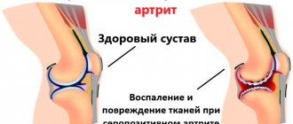
Achalasia cardia (achalasia of the esophagus) is a disease characterized by the lack of opening of the lower esophageal sphincter in response to the passage of food or liquid through the esophagus, due to a violation of the conduction of nerve impulses to the muscles of the esophagus and cardia. It is necessary to immediately clarify the correct name of the disease, because people call both achalasia of the cardia and achalasia of the esophagus. Previously, some doctors believed that it was correct to say achalasia of the esophagus, but the international classification of diseases of the 10th revision put an end to these disputes; it indicated achalasia of the cardia, which means this is the correct name for this disease. Achalasia affects equally often both men and women, more often only at the age of 30 - 45 years. This disease occurs in 3 - 20% of all diseases of the esophagus. Previously, it was believed that achalasia occurs in 1 person per 100,000 population, but with improved diagnosis and accumulation of data, reports began to appear that it occurs with a frequency of 5 and some authors believe that even 10 people per 100,000 population
Cause of achalasia cardia
It must be said right away that the exact cause of achalasia cardia is not known. There are various theories about the origin of this disease. The genetic theory is based on the fact that patients with esophageal achalasia have changes in the same gene loci, in addition, the risk of developing achalasia in children with Down syndrome is 200 times higher than in the population. Infectious hypothesis - based on the fact that when infected Trypanosoma cruzi, which causes Chagas disease, also causes dysphagia, which is similar in mechanism to cardial achalasia. In addition, the possibility of developing achalasia against the background of infection with the Herpes zoster virus and measles virus is being considered. Autoimmune theory is based on the fact that autoimmune diseases often occur in patients with esophageal achalasia. Thus, in patients with achalasia cardia, the risk of developing such diseases as increases: type 1 diabetes mellitus by 5.4 times, hypothyroidism by 8.5 times, Sjogren's syndrome by 37 times, systemic lupus erythematosus by 43 times, uveitis by 259 times . But none of these theories has received convincing confirmation to date, so it cannot be said that the cause of this disease is known.
What contributes to the disease achalasia
Experts do not name the specific cause that causes the development of the disease. It is associated with infections, compression of the esophagus, inflammation, tumors of malignant origin, infiltrative lesions, etc. In childhood, esophageal achalasia is usually detected after five years of age.
The first manifestations of the disease go unnoticed, so it is diagnosed late. If a child has dysphagia and starts vomiting after eating, you need to sound the alarm. These are common symptoms characteristic of achalasia.
Classification of achalasia cardia
The classification of achalasia cardia is important, since depending on the degree of the disease, a decision is made on the choice of one or another treatment tactic. There are 4 stages in total:
Stage I (early) - transient disturbances in the passage of food, the diameter and peristalsis of the esophagus are not changed outside of an attack; Stage II - persistent disruption of the passage of food, peristalsis of the esophagus is enhanced, the esophagus is not significantly dilated; Stage III – a stricture of the cardiac part of the esophagus is revealed, the latter is significantly dilated, peristalsis is sluggish and superficial. There is fluid in the esophagus on an empty stomach; Stage IV – stage of complications with ulcerations and erosions of the esophagus. The latter is expanded to 10 cm, S-shaped, peristalsis is either completely absent or multidirectional, superficial.
Treatment
At the initial stage, when problems with swallowing are minor, conservative therapy can be used; the drugs used are aimed at eliminating spasm and alleviating the patient’s condition. Endoscopic bougienage may also be performed to widen the narrowed area in the esophagus. To obtain the result, several procedures will be required, but the effect of dilatation is short-lived.
A very effective way to treat esophageal achalasia is esophagomyotomy - a surgical procedure during which part of the wall of the esophagus is dissected, but only to the submucosal layer - without opening the lumen, thus restoring normal swallowing. If there are contraindications to surgical treatment, as well as in elderly patients with recurrent disease who have previously undergone surgery, endoscopic dilatation is indicated.
If there are contraindications to surgery and endoscopic cardiodilation, endoscopic administration of an intrasphincteric injection of botulinum toxin A is possible. The procedure is safe and suitable even for patients with severe diseases of the cardiovascular and respiratory systems. But the effect lasts only 2-3 months, so repeated procedures may be required.
In our clinic, for each patient, a technique is selected that will be most effective in his or her case. If in the area of narrowing there are no traces of cardiodilation performed in the past, and there is also no depletion of the muscular wall of the esophagus, surgery is recommended; its timely implementation leads to recovery. Surgical intervention is performed by laparoscopy and even with a stretched esophageal wall leads to good results.
In this case, the operation itself is performed using video endoscopic equipment, which allows the surgeon to perform all manipulations with maximum precision, excluding damage to structures located in this area (vessels, nerves, fascial spaces). Our specialists have improved the technique, so patients can count not only on a shorter recovery period, but we have managed to minimize the incidence of intraoperative complications and relapses. Today this figure does not exceed 0.2%.
Clinical picture of achalasia cardia
Most often, it will not be difficult for an experienced gastroenterologist or surgeon to suspect achalasia of the esophagus based on the patient’s complaints. Most often, patients complain of dysphagia, difficulty or complete impossibility of food passing through the esophagus. So-called paradoxical dysphagia often occurs. when solid food passes more easily than liquid food. Over time, dysphagia progresses and in particularly severe cases, patients lose the ability to eat through the mouth, which leads them to exhaustion. A common symptom of achalasia cardia is the so-called wet pillow symptom, when the patient experiences reflux of the contents of the dilated esophagus into the mouth, and if the person is sleeping, the contents flow out onto the pillow, this can be either sputum or food debris. Often the pain intensifies against the background of stressful situations, or when the patient is nervous, which often leads patients with achalasia cardia to see a psychiatrist instead of a surgeon.
Often this disease is accompanied by quite intense pain. At the beginning of the disease, the pain is spastic, paroxysmal, and as the disease progresses, the pain becomes less intense, but almost constant.
Another common symptom of achalasia cardia is regurgitation, which is the reflux of the contents of the dilated esophagus into the mouth, the so-called esophageal vomiting. This condition actually resembles vomiting, but unlike it, the patient does not experience nausea, and it occurs most often when bending over or lying down.
A common symptom of esophageal achalasia is also a cough, and the development of bronchitis, and sometimes pneumonia. The problem is that during regurgitation, aggressive and infected contents enter the respiratory tract, which causes the above complications.
Common symptoms
The main symptom of achalasia is dysphagia, which occurs in almost all patients. The time between the appearance of the first symptoms and a visit to the doctor can range from one to ten years. The second most common symptom is congestion in the esophagus. Manifested by coughing and attacks of suffocation at night.
Heartburn and chest pain can also be signs of achalasia. Localized mainly behind the sternum, squeezing or squeezing pain is felt in the lower jaw, neck and back. Achalasia is often mistaken for gastroesophageal reflux or another disease accompanied by heartburn. If it does not occur from food and does not go away after taking antacids, there is a possibility that the problem is related to esophageal achalasia.
Diagnosis of achalasia cardia
Diagnosis of achalasia cardia is most often not difficult. FGDS (gastroscopy) - it is necessary to begin the examination with this procedure. When performing an FGDS, a narrowing of the esophagus is detected at the point where it enters the stomach; endoscopically, the device passes into the stomach with force. With achalasia grades 3 and 4, the esophagus is dilated, contains food debris on an empty stomach, and ulcers and erosions of the esophagus are often detected.
Barium X-ray of the esophagus - allows you to evaluate the speed of passage of contrast through the esophagus in patients with esophageal achalasia. The transverse size of the esophagus, peristalsis, and the presence of bends are also assessed. It is on the basis of radiography that the degree of achalasia of the cardia is determined.
CT examination - performed to assess the presence or absence of inflammation in the surrounding tissues, the presence of bronchitis or pneumonia. Motility of the esophagus - is considered the “gold standard” for diagnosing achalasia cardia, however, the equipment for its implementation is extremely expensive, and its use is quite limited. In this regard, several clinics, mainly research ones, have such equipment.
What are the complications?
With achalasia, irreversible changes occur throughout the body. Among the most common complications associated with esophageal disease are:
- squamous cell carcinoma;
- bezoars;
- volumetric formations on the neck;
- exfoliating submucosal layer;
- distal diverticulum;
- phlebeurysm;
- lung disease;
- purulent pericarditis;
- pneumopericardium, etc.
With prolonged progression of the disease, the esophagus expands and its walls become thinner, which becomes the cause of complications. In most cases (85%) those suffering from achalasia experience noticeable weight loss.
Doctor's comment
If the symptoms characteristic of achalasia cardia cause physical and psychological suffering, you have the opportunity to get rid of the disease quickly and painlessly, at the same time giving up many medications that bring only temporary relief. There are several ways to treat this disease; we will choose the one that will be effective in your case. The low-traumatic operation is performed using endoscopic equipment, which allows all manipulations to be performed as delicately as possible. Moreover, the improved technique allows surgical intervention without the risk of further relapses. Do not wait for complications to develop and do not waste time on ineffective conservative therapy; timely treatment gives every chance of a quick recovery!
Head of the surgical service at SwissClinic Konstantin Viktorovich Puchkov
Modified laparoscopic cardiomyotomy – maximum effect with minimal risk
Professor Konstantin Viktorovich Puchkov, who heads the Swiss University Clinic in Moscow, is one of the best surgeons in Europe, a brilliant specialist in the field of classical and laparoscopic surgery with 25 thousand years of experience in operations, and has improved the Geller technique. The fundamental differences of its modification:
- minimally invasive surgery through several punctures and incisions of the abdominal wall 5-10 mm long;
- thorough inspection of the bottom of the esophageal incision and suturing of microdamages with atraumatic needles and absorbable suture material;
- duplication of the bottom of the esophageal incision, the anterior wall of the stomach, suturing along the entire perimeter of the defect. This stage of the operation is very complex, so it is not practiced by most surgeons. But Professor K.V. Puchkov always performs it, thereby minimizing the risk of complications and relapse of the disease;
- maximum preservation of the anatomical relationship of the esophagus, stomach and diaphragm;
- strengthening of the cardiac sphincter of the esophagus by fundoplication at 120°. This is the name given to the fold-cuff formed from the bottom of the stomach, which prevents sphincter insufficiency and reflux (reflux) of stomach contents into the esophagus;
- the use of electrosurgical instruments on the LigaSure platform, which allows you to work bloodlessly, accurately and as sparingly as possible for tissues;
- the use of modern anti-adhesive agents to prevent adhesive disease;
- postoperative period without installation of a gastric tube. Self-administration of liquid food the very next day.
Professor K.V. Puchkov is convinced that surgical treatment should be resorted to without wasting time on ineffective and unsafe conservative methods.
Having specializations and vast practical experience in the field of surgical oncology, gynecology, urology, coloproctology, Professor K.V. Puchkov often performs simultaneous (combined) operations. They allow surgical treatment of other diseases of the abdominal cavity and pelvis, in addition to achalasia cardia, in the process of one intervention and one-time anesthesia. This eliminates the need for patients to undergo sequential operations with an interval of 1-1.5 months, each time undergoing a preoperative examination, undergoing anesthesia and a postoperative recovery period.
Professor K.V. Puchkov always answers questions from patients that come to him remotely. Every day he devotes several hours to such communication, despite regular operations in Moscow, Geneva, Freiburg and other cities of the world, numerous master classes in Russia and abroad, leadership of the Swiss University Clinic and specialized departments at the Academy of Postgraduate Education of Physicians.
We forward all requests received through application forms or a consultant in our chat to Professor K.V. Puchkov. You can get detailed recommendations from him or a second opinion on the treatment tactics for achalasia cardia proposed by other doctors, and agree on a time for a remote video consultation or an in-person appointment in Moscow.
Achalasia cardia is a neuromuscular disease of the esophagus (NMSD), characterized by the absence of reflex opening of the cardia during swallowing, accompanied by impaired peristalsis and decreased tone of the thoracic esophagus [1-4]. Together with diffuse esophagospasm and other motor disorders, it is classified as a group of neuromuscular diseases of the esophagus [5].
This disease was first described by the English physician T. Willis in 1672 in a patient with constant progressive vomiting; It was possible to restore the patency of his esophagus using a sponge attached to a whalebone [5-7].
The prevalence of achalasia cardia, according to world statistics [7-9], is 0.6-2 cases per 100,000 population (regardless of gender), in the USA - 1 case per 100,000 population. The share of esophageal achalasia in the structure of all diseases of this organ is 3-20%.
Over the entire period, more than 25 classifications of achalasia cardia have been adopted [10–13], for example, the classification of T.A. Suvorova (1966), in which two types of esophageal achalasia were identified. A three-stage classification of cardiospasm was proposed by G.D. Vilyavin in 1978. In 1987, the classification of T.A. Suvorova was changed and supplemented by A.L. Grebenev [7]. In our country, the most common classification is B.V. Petrovsky (1962), according to which four stages are distinguished: I (initial) - the esophagus is not dilated, the cardia opening reflex is preserved, but the motility of the esophagus is strengthened and discoordinated; II - the cardia opening reflex is absent, the esophagus is dilated to 4-5 cm; III - significant dilation of the esophagus up to 6-8 cm, retention of fluid and food in it, lack of propulsive motility; IV - sharp expansion (more than 8 cm), elongation and s-shaped curvature of the esophagus with atony of the walls, prolonged fluid and food retention [2, 5, 7, 12]. This classification is based on the results of radiological and endoscopic examination methods.
In X-ray contrast examination of the esophagus, typical signs of achalasia cardia are expansion of the lumen of the esophagus, absence of a gas bubble in the stomach, delayed release of the esophagus from the contrast agent, absence of normal peristaltic contractions of the esophagus, narrowing of the terminal esophagus (“candle flame”) [9, 10].
During an endoscopic examination, first of all, attention is paid to the degree of dilatation of the lumen of the esophagus, the presence of food debris, liquid and mucus in it. At the same time, the condition of the mucous membrane of the esophagus is assessed, its thickness, color, shine, and the presence of peristalsis is determined. Typical signs during esophagogastroduodenoscopy: weakened peristalsis of the esophagus, lack of adequate relaxation of the lower esophageal sphincter (LES), narrowing of the esophagus in the area of the LES and its suprastenotic expansion; slight resistance when passing the endoscope through the LES [14, 15]. In the case of esophagitis, thickening of the folds, hyperemia of the mucous membrane, erosion and ulceration are observed.
Currently, the Chicago classification of esophageal motility disorders, first proposed in 2008 and revised in 2011 and 2014, is generally accepted throughout the world. (SS v3.0) [4, 16-18]. This classification is based on the interpretation of objective data recorded by High-Resolution Manometry (HRM). HRM is now the generally accepted standard for diagnosing esophageal motor function.
According to this classification, three types of achalasia are distinguished depending on the predominance of certain motor disorders of the esophagus, such as LES hypertension, diffuse esophageal spasm, nonspecific or ineffective esophageal motility (see figure).
Manometric picture of achalasia cardia. a - type I; b - type II; c - type III. In type I achalasia cardia (classical achalasia), in 100% of wet swallows there is no peristalsis of the thoracic esophagus - this is severe hypokinesia. In type II, there is no normal peristaltic wave of contraction, but there is a uniform spasmodic contraction of moderate intensity (>30 mmHg) along the entire length of the esophagus from the upper to the LES in more than 20% of wet swallows. In this type, peristalsis of the thoracic esophagus is preserved. Type III is characterized by the absence of a normal peristaltic wave, the presence of isolated episodes of peristalsis in the distal esophagus, or premature spasmodic contractions (distal esophagospasm) recorded in more than 20% of wet swallows - this is a hyperkinetic type of achalasia cardia. With it, non-relaxation of the LES is accompanied by an extremely powerful contraction of the thoracic esophagus [4, 19-21].
Clinical manifestations of achalasia cardia are most often observed between the ages of 20 and 40 years [1, 5, 7]. The main symptoms of the disease are progressive dysphagia, regurgitation and chest pain associated with incomplete emptying of the esophagus and chronic esophagitis [22-24]. Other symptoms of the disease include weight loss, hiccups, nausea, increased salivation, bad breath, and occasionally heartburn [9].
Patients with achalasia cardia may experience the above clinical symptoms for a long time, and the symptoms may be wave-like. Periods of increased clinical manifestations can be abruptly replaced by periods of satisfactory well-being.
Despite the fact that achalasia cardia is a relatively rare disease, it carries a risk of severe consequences, such as, for example, adenocarcinoma of the esophagus [11, 25, 26]. Achalasia of the cardia, according to various authors [25, 27], can be classified as a precancerous disease, since it is known that cancer develops in 3-8% of patients with cardiospasm and achalasia of the cardia and the likelihood of its occurrence increases with increasing duration of the disease, and not only in the cardia, but also in the altered esophagus. In this regard, its timely detection and treatment is necessary.
Conservative treatment for achalasia cardia includes drug therapy with diet correction [7, 12]. Pharmacological therapy as a stand-alone method is the least effective, rarely leads to long-term and lasting positive results, does not reduce symptoms and is often accompanied by side effects, so it is now receiving less attention than other treatment methods [3, 28].
Over the past 40-50 years, at the initial stage of treatment of patients with achalasia cardia, it was considered advisable to use non-operative methods, and the main and most accessible of them has always been step-by-step balloon pneumatic dilatation (PD) under fluoroscopic control [7, 12].
After dilatation and to monitor the condition after treatment, which must be carried out once a year, patient complaints can be assessed using the Eckardt scale (see table)
Eckardt scale Note. * 0-3 points - remission, more than 4 points - ineffectiveness of PD. [29, 30].
In addition to regression of the clinical manifestations of the disease, a prognostic indicator of long-term remission after PD is the achievement of an LES pressure of less than 10 mm Hg, measured at the end of the procedure. After P.D. It is necessary to conduct a control X-ray examination of the esophagus with contrast.
In recent years, with the development of endoscopic technologies, many authors [7] propose to refrain from using PD to prevent the formation of scars in the area of the cardioesophageal junction, which subsequently complicates the performance of endoscopic operations.
The first reports in foreign literature [31] on the use of botulinum toxin A for the endoscopic treatment of cardiospasm and achalasia cardia appeared back in 1994. The procedure is based on intramural endoscopic injection of botulinum toxin A into the LES at a dose of 80-100 units, with 1 ml The drug (20-25 units) is injected into each of the four quadrants of the LES under visual control. The effectiveness of therapy is about 80% during the 1st month of observation, 70% after 3 months, 50% after 6 months and about 40% after 1 year, and therefore sometimes requires a second injection [7].
Over the entire history of treatment of patients with NMZP, more than 60 surgical techniques have been developed, for example, operations by J. Mikulicz-Radeck, G. Marwedel and W. Wendel, anastomosis of the esophagus with the stomach as modified by N. Geyrovsky, S.S. Yudina, E.L. Berezova and others, which, due to the occurrence of frequent relapses and the development of severe postoperative complications (reflux esophagitis, esophageal stricture), are practically not performed in our time and are of only historical interest [13]. The unsatisfactory results of such operations contributed to the search for new approaches to the surgical treatment of achalasia.
The idea of myotomy for the treatment of NMCD was first proposed by G. Gottstein in 1901 [32, 33]. In 1913, E. Heller [34] performed the first such operation and described it in his article “Extramucosal cardioplasty for chronic cardiospasm with esophageal dilatation”; the operation received worldwide recognition and became the basis for most surgical interventions performed [32]. Esophagocardiomyotomy according to E. Heller, which consists of dissecting the muscular membrane of the lower end of the dilated segment of the esophagus and the cardial part of the stomach along the anterior and posterior walls, was complicated by the development of gastroesophageal reflux disease in 15% of cases [7].
Many modifications have been proposed, for example, an organ-preserving cardioplastic operation developed at the Russian Scientific Center for Surgery of the Russian Academy of Medical Sciences, the basis of which is esophagocardiomyotomy according to E. Heller with incomplete fundoplication [5]. Due to the fact that organ-preserving operations could not always be performed, the development of complications in the form of recurrent reflux esophagitis and peptic strictures of the esophagus forced subtotal resection of the esophagus with simultaneous esophagoplasty with an isoperistaltic gastric tube with anastomosis in the neck [35].
Proposed in 1972 by B.V. Petrovsky’s method of plasty of the esophagogastric junction with a diaphragm flap on a pedicle from a left-sided transthoracic approach often led to relapse of dysphagia caused by the development of a “scar block” around the esophagus and severe peptic reflux esophagitis [36]. To avoid unwanted complications, students of B.V. Petrovsky - A.F. Chernousov and E.N. Wangqian proposed pneumatic stepwise cardiodilatation (as the main method of treatment of achalasia cardia stages I-II), and in patients with stages II and III of achalasia cardia - organ-sparing cardioplastic surgery, which is based on a modified cardiomyotomy according to E. Heller with incomplete fundoplication, which helps prevent reflux -esophagitis and formation of esophageal diverticulum [37]. However, after this operation, some patients may develop residual dysphagia associated with “hyperfunction” of the fundoplication valve.
Progress in the treatment of achalasia cardia is associated with the introduction of laparoscopy and thoracoscopy into clinical practice. Laparoscopic methods in modern surgery have taken one of the leading places in helping patients with NMCD [2, 38]. In terms of effectiveness, minimally invasive interventions are not inferior to open surgical operations - 94 and 84%, respectively, and the incidence of postoperative complications is lower when they are performed [7].
A meta-analysis of the results of treatment of patients with achalasia cardia, performed by W. Hoogerwerf and P. Pasricha [39] in 2002, showed the greatest effectiveness of laparoscopic myotomy among all methods of treating this disease.
In the terminal stage of the disease, extirpation or subtotal resection of the esophagus is performed with simultaneous gastric tube plasty, including the use of laparoscopic and thoracoscopic techniques [40]. Indications for choosing a method of surgical intervention are determined individually [41]. At the same time, in patients with contraindications for radical surgery, organ-preserving techniques are possible [42].
The active development of minimally invasive intraluminal endoscopic surgery has led to the creation, based on two techniques - myotomy and endoscopic dissection in the submucosal layer - of a new minimally invasive method for the treatment of achalasia cardia - oral endoscopic myotomy (POEM). Since the 2000s, the submucosal layer of the gastrointestinal tract has become a working space for operative endoscopy [43]. The next step, which made it possible to get closer to POEM, was the development of the method of transluminal endoscopic surgery (Natural Orifice Translumenal Endoscopic Surgery (NOTES) - intraluminal endoscopic surgery through natural openings) - exit from the wall of the gastrointestinal tract using a tunnel displacement of the entry point for safe exit into the cavity body, in this case the overlying mucosal flap served as a flap as a protective sealant [44]. This technique is based on the formation of a submucosal tunnel with access from the lumen of the esophagus through a small incision in the mucous membrane of the thoracic region to the area of increased muscle tone and subsequent dissection of the circular muscle fibers of the distal esophagus, LES and cardia of the stomach [45, 46].
In 2007, P. Pasricha et al. [47] proposed and successfully tested the technique of endoscopic myotomy in an experiment on animals - control manometry as a result of the operation revealed a significant decrease in pressure in the area of the LES, and when observed for 7 days, no signs of peritonitis and mediastinitis were noted. Further studies [48] showed that the effect on LES pressure was comparable to any previously used treatment for achalasia cardia.
In humans, the endoscopic myotomy technique was first developed and performed in 2008 by H. Inoue and colleagues (Yokohama, Japan). He also proposed a name for the new technique: POEM [45]. At the initial stage, POEM was performed in 17 patients with achalasia cardia. The technique consisted of creating a long submucosal tunnel (average length about 10-12 cm), followed by endoscopic myotomy of circular muscle fibers 8 cm long (6 cm in the distal esophagus and 2 cm in the cardia of the stomach). As a result, the symptoms of dysphagia were relieved in all patients and the resting pressure of the lower esophageal sphincter decreased on average from 52.4 to 19.9 mm Hg. [49].
Currently, POEM is used in all patients with symptomatic achalasia of all types [50]. Initially, the use of POEM was limited to patients under 18 years of age, but it has now been successfully used in patients over the age of 3 years [51].
The results of the first operations using the POEM method indicate that the new technique can be effective and be an alternative for the treatment of achalasia cardia with classical methods, however, its role in patients with previous operations, age restrictions and concomitant diseases has not yet been fully studied [52].
The main advantages of this technique are the absence of the risk of uncontrolled perforation of the esophagus, minimal invasiveness and low trauma of the surgical operation, preservation of the ligamentous apparatus of the esophagus, short recovery and rehabilitation periods [53].
In 2015, H. Inoue [54] published a report on the largest current experience of using POEM - 500 patients with achalasia cardia, with the following results: endoscopic myotomy was successfully performed in all patients, side effects of the operation were noted in 3.2% of cases , 2 months after POEM, when questioning patients, a significant decrease in scores on the Eckardt scale was recorded - from 6 to 1 point, LES pressure from an average of 25 mm Hg. Art. before surgery it decreased to 13 mmHg. Art. Gastroesophageal reflux disease was detected in 16.8% of patients. Enrollment has been completed for a randomized controlled trial comparing endoscopic and laparoscopic myotomy for achalasia. It includes 240 patients from the Czech Republic, Germany, Italy, the Netherlands and Sweden. The expected completion date of the study is December 2021. There are currently several systematic reviews and meta-analyses based on retrospective data published in recent years. R. Talukdar et al. [46] concluded that there is no difference in Eckardt score between POEM and laparoscopic myotomy. However, it was noted that the duration of the operation was significantly reduced with POEM.
In 2021, the Mayo Clinic (Rochester, USA) shared its experience of successfully treating 28 patients between April 2012 and May 2015 using a combined method that included POEM and POEM supplemented with laparoscopic E. Heller myotomy with incomplete fundoplication . The authors noted a decrease in Eckardt scores from 6 to 0, and only 3 patients continued to complain of dysphagia in the postoperative period [55].
L. Marano et al. [56] systematically analyzed data from 486 patients (196 in the POEM group and 290 in the laparoscopic myotomy group). The study showed that the difference in postoperative clinical manifestations, duration of surgery and side effects was not significant. Patients' length of hospital stay was significantly shorter with POEM.
In Russia, POEM is just beginning to be actively introduced into clinical practice; data on its use are extremely scarce. In 2015 E.D. Fedorov [2] presented the results of using POEM in 4 patients with achalasia cardia and concluded that this method was immediately effective and relatively safe in the early stages. In 2021 K.V. Shishin published the results of the successful use of the method in 15 patients with achalasia cardia, indicating its effectiveness and safety.
Despite the positive reviews about the new method of treating patients with achalasia cardia, there are a number of problems and questions regarding the features of the operation [57]. In the process of performing POEM, such technical aspects must be resolved as the choice of the level of access in the esophagus for the formation of a submucosal tunnel, identification of the LES in the formed tunnel, the length of the myotomy, the issue of the full-thickness of the myotomy - limit ourselves to only dissecting the internal circular layer or perform myotomy and external longitudinal muscle fibers [ 58].
Thus, the improvement of technical aspects, the emergence of a minimally invasive surgical technique of POEM, the development of indications and contraindications for this, and the assessment of immediate and long-term results of the intervention are promising areas for further scientific research.
The authors declare no conflict of interest.
The authors declare n conflicts of interest.
Information about authors
Gasanov A.M.
. — https://orcid.org/0000-0002-1994-2052
Aliev N.A
. — https://orcid.org/0000-0002-9772-1351
Danielyan Sh.N
. — https://orcid.org/0000-0001-6217-387Х; e-mail
Gasanov AM
— https://orcid.org/0000-0002-1994-2052
Aliev N.A.
— https://orcid.org/0000-0002-9772-1351
Danielyan Sh.N.
— https://orcid.org/0000-0001-6217-387Х
Advantages of treatment at the Swiss University Hospital
- The clinic is equipped with the latest generation equipment, which allows the operation to be performed without damaging nearby structures, the muscularis mucosa is dissected without damaging the mucosa, intraoperative complications are also excluded, etc.
- The treatment method is selected for each patient individually, taking into account his state of health. Great importance is given to preserving the natural anatomical location of the esophagus, diaphragm and stomach, which avoids the occurrence of reflux esophagitis and sphincter insufficiency in the future.
- When treating patients, the clinic uses a technique that makes it unnecessary to leave a transgastric tube after surgery, which makes the postoperative course easier. Thanks to the original method used, the risk of further relapse is minimal; in more than 97% of patients, good results were achieved, confirmed by fibroesophagogastroduodenoscopy, X-ray examination and esophagomanometry.
- In our clinic, patients with several diseases requiring surgical treatment can undergo simultaneous surgery. Performing several surgical interventions during one anesthesia reduces the burden on the body, hospitalization time, and recovery, compared to conventional surgery, takes much less time.