Parasitic diseases do not manifest themselves clearly for a long time, causing irreparable harm to health. Echinococcus is a helminth that causes irreversible changes in the liver, kidneys, lungs, heart and toxic damage to the body as a whole. Its identification is difficult, and prevention and treatment require an integrated and systematic approach. However, knowing the symptoms of echinococcosis in humans, the characteristics of the development and course of the disease, the most progressive ways to identify the parasite and restore health, you can avoid the enormous harm that it causes.
Helminths are an unobvious danger
The World Health Association has collected and systematized data on human infection with helminths, finding that 10 million people worldwide become victims of helminths every year. In Europe, parasitic worms, including echinococcus, are present in the body of every third person. Russia registers about 2 million such diseases a year, but experts express the opinion that this number is significantly underestimated. The problem of diagnosing echinococcosis, its treatment and prevention is acute and is complicated by difficulties in detection. Up to 95% of patients are not even aware of the danger that directly threatens them, and doctors do not consider helminths as the source of symptoms.
Echinococcus photo
In the human body, echinococcus (the causative agent of the disease) and other helminths have been implicitly present for a long time, and people may complain for years about poor health, unaware of the true causes of the problems. “Disguising themselves” as seasonal colds, allergic reactions, inflammations, parasites are not detected during traditional studies. Getting tested for echinococcus is an obvious idea, but the problem is that not a single conventional laboratory method will give a guaranteed reliable result. This is due to the specificity of the penetration of helminths into the human body. Although humans are the intermediate host of echinococcus, the parasite’s infestation causes enormous harm to health—mechanical and chemical. Helminth:
- finds ways to penetrate the body and reproduce in it (the fertility of echinococcus is high);
- localized and fixed in organs most suitable for growth, reproduction and parasitism of echinococcus;
- goes through several stages of development, causing echinococcosis in various forms - what makes echinococcus dangerous for humans;
- removes eggs and larvae from the body naturally - this is how one person becomes the cause of infection for others.
echinococcus in meat
Diseases associated with echinococcus initially manifest themselves as illnesses of a completely different medical “profile”. Therefore, doctors detect a maximum of 20% of cases of helminth infection. In the remaining 80%, the parasite is not detected, causing toxic-allergic damage to organs of vital importance. Diseases caused by echinococcus, according to WHO, are misdiagnosed in 70% of cases, in addition:
- traditional laboratory tests do not detect echinococcus in humans promptly and reliably, since the parasites have multidirectional harmful effects;
- the identified symptoms of echinococcus in 90% of cases are diagnosed as oncology and efforts are directed in the “opposite” direction, without eliminating the true causes of the disease;
- most practicing doctors do not have experience in treating echinococcosis;
- Even with a successful diagnosis of echinococcus, a single dose of anthelmintic drugs does not completely rid the body of them - cysts and other forms remain in the body, causing new outbreaks of the disease.
Disturbances in metabolic processes, the immune system, weakening of protective functions, and sugar deficiency due to echinococci can lead to unprecedented consequences. Death is no exception. Echinococcosis is especially dangerous in children, especially since parents think of parasites as the last cause of problems. Therefore, it is very important to know about the symptoms and methods of getting rid of the parasite. Let's consider the cycle of echinococcus and the mechanics of helminth infection.
photo of the worm Echinococcus
Prevention
Preventive measures for infection with echinococcosis include a set of measures:
- Regular deworming of personal dogs at least 4 times a year.
- Wash your hands promptly and thoroughly, especially after contact with animals, after caring for livestock, working with land, contact with sheep wool, and cutting the skins of wild carnivores.
- Do not drink water without boiling from natural sources (springs, wells).
- Thoroughly wash vegetables, fruits, herbs and berries consumed raw.
- Do not consume unwashed wild berries or raw milk.
Echinococcus and the diseases it causes
2.1 Echinococcus as the causative agent of the disease
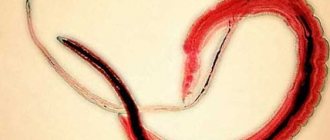
Echinococcus is one of the small parasitic worms. This is a pathogen resistant to the external environment of a dangerous disease - echinococcosis. The type and class of echinococcus have been determined a long time ago; it is familiar to biologists, but, unfortunately, little known to doctors. The parasite is a type of tapeworm that can be seen with the naked eye. The size of echinococcus is up to several millimeters; eggs and cysts are much smaller. Sometimes echinococci up to a centimeter long are found, but even in this case they are not easy to identify. The parasite belongs to the tapeworm and, having penetrated the body, affects many key organs, from the liver to the brain. The worm consists of:
- scolex echinococcus is the head on which four suckers and hooks are located in two rows;
- necks;
- segments of varying degrees of maturity - the most mature ones contain the uterus and eggs of echinococcus.
Echinococcus mri
The life cycle of echinococcus is sequential development from egg to adult. At each stage, the worm causes harm to the human body. The eggs contain Echinococcus oncospheres, in which the parasites develop into larvae. Formations die under the influence of bright sunlight; extreme temperatures and aggressive acidic and alkaline environments have little effect on them.
The larval stage of development is called the fin or larvocyst of echinococcus. It is this that causes disease.
In pathology, the resulting fins of echinococcus, namely Echinococcus granularis, are of key importance. They cause echinococcosis in the hydatid form. The variety Echinococcus multilocularis is the cause of the alveococcosis form of the disease. The pathogens have morphological and biological differences (although the life cycle of echinococci is the same), they develop in different organs and tissues:
- the hydatid form provokes echinococcosis of the liver, lungs, and other organs;
- alveococcosis varieties primarily affect adjacent tissues.
Alveococcosis (a disease caused by a pathogen) is less common than hydatid forms, but the methods of infection with echinococcus are identical in both cases. This is direct transmission from carriers to healthy people and animals. In this case, the manifestation depends on the lesion. Having found out which symptoms in a person are caused by echinococcus in the liver, and which, for example, in the lungs, you can develop the right strategy to combat it.
life cycle of echinococcus
2.2 How echinococcus is transmitted from carriers to humans
Domestic and wild animals - those who are the main hosts for echinococcosis - carry the pathogen in the intestines, transmitting it to people. Even at the beginning of development, single-celled echinococcus can be transported from key carriers to non-definitive ones. Among others, the intermediate host of echinococcus is humans, as well as domestic animals. The main carriers of the parasite are canines, in which the helminth lives in the form of adult echinococci up to 5-10 mm in size. These include wolves and dogs. Echinococcus is found in foxes and jackals, as well as in:
- felines - domestic cats, lynxes, lions;
- rodents are intermediate hosts of echinococcus;
- small and large livestock - the causative agent of echinococcosis may be contained in its meat;
- wild deer, elk and other artiodactyls.
echinococcosis in humans
The definitive host of echinococcosis is usually a representative of the canine family. However, the greatest harm to people is caused by intermediate carriers of cestodes - rodents and livestock. Is it possible to become infected with echinococcus from a sick person who has unwittingly become the “host” of the worm? Yes, and this is a separate large-scale problem. Infection with echinococcus can affect a family or social group, so regular prevention is of paramount importance.
It was revealed that the causative tapeworm is localized, as a rule, in the countries of Africa, Central Asia and the Middle East. Cases of human infection with echinococcosis are not uncommon in the southern Russian regions, as well as in Siberia and Yakutia. Foci of pathology were observed in Australia and New Zealand, Latin America. Considering the transmission routes of echinococcus, we can talk about the planetary scale of the problem.
Echinococcus color
2.3. Mechanism and routes of infection with echinococcus
The larva of echinococcus enters the organisms of carriers mainly through alimentary and household contact. Invasion occurs when:
- close contact with sick animals - often Echinococcus parasitizes the body of the human host;
- walking in the habitat of infected wild animals - spheres, echinococcus cysts can enter the body when collecting mushrooms, fruits and berries, eating them without washing;
- working at a slaughterhouse, in a carcass processing workshop - echinococcus is often found in meat;
- Eating dishes (kebabs, liver) without sufficient heat treatment is a common way of transmitting echinococcus.
At all stages of the development cycle of echinococcus, the parasite poses a danger. Mechanical infection of the intermediate host usually occurs when oncospheres are ingested. The structure of the echinococcus is such that the segment with mature eggs is torn off from the individual and their shells are destroyed. The embryos that form from oncospheres penetrate the gastrointestinal mucosa (intestines) and the portal circulatory system. Therefore, when detecting echinococcosis, ultrasound of the liver, kidneys and other organs will not give a 100% reliable result.
Echinococcus larva
Some of the embryos enter the systemic circulation. From where they penetrate into vital organs, causing diseases of the lungs, liver, and also:
- echinococcosis of the spleen;
- echinococcosis of the pelvic organs;
- echinococcosis of the brain;
- echinococcosis of the intestines, omentum, kidneys, and so on.
Echinococcus liver most often affects the lungs. However, not only these organs suffer, but also health in general. When localized in the human body, echinococcus affects it in different ways.
adult echinococcus
Treatment for echinococcus in EUGENA without surgery.
Our specialized clinic offers all those infected with echinococcus to undergo treatment without surgery , based on the medication method. The method was developed by our chief physician and part-time director of our clinic, Evgeniy Li.
A unique technique allows you to get rid of the parasite quickly and effectively! The method consists of a targeted effect on the affected organs and tissues, which allows for the targeted removal of echinococcus.
The method was discovered by Eugene Lee in 1997 and has since developed and improved. At the moment, over ten years of use, the principle of treatment has helped many people get rid of parasites, who are still healthy and are periodically observed in our clinic for preventive purposes.
Take care of your health and follow the rules, thanks to which you will be able to avoid infection with such a parasite. And if it suddenly turns out that the parasite took you by surprise, we will become your reliable assistant in getting rid of it!
Echinococcosis and causes of damage to the body
The disease, known as echinococcosis, is caused by Echinococcus parasites in the larval stage. Because of them, cysts are formed, multi-chamber foci of alveolar damage growing in the tissues. If the larvae are not phagocytosed or destroyed, they form cysts. They are single-chamber echinococcal formations, which contain:
- outer layered cuticle made of chitin, inside which the development of echinococcus occurs;
- the inner germinal layer, where echinococcus worms “float” in the embryonic state;
- clear liquid (succinic acid without protein) filling the cyst.
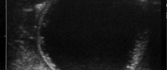
Hydatid echinococcosis causes unilocular formations. This disease is more common than alveolar disease and primarily affects the liver. In organs and integuments of the body, echinococcus forms capsules around cysts, building up connective tissue. The amount of liquid in the bubbles gradually increases. As a result of this, they grow, sometimes to enormous sizes, provoking cystic echinococcosis. Scolexes develop in the blisters - the so-called hydration sand. It can be detected by CT scans for echinococcosis. Subsequently, daughter and grandchild cysts appear, completing the life cycle of the parasite. Reproduction of echinococcus and its transmission from the body of an infected person is possible if the integrity of the blisters is violated.
Multichamber cysts have a different structure. If a single-chamber echinococcus develops inside a person in the form of a single bubble, then the alveolar form produces multiple foci with necrosis around them. Cysts grow by budding not inside the maternal formations, but outside. The clinical picture of the spread of echinococcus in this case is similar to the appearance of malignant neoplasms. Echinococcosis, as an alvear-type disease, develops due to the movement of parasites along the bloodstream and lymphatic ducts, like a metastasis. Cysts are not localized in the liver and other organs, like single-chamber formations, but form clusters of individual blisters. In this case, the alvear form of the disease occurs, just as a person becomes infected with echinococcosis of the hydatid type.
Echinococcus on ultrasound
The disease, according to available data, previously occurred predominantly in women. However, in the last two decades the picture of echinococcal lesions has changed. Diseases now affect both sexes in equal proportions. The growths can grow over decades, manifesting symptoms that doctors do not associate with the presence of parasites. Recurrent stages of echinococcosis caused by residual and intermediate forms of the helminth are very dangerous.
The rate of increase in lesions is influenced by the state of immunity, therefore echinococcus in a child or an elderly person is especially dangerous. Also, the scale of helminth damage increases the presence of chronic diseases. The pathogenic effect of echinococci manifests itself clinically in different ways.
Hydatid cyst
Finna in the affected organ (usually the liver) grows and after 5 months turns into a vesicle about 5 mm in diameter. Further, the echinococcal cyst grows extremely slowly, but over many years it can reach enormous sizes. Cases of cysts weighing 12 kg or more have been described. An echinococcal cyst is a round, densely elastic white formation, covered on the outside with a dense fibrous membrane (capsule).
As the cyst grows, it compresses the surrounding tissue. The parasite feeds on nutrients that are extracted from the blood and interstitial fluid of the host.
Fluid filling the cyst
The cyst cavity is filled with a yellowish liquid, neutral reaction, containing protein, sodium chloride, tyrosine, grape sugar, succinic acid and chitin.
Clinical manifestations of echinococcosis
4.1 Stages and features of echinococcosis
There are the following stages of echinococcosis:
- latent, in which a person becomes infected with echinococcus - it occurs without symptoms and ends in the formation of cysts;
- the occurrence of individual symptoms, complaints of deterioration of health - signs of echinococcus in humans are poorly expressed;
- pronounced manifestation of the clinical picture - the most “noticeable” stage of the development cycle of echinococcosis;
- complications of echinococcosis.
The latent period of the disease, which lasts 5-20 years, may manifest itself with heterogeneous symptoms or not be monitored at all. When hydatid echinococcosis affects the liver of the lungs, in 91% of cases the parasites are found by chance. With the increasing number of means of detecting and preventing echinococcus, it is detected at earlier stages than two or three decades ago.
liver echinococcosis
The weight of the bubbles that the worms form sometimes reaches 16 kg; they put pressure on the surrounding tissues, changing them. At the same time, the clinical manifestations of echinococcosis are not expressed, which complicates diagnosis. There are many individuals inside the cysts, and the fluid that penetrates the circulatory system has a depressing effect on all functions of the body. Without treatment, echinococcal diseases lead to complete depletion and, in some cases, death. Death occurs when large cysts rupture, while the patient may not notice the parasites until the last moment. Signs of echinococcosis in humans, as well as the clinical manifestations of the disease, depend on the location of the helminths and the size of the cysts. Using them, you can identify characteristic symptoms and determine that a particular organ is affected by helminths.
4.2 Liver echinococcosis
Hepatic echinococcus is the most common case of the presence of the parasite in the body. Echinococcosis of the liver as a disease is known more than other lesions of this helminth. The organ increases in size, an abscess and secondary infections caused by suppuration of cysts may develop.
Echinococcus, which has settled in the human liver, forms tumors that compress the bile ducts, which leads to yellowing of the skin, which is accompanied by itching. It manifests itself especially painfully if the parasite has infected the child’s body, but liver echinococcosis in children is dangerous not only for this reason. Due to cysts, fluid accumulates in the abdominal cavity, and their rupture provokes anaphylactic shock. In the organ, echinococcosis most often occurs of the hydatid type, rather than the alveolar type.
Treatment of echinococcus in the liver is complicated by the fact that examination and palpation can only identify painful areas, and diarrhea, nausea and weakness are not associated with the presence of helminths. Without intervention, the disease leads to the accumulation of bilirubin, increased frequency of fainting and other symptoms of liver echinococcosis. If the problem is not eliminated, the situation will lead to surgical intervention. Surgeries due to echinococcus in the liver may be necessary if the contents of the blisters enter the abdominal cavity and develop peritonitis.
4.3 Pulmonary echinococcosis
Echinococcosis as a lung disease is not much less common than liver damage. Cysts deform the chest and impair breathing. However, the diagnosis and treatment of echinococcus is difficult due to the similarity of the disease manifestations with the symptoms of tuberculosis. Bloody spots in the sputum, shortness of breath, chest pain, and cough are often mistaken for oncology. Without surgical treatment of echinococcosis, pleurisy, suppuration and rupture of cysts, and other complications may develop. If formations break into the pleura, anaphylactic shock occurs and in most cases the patient dies. Pulmonary echinococcosis in children can be more severe than in adults and develop to dangerous clinical stages more quickly.
pulmonary echinococcosis
4.4 Brain echinococcosis
Echinococcus in the brain often gives a clinical picture similar to the manifestations of schizophrenia. The growth of the bladder activates general and focal neurological manifestations. Symptoms of cerebral echinococcosis are rarely “deciphered” as the presence of parasites, but the disease has its own clinical picture. In 90% of cases, due to echinococcosis of the brain, hypertension syndrome develops with dizziness, pain, and even epileptic seizures. Among the mental disorders caused by the parasite are delusional states, severe depression, and dementia. Echinococcus in the brain provokes seizures and paresis. The clinical picture of the lesion is severe, the disease progresses rapidly. When decoding a blood test for echinococcus in the brain, eosinophilia is almost always detected.
4.5 Renal echinococcosis
A feature of echinococcus in the kidneys is the formation of cysts in the cortex. The route of transmission of echinococcosis is through arterial blood flow. The presence of the parasite causes intoxication, urinary disorders, and colic. Helminths in the kidneys are one of the causes of echinococcosis of the genitourinary system, with which the organ is associated. Without treatment, the patient may experience worsening pyelonephritis and constant pain in the hypochondrium and lumbar region. Scraps of scolex are sometimes found in urine, but in general, the symptoms of infection with echinococcus are often confused with other diseases.
Finns echinococcus
In other organs - the heart, the spleen - worms are localized much less frequently, but these cases cannot be excluded. Without treating echinococcus with drugs that provide a complex effect, you may encounter cyanosis of the extremities, pulmonary embolism and other deadly ailments. Selecting funds is a difficult problem. Reviews about the treatment of echinococcosis with folk remedies are contradictory and, as a rule, negative, and traditional medicine often “throws its energy” at eliminating symptoms, considering them manifestations of other diseases. However, by infecting the body, the parasite gives a fairly clear “picture” by which its presence can be detected.
Classification
Depending on the location of the hydatid cyst, echinococcosis of the liver, lungs, biliary tract, brain (brain/spinal), spleen bones, etc. is distinguished.
According to the pathogenetic principle:
- Primary isolated liver damage.
- Recurrent: secondary disseminated; secondary recurrent; secondary metastatic (combined lesion with other organs).
- Residual multiple echinococcosis of various organs.
By stages of clinical course:
- asymptomatic stage;
- stage of initial manifestations;
- stage of pronounced manifestations;
- stage of complications.
Characteristic symptoms of echinococcosis and methods for identifying the parasite in the body
5.1 Determining the presence of echinococcus in the body by symptoms
For a person thinking about the problem of helminths, the easiest way to diagnose diseases is to analyze the symptoms. Of course, you can turn to laboratory diagnostics of echinococcosis, but you need to be prepared for a false negative result. Analytical power to date does not clearly determine whether there are parasites in the body or not. Having donated blood for echinococcus, the patient does not receive all the necessary information.
Echinococcus eggs
Numerous observations of caring scientists and medical workers made it possible to create a reliable and informative list of signs characteristic of echinococcus. These include:
- Increasing incidence of acute respiratory infections and chronic diseases lasting for years. These are common symptoms of echinococcus in humans and treatment of ailments should include not only the “physical” elimination of parasites, but also restoration of the balance of microelements. Its violation causes a systemic deterioration in well-being.
- Increased fatigue, a noticeable and “unreasonable” decrease in performance. Echinococcus flatworms, penetrating the body, poison it with toxic products of their vital activity, which entails these symptoms. Micronutrient deficiencies caused by the presence of the parasite worsen the problem.
- Deteriorated appearance. Acne, papillomas, seborrhea, rash due to echinococcus occur very often. When the gastrointestinal tract is infected with parasites, nails and hair become brittle, cracks appear on the heels, and the skin dries out. When echinococcus penetrates the body, the integument becomes rougher, and premature wrinkles become more noticeable.
- Inflammatory infections - frequent tonsillitis, tonsillitis, sinusitis. Toxic secretions prevent the immune system from dealing with the problem. Therefore, when identifying symptoms of echinococcosis in humans and treating the detected disease, attention should be paid to restoring the immune system.
- Problems with the gastrointestinal tract. Echinococcus helminths cause bloating, stool disorders (constipation, diarrhea), and gastrointestinal syndromes.
- Allergies are primarily dermatic manifestations. Carriers of echinococcosis reduce the protective abilities of the immune system, which provokes rashes, increased sensitivity to allergens, and a more severe course of hay fever and urticaria.
- Anemia, sleep disturbances, neuroses, pain in joints and muscles and other symptoms that are caused by echinococcosis at different stages.
After a person becomes infected with echinococcus, he begins to experience a general malaise, against which specific symptoms develop. In addition to anemia, acute respiratory infections, and rashes with echinococcosis, nausea and vomiting occur after heavy and spicy foods. Abdominal pain also appears with a shift to the right hypochondrium - these are signs of hydatid echinococcosis of the liver.
When the lungs are affected, the hacking cough intensifies, often with an unpleasant odor. In the later stages, echinococcosis causes severe pneumonia. The rupture of large cysts in any organ entails a state of shock and, in some cases, death. Treatment of echinococcus with medications should be aimed not so much at eliminating the symptom, but at comprehensive recovery. Eliminating the destructive effects of worms in the body is as important as “removing” them from it.
structure of echinococcus
5.2 Progressive instrumental diagnosis of echinococcosis
The presence of parasitic Echinococcus worms in the body can be detected using progressive techniques and the latest equipment - “IridoScreen” from. Scanning the iris of the eye provides comprehensive information about the functional state of internal organs, the central nervous system, and the gastrointestinal tract, and in general, the results of the study allow:
- determine the level of toxic load on the body: an increase in its level is one of the most striking symptoms of echinococcosis and treatment should be prescribed immediately;
- assess the degree of deviation of microelements from the norm - by destroying the key “building” material, echinococcus causes weakened immunity and many dangerous diseases;
- learn about the condition of the liver, heart, kidneys, lungs and other organs and systems of the body.
Echinococcus in the liver
It has been proven that any infection by echinococcus or other parasite causes toxic-allergic irritation, activating the flow of reflex impulses through the central nervous system. They are transmitted to the iris of the eye - to a certain zone, which is a projection of the affected organ. Research using iridological methods allows you to accurately determine the degree of changes in the body. This is an effective measure for the prevention of echinococcosis, detection and control of the parasite.
Diet
Diet for liver cysts
- Efficacy: no data
- Terms: 1-6 months/lifetime
- Cost of products: 1500-1600 rubles. in Week
Diet 0 table
- Efficacy: therapeutic effect after 21 days
- Terms: 3-5 months
- Cost of products: 1200-1300 rubles. in Week
For echinococcal liver cysts outside the surgical period, the Diet for Liver Cysts . In the preoperative period (on the eve of the operation) dietary Table No. 15 ; after surgery – Diet 0 table ; in the early postoperative period, Dietary Table 1A , 1B , and in the late postoperative period - Treatment Table No. 5 . For echinococcal cysts in the lungs outside the surgical period - Diet 15 , in the postoperative period on days 1-2 Diet No. 0a , and from day 3 - No. 1 - surgical, from day 5 dietary Table No. 11 or 13 .
List of sources
- Sergiev V.P., Legonkov Yu.A., Musaev G.Kh., Poletaeva O.G. Cystic echinococcosis (unilocular): clinical picture, diagnosis, treatment, prevention. M.: Vector-Best; 2008.
- Shodmonov I.Sh., Razikov Sh.Sh. EPIDEMIC SIGNIFICANCE OF ECHINOCOCCOSIS // Modern problems of science and education. – 2015. – No. 2-1.
- Deineka I. Ya. Human echinococcosis. - M.: Medicine, 1968. - 376 p.
- Trishin, M.V. Epidemiology and diagnosis of echinococcosis in the child population of the Orenburg region in 1994-2012 / M.V. Trishin, P.V. Gureeva, I.A. Sim // Bulletin of the Orenburg Scientific Center of the Ural Branch of the Russian Academy of Sciences (electronic journal). - 2014. - No. 1. — P. 4-7.
- Dadvani S.A., Strelyaeva A.V., Gostishchev V.K. and others. Minimally invasive surgical interventions and chemotherapy for echinococcosis // Annals of Surgery. - 2000. - No. 4. - P. 38-41.