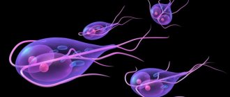
Opisthorchiasis is one of the most dangerous helminthiases. The causative agent of this helminthic disease is the cat, or Siberian, fluke (Opisthorchis felineus).
The name is not accidental: this worm lives in water bodies and is transmitted, among other things, through cats, to whom fishermen often give raw small fish.
With opisthorchiasis, the liver and pancreas are damaged by worms - liver trimatodes.
The source of opisthorchiasis infection for both people and animals is fish that has not been properly processed: insufficiently boiled, fried, salted or dried.
In addition, infection with opisthorchiasis can occur due to poorly washed knives and cutting boards, on which remains of fish with infective larvae remain if food that is not subject to heat treatment is cut into them. Most often, opisthorchiasis is diagnosed in places where river fish are actively caught.
Symptoms of opisthorchiasis
Often the disease is asymptomatic and in this case it is very difficult to diagnose it - the patient does not experience pain or discomfort, helminths are detected by chance during a routine or emergency examination. If the clinical picture is pronounced, then the first symptoms of opisthorchiasis appear 2-4 weeks after infection:
- temperature rise, up to critical levels;
- severe weakness, excessive sweating;
- nausea turning into vomiting (regardless of food intake);
- regular headaches;
- pain, aches in muscles and joints.
If the disease is severe, there will be blurred consciousness and delirium - this is how the body reacts sharply to the introduction of helminths into it. In most patients, the liver and spleen are significantly enlarged. If left untreated, the bile ducts begin to become clogged with opisthorchiasis, which leads to the rapid development of hepatitis and jaundice.
Signs of opisthorchiasis in childhood manifest themselves intensively, and the disease is severe. In addition to the above symptoms, there may be severe pain in the right hypochondrium, which intensifies even with minor physical exertion.
Opisthorchiasis - symptoms and treatment
Opisthorchiasis (Ob disease) is an extraintestinal parasitic disease with a fecal-oral transmission mechanism, caused by parasitism in the human hepatobiliary system and pancreatic ducts of flat parasitic worms of the genus Opisthorchiasis. Clinically characterized by inflammatory changes in the bile ducts and toxic-allergic reactions of varying severity.
Why is opisthorchiasis dangerous?
In the absence of adequate treatment, the risk of developing cancer of the biliary system of the liver, pancreas and immune-mediated pathologies of the bronchopulmonary system increases sharply.
Etiology
Type - Plathelmintes (flatworms)
Class - Trematodae (fluke trematodes)
Squad - Fasciolata
Family - Opisthorchidae
Genus - Opisthorchis (flukes)
Species - Opisthorchis felineus (cat/Siberian fluke), Opisthorchis viverrini (civet/squirrel fluke) + about 30 more species of little significance in human pathology.
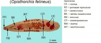
The structure of opisthorchis:
The pathogen was first discovered in cats (hence the species name) and described in 1884 by the Italian scientist Sebastiano Rivolta. In 1891, Professor Konstantin Nikolaevich Vinogradov also described and determined the taxonomic affiliation of the parasite.
Adults parasitize the hepatic bile ducts (in 100% of cases), the gallbladder (up to 60%), and the pancreatic ducts (less than 40%) of humans and carnivores. Their nutrition is provided by surrounding tissues, secretions and blood.
Opisthorchis (for example, Opisthorchis felineus) are small lancet-shaped flatworms up to 20 mm long and up to 4 mm wide. The body is covered with a skin-muscular sac, there is no cavity. There are two suckers (oral and abdominal), with the help of which the parasite attaches to the host tissues.
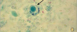
The circulatory and respiratory systems are absent. The organs of the nervous system and secretions are poorly developed. They are hermaphrodites (they combine the male and female reproductive systems in one organism). They produce up to 900 eggs per day (much less than other parasitic worms, which makes diagnosis much more difficult). The eggs of the parasite are released into the external environment with feces and contain a viable larva.
The eggs have a faint yellowish color, a double-contour shell, externally reminiscent of the surface of a melon, with a cap at one pole and a thickening at the other pole. Their dimensions are 0.010-0.019 x 0.023-0.034 mm. Eggs can be stored in soil for up to one month, in latrines for up to seven months, in winter in the air for no more than 2-3 days, in fresh water for up to 1.5 years.
The development of larvae is possible only when the eggs are exposed to favorable conditions in a freshwater body, where they become food for bithinid mollusks (the first intermediate host). In their intestines, a larva emerges from the egg - miracidia. It penetrates the body of the mollusk, develops and turns into a sporocyst, a redia (a sac-like formation with a large pharynx and intestines), which, migrating into the hepatopancreas of the mollusk, gives rise to the formation of a cercarium (tailed larva). Then, under favorable conditions (water temperature not lower than + 20°C), as they mature, the cercariae leave the body of the mollusk into the reservoir. There, within 30-60 hours (the period of free-living activity is after death), they attach to the body of fish of the carp family (an additional host is ide, chebak, carp, gudgeon, rudd and others).
An interesting mechanism is that cercariae “recognize” the desired fish species, which is realized through receptor hairs on their body that have an affinity for the chemical and biological properties of fish mucus. Over the entire life of a fish, up to several tens of thousands of parasites can attach to it. After attachment, the cercariae cast off their tail, penetrate into the muscles, subcutaneous tissue and ligaments of the fish, where they evolve to a metacercaria (located in a two-layer cyst, size 0.17-0.21 mm) and after six weeks of development they reach the phase invasive for humans - active a larva with formed suckers, whose lifespan can reach eight years.
It is noteworthy that even when a fish is massively infested with metacercariae, the latter, as a rule, do not cause obvious inconvenience to it. The duration of development of the parasite in mollusks is about two months, and the entire life span from egg to human infection is about four months.
When a person consumes infected, insufficiently processed fish, metacercariae enter the human digestive system, which, under the influence of gastric enzymes, lose their outer shell, and then, under the influence of duodenal secretions, are completely freed from the protective walls (length 0.44-1.36 mm, width 0. 15-0.30 mm) and migrate through the ampulla of the duodenal papilla into the common bile and intrahepatic ducts of various calibers. Sometimes they enter the pancreatic ducts. The number of parasitic worms in humans is unlimited and can reach several tens of thousands of individuals. After 3-4 weeks, the parasites reach sexual maturity and begin to produce eggs. The lifespan of opisthorchiasis in humans is calculated in tens of years.[1][3][5]
Epidemiology
The infection is natural and focal. Distribution area:
- Opisthorchis felineus - mainly river basins of Western Siberia, the Ob-Irtysh basin, where in some areas the incidence of the population is up to 90% (for example, in the Tomsk region the average incidence rate is 841 cases per 100 thousand population, and this is only a small part of the identified cases) , less intense foci are located in the Urals, in the European part of Russia, Kazakhstan (O. felineus arvicola) and Europe;
- Opisthorchis viverrini - Southeast Asia (mainly in Thailand due to consumption of raw Koi pla fish), South Asia, possible imported cases in other countries.
Sources of infection are sick people and carnivorous mammals.
The mechanism of infection is fecal-oral, food route, rarely contact household (when cutting utensils are infested with larvae of the parasite). The factors of invasion are insufficiently heat-treated, under-salted, under-smoked, raw freshwater fish of the carp family (ide, dace, chebak, roach, rudd, gudgeon, minnow and others).
Opisthorchiasis cannot be infected by any other means (for example, through water, meat, grass, soil, from humans, etc.).
Immunity does not develop after an illness; on the contrary, specific sensitization of the body occurs, and repeated infections can occur with more severe symptoms.[1][2][5][7][8]
Diagnostic measures
The standard diagnostic procedure includes:
- ultrasound examination of the liver and gallbladder with ducts;
- general blood analysis;
- blood chemistry.
The doctor will definitely interview the patient and find out the level of risk of infection with the type of helminth in question. Additionally, a computed tomography may be prescribed, which will show an enlarged liver and spleen, pancreas, dilated bile ducts and their blockage.
The most reliable test will be for opisthorchiasis, which consists of identifying helminth eggs in feces and duodenal contents.
Diagnostics
It is necessary to determine whether the person lives in the territory of endemic foci of opisthorchiasis.
It is important to find out when the fish was last eaten, how it was cooked, or whether it was completely raw. Blood tests often show elevated levels of bilirubin, amylase and lipase. Instrumental studies show a characteristic picture for diarrhea, cholecystitis, hepatitis, pancreatitis and some other diseases of the digestive system.
To detect parasite eggs, stool analysis is performed, as well as the contents of the gallbladder. The doctor may also advise you to undergo an FGDS, ultrasound, CT or MRI of the abdominal organs.
Treatment of opisthorchiasis
After confirming the diagnosis, the doctor will prescribe therapy, the goal of which is to completely get rid of helminths. Treatment of opisthorchiasis involves the administration of the following medications:
- Hepatosol, Florenta, Achillan are a preparatory stage of therapy that ensures cleansing of the liver and gallbladder;
- Ecorsol, Toxidont-may, Populin, Gepatosol - directly affect helminths;
- Ahillane, Vitamix, Shirline - a recovery stage, during which the functionality of the liver and neighboring organs is normalized.
If the patient complains of severe pain in the right hypochondrium, which indicates spasms of the gallbladder, then he is additionally prescribed No-shpa, Platyfillin. At the same time, antihistamines (anti-allergic) drugs, calcium salts and vitamin-mineral complexes are necessarily selected to strengthen the immune system and support the body.
Treatment should be carried out under the supervision of a doctor, because specific tests will need to be periodically carried out to determine the success of therapy. Efficacy is assessed by the complete absence of helminth eggs in feces and bile. The conclusion about complete recovery is made after 3-4 studies.
Why is opisthorchiasis dangerous?
When infected with opisthorchiasis, a reaction first occurs to the release of parasitic toxins:
- increased vascular permeability;
- swelling of soft tissues and internal organs;
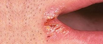
- rashes and skin itching;
- allergic reactions.
Since the main site of parasitism of the liver fluke is the gallbladder, the bile ducts, to which the helminth is attached, inflammation develops in their walls (cholecystitis, cholangitis), the movement of bile slows down and is blocked. There may be blockages of small ducts by helminths and the formation of stones.
At the same time, the outflow of pancreatic juice from the pancreas is disrupted with the expansion of its ducts and the proliferation of connective tissue (fibrosis). The most severe forms occur with destruction and cirrhosis of the liver, peritonitis (inflammation of the peritoneum). The long-term, long-term presence of the liver fluke in the biliary system can result in liver cancer, gall bladder cancer, and pancreatic cancer.
The causative agent of opisthorchiasis and its life cycle
Life cycle of the cat fluke
Cat or Siberian flukes (Opisthorhis felineus) are also called opisthorchid flukes. They look like a small parasite, 8-15 mm in length and no more than 2 mm in width. The parasite is yellow-brown in color, flat with suction cups at the base, with which it clings to the host’s body. The worm is a hermaphrodite, like others from the class of flukes, which allows it to lay eggs in the body of the final host without special conditions.
Going through a full life cycle, opisthorchia changes more than one host. Feces infected with parasite eggs end up in the reservoir. There is a settlement of freshwater snails. And for the next couple of months, development takes place inside this mollusk. The eggs will then hatch into cercariae, which wait to be swallowed by the fish. Mostly cyprinids are infected. But the most interesting thing is that the exception is the carp itself.
Predatory fish can become infected if they swallow fish with opisthorchiasis. From unprocessed, thermally contaminated fish, the cat fluke ends up in the final host - a person or a piscivorous animal. Then the cycle begins again.
This worm has a rather long development period. First, time passes until the larvae form special suckers while still inside the fish, with which they later cling to the mucous membrane of the internal organs. It is the suckers that lead to damage to the walls of organs and the development of bacterial infection. Sometimes a rash appears on the patient's skin as an allergic reaction.
In just one month, helminths mature inside the human body and begin to reproduce. In one week, 5000-10000 eggs are produced by just one individual. Eggs accumulate in the body, which leads to stagnation of bile.
Prevention of helminthic diseases
- cook the fish for 15-25 minutes;
- cooking cutlets, meatballs, etc. - 15-25 minutes;
- baking pies with fish for at least 45-60 minutes;
- cold smoking of fish must be carried out either after pre-salting for 2-3 days, or after freezing;
- fry large pieces of fish spread out and always in fat for at least 20 minutes, fry small fish whole for 15-20 minutes;
- salt the caviar (at a temperature of 5-6°C), the ratio of the amount of salt is 6% to the weight of the caviar for 12 hours, for example: 60 g of salt per 1 kg of caviar, or salting the caviar in a 5% solution (50 g of salt per 1 kg of caviar ) at least 2 days with periodic mixing of caviar;
- salt: small fish for 14 days, large fish (over 25 cm) for 40 days with the addition of 2 kg of salt per 10 kg of fish;
- drying of fish for at least 3 weeks with preliminary salting for 2-3 days; d freezing: at 40°C-7 hours, at 35°C-14 hours, -28°C-32 hours.
The danger of contracting opisthorchiasis and diphyllobothriasis does not disappear all year round.
Prevention
Watch a video on how to protect the liver from opisthorchiasis:
To avoid infection, you should remember a few simple rules:
- Do not buy fish from private individuals who do not check the product for parasites.
- Cook the fish for 20 minutes after boiling.
- To ensure that the fish is cooked well, it needs to be fried in small pieces rather than whole.
- Fry the fish only under a tightly closed lid for at least 20 minutes.
- Bake fish in the oven for at least one hour.
- It takes 2-5 weeks to salt the fish, depending on its size.
- For drying, you first need to salt the fish, and only then dry it for at least three weeks.
- The larvae die at a temperature of -40 in just a few hours. Or when frozen at 30 degrees below zero for more than two days.
- It is dangerous to eat raw and lightly salted fish.
- It is not recommended to eat prepared fish dishes in dubious catering establishments.
- Knives and cutting boards should be thoroughly disinfected after fishing.
- Before eating, you should wash your hands well for at least 30 seconds.
- Vegetables from the garden beds also need to be processed well.
Treatment for opisthorchiasis can only be prescribed by a specialist. Do not try to remove parasites yourself. This is not a simple disease that requires competent reduction of the level of intoxication and prevention of possible complications.
Immunity to opisthorchiasis is not developed, so you can become infected again. So be sure to follow the rules given above.
Why should you be careful with sushi?
Japanese cuisine is gaining more and more popularity, and the number of restaurants that offer sushi and rolls is growing every year. Therefore, many people are interested in whether sushi is dangerous for health? With all the obvious benefits of sushi, they are also fraught with danger. To prepare Japanese cuisine, raw fish is used, which means it may contain opisthorchid larvae.
Parasites do not die even when frozen and can live for up to a week. Since in standard freezers the temperature is not lower than -18 degrees, and to destroy the larvae requires freezing to -40 degrees. Therefore, sushi can cause opisthorchiasis.
How is the disease transmitted? Can you get infected from a person or a pet?
Wash vegetables with baking soda
When to be wary of infection and when not to:
- Fish eaten with opisthorchiasis that has not been properly processed is contagious.
- Infection does not occur in everyday life, through airborne droplets or through intimate contacts.
- Also, a sick animal is not contagious. However, opisthorchid larvae may remain on the hands or nails after contact with a pet. And from there it gets into the digestive tract. So always wash your hands thoroughly before eating anything.
- The larvae may end up on vegetables from soil that has contained feces from infected animals. If food is not processed before consumption, you can get opisthorchiasis.
- Also, parasites can end up in the body in accidentally swallowed water when swimming in ponds, lakes, and rivers.
To summarize, we can say that by adhering to basic hygiene rules, infection from a person or animal is excluded.
How and with what to treat opisthorchiasis in adults and children?
Hospitalization for opisthorchiasis is indicated:
- children under 3 years of age with any form of opisthorchiasis;
- children over 3 years of age with severe forms of acute and chronic opisthorchiasis;
- children of any age with dysfunction of the liver, pancreas, or impaired bile outflow.
Treatment of opisthorchiasis in adults includes:
- Praziquantel is a basic drug in the treatment of opisthorchiasis. Has a paralyzing effect on the parasite.
- Antiallergic drugs - used for allergic manifestations of the disease.
- Choleretic, anti-inflammatory drugs - eliminate stagnation of bile and inflammatory manifestations in the bile ducts.
An effective means of pathogenetic therapy for opisthorchiasis is the drug Ecorsol. The drug is especially indicated when other anthelmintic drugs are ineffective. The components of the drug provide:
- antiparasitic effect;
- anti-inflammatory effect – promotes the healing of erosions of the biliary tract;
- choleretic effect – stimulates the flow of bile through the ducts;
- antimicrobial effect – prevents the attachment and reproduction of pathogenic flora.
Ecorsol contains extracts of aspen bark and Solyanka holmovoi. What other herbs are used by traditional medicine to treat the disease, watch the video.
How does opisthorchiasis become infected?
Penetration of the pathogen into the body occurs due to insufficient cooking of fish. This problem is widespread among the indigenous inhabitants of endemic areas, since their main source of food is river fish. At the same time, the incidence in some areas of the Ob and Irtysh basin reaches 100%.
How does the pathogen parasitize the body?
The helminth larvae, having reached the duodenum, spread through the hepatobiliary system where they reach puberty and reproduce.
The pathogenic effect of the parasite on the liver and biliary system consists of:
- In the interaction of parasite antigens and body antibodies - the development of autoimmune allergic reactions.
- In the mechanical effect of helminths on the walls of the bile ducts, their inflammation, the development of dystrophic changes.
- In the blocking of the lumen of the biliary tract by opisthorchiasis and disruption of the outflow of bile.
- In the development of dystrophic changes in the liver and pancreas.