General information
Due to the high pace of life, working with equipment, driving vehicles, constant migration and active travel of people, the risk of injuries, including severe ones, increases.
In case of severe injuries and polytrauma, victims develop a traumatic disease - this is a set of changes and reactions that occur in the body from the moment of injury until its outcome. Traumatic illness occurs in several periods. The first of these is traumatic shock (ICD-10 code T79.4). This is a severe, critical condition, characterized by severe disturbances in vital functions, especially blood circulation - a critical decrease in blood flow in the tissues develops. Pulmonary and tissue gas exchange and metabolism in tissues are also disrupted, and endotoxicosis (poisoning of the body with tissue decay products) increases.
Hemodynamic disturbances arise due to hypovolemia (decreased circulating blood volume) that develops against the background of blood loss. In patients with severe shock, the volume of the liquid part of the blood also decreases due to its transfer into tissues (interstitial edema). As a result, the blood thickens sharply, the pressure decreases, and under these conditions the function of the heart as a pump decreases almost twice (small output syndrome). Thus, in shock, hypocirculation syndrome with impaired tissue perfusion in response to mechanical damage comes first. A type of traumatic shock is burn shock . They have much in common in pathogenesis and basic approaches to treatment.
The period of acute shock is followed by a period of multiple organ failure lasting 3-7 days. This is followed by a period of infectious complications (up to 1 month) and a period of delayed convalescence. Thus, disturbances in important body functions caused by injury are long-lasting. If in case of shock all measures are aimed at saving life (eliminating the causes of shock, correcting blood circulation and breathing), then in the future the traumatic disease requires long-term treatment until the outcome.
The processes occurring in later periods are associated with the initial hours of the disease. The relevance of the problem is that the state of shock not only poses a threat to life if assistance is provided incorrectly and untimely, but also has adverse consequences regarding the physical and mental health of a person.
Symptoms of traumatic and hemorrhagic shock
Injured patients are initially agitated, restless, and complain of intense pain in the area of damaged tissue. With severe pathology, depression of the nervous system activity is observed (lethargy, coma). Breathing is frequent and deep; auxiliary muscles are actively involved in it. As the central nervous system is depressed, breathing becomes weakened and shallow. In comatose patients, obstructive breathing disorders may occur (retraction of the tongue, accumulation of sputum and blood in the mouth and throat, and the flow of gastric contents).
The skin of patients becomes bluish in color and becomes “marbled” as the shock lasts. This is especially noticeable above the knees and on the hips. With significant blood loss (more than 30% of the blood volume), the skin is pale and moist. Due to impaired microcirculation in patients, peripheral body temperature decreases. The difference between rectal temperature and skin temperature is more than 3 degrees Celsius. A symptom of a “white spot” appears (when you press on the skin, a white spot appears, which normally disappears after 3 s. A significant prolongation of the time of existence of the spot indicates microcirculatory disorders).
Loss of blood and tissue fluid leads to a decrease in the turgor of subcutaneous tissue, the tone of the eyeballs, and the blood supply of the saphenous veins. The pulsation of peripheral arteries is weak; the pulse becomes frequent, soft, thread-like. Blood pressure and central venous pressure decrease. Due to hypotension and compensatory spasm of the renal arteries, renal blood flow (renal perfusion) is reduced. Oliguria develops; the hourly urine output rate is less than 0.5 ml/kg body weight.
Classification of blood loss (1998 , USA )
| Class | Symptoms | Volume of blood loss (% of total blood volume) |
| I | Orthostatic tachycardia | 15 |
| II | Orthostatic hypotension | 20-25 |
| III | Hypotension in the supine position, oliguria | 30-40 |
| IV | Impaired consciousness, collapse | More than 40 |
In traumatic shock, erectile and torpid phases are distinguished. The erectile phase does not always occur. It manifests itself at the prehospital stage, lasts a short period (up to several minutes) and is manifested by excessive agitation of the patient. The torpid phase is manifested by inhibition of the body's life-supporting systems.
To identify the volume of blood loss and the degree of shock depth, it is advisable to use the diagnostic indicator of the Algover shock index (the ratio between the heart rate and the value of systolic blood pressure). Normally, this indicator in healthy people is 0.5 - 0.7 (for example, with a heart rate of 60 per minute and a blood pressure system of 120 mm Hg, the shock index will be 60: 120 = 0.5). A decrease in blood pressure and compensatory tachycardia in patients in shock increases the shock index.
The relative simplicity of the study allows you to quickly determine the degree of shock and the amount of blood loss to adequately restore the deficit of blood volume. The following calculations are used.
Determination of the volume of blood loss using the shock index
| Shock index | Volume of blood loss (% of blood volume) | Shock level |
| Up to 1 | Up to 20 | 1 |
| 1,1-1,7 | 20-40 | 2 |
| 1.8 and higher | More than 40 | 3 |
For example, the victim’s body weight is 80 kg, hemodynamic parameters: blood pressure - 80 and 50 mm Hg, heart rate - 120 beats / min. The proper circulating blood volume for him is 70 ml/kg body weight, i.e. 70 x 80 = 5600 ml.
Shock index = 120: 80 = 1.5.
Degree of shock depth (according to the table) = 2.
With this value of the shock index, blood loss will be about 30% of the bcc. So, the patient lost 30% of 5600 ml, or
5600 x 0.3 = 1680 ml of blood.
As the shock lasts, even when the bleeding has stopped, the BCC continues to decrease (the so-called relative blood loss, caused by the stasis of red blood cells in the microcirculation vessels, their aggregation, rejection and exclusion from the general circulation).
When blood loss occurs, the compensatory mechanism of water entering the vascular bed from the intercellular space is activated and the phenomenon of “dilution” of blood occurs. Laboratory studies reveal a decrease in hemoconcentration parameters (hematocrit, hemoglobin, red blood cells and protein). This mechanism begins to work within the first minutes after injury. However, it is possible to finally assess the volume of lost blood based on hemoconcentration parameters only after 12-24 hours.
An immediate threat to life is rapid massive blood loss (more than 30% of the blood volume within an hour); with chronic anemia, the body is able to exist even with a deficiency of 70% of erythrocytes and 30% of plasma.
Clinical signs of hemorrhagic shock (according to G.M. Susla et al., 1999)
| Index | Blood loss | |||
| I class | II class | III class | IV class | |
| Blood loss (% of bcc) | <15 | 20-25 | 30-40 | More than 40 |
| Pulse | <100 | >110 | >120 | >140 |
| Arterial pressure | N | N | Reduced | Reduced |
| Pulse pressure | N or increased | Reduced | Reduced | Reduced |
| White spot test | N (2 s.) | Positive (more than 3 s.) | Positive | Positive |
| Respiration rate/min. | 14-20 | 20-30 | 30-40 | More than 40 |
| Hourly diuresis (ml/hour) | >30 | 30-20 | 15-5 | Expressed oliguria |
| Psychostatus | Minor concern | Moderate anxiety | Marked restlessness | Fainting, coma |
Pathogenesis
The pathogenesis of traumatic shock can be represented as follows: a deficiency of circulating blood and plasma due to blood loss and the release of the liquid part of the blood into the tissue, which leads to a decrease in venous return and a decrease in the stroke volume of the heart with the development of changes in vital organs and disruption of their function.
The trigger for any shock is the discrepancy between the volume of circulating blood and the volume of the vascular bed. As a result, the pressure decreases, to which the baroreceptors of the vascular wall react. Impulses from them enter the brain and the sympathoadrenal system is activated: adrenaline and norepinephrine and accumulate in them. Catecholamines , affecting the heart and blood vessels, cause increased frequency and intensification of heart contractions, vascular spasm (skin, kidneys, gastrointestinal tract). Centralization of blood circulation occurs. Catecholamines do not affect the blood vessels of the brain. This mechanism maintains pressure at a sufficient level until the supply of catecholamines is exhausted. These phenomena correspond to the erectile phase of shock.
When catecholamine reserves are depleted, the second phase of shock (torpid) develops. All spasmodic vessels lose their tone (dilate) and the cardiac output decreases. By this time, non-oxidized products have accumulated in the damaged tissues, which enter the bloodstream, which worsens the situation. , multiple organ failure quickly develops .
Causes and mechanisms of development
What is important for the development of traumatic shock is not so much the absolute amount of blood loss as the rate of blood loss. With rapid blood loss, the body has less time to adjust and adjust, and shock is more likely to develop. Therefore, shock is more likely when large arteries, such as the femoral artery, are injured.
Severe pain, as well as neuropsychic stress associated with injury, undoubtedly play a role in the development of the shock state (although they are not its main cause) and aggravate the severity of shock.
Factors leading to the development of traumatic shock or aggravating it are also injuries with damage to particularly sensitive areas (perineum, neck) and vital organs (for example, a wound to the chest, rib fractures with impaired external respiration function, traumatic brain injury). In such cases, the severity of shock is determined by the amount of blood loss, the intensity of the pain syndrome, the nature of the injury and the degree of preservation of the function of vital organs.
On this topic ▼
Shock
Types, phases, degrees and treatment
Rapid and massive blood or plasma loss leads to a sharp decrease in the volume of circulating blood in the victim’s body. As a result, the victim’s blood pressure quickly and severely drops, the supply of tissue with oxygen and nutrients deteriorates, and tissue hypoxia develops. Due to the lack of oxygen in the tissues, toxic under-oxidized metabolic products accumulate in them, metabolic acidosis develops, and intoxication increases. Lack of glucose and other nutrients in tissues leads to their transition to “self-sufficiency” - lipolysis (fat breakdown) and protein catabolism increase.
The body, trying to cope with blood loss and stabilize blood pressure, reacts by releasing various vasoconstrictor substances into the blood (in particular, adrenaline, norepinephrine, dopamine, cortisol) and spasm of peripheral vessels. This may temporarily stabilize blood pressure at a relatively “acceptable” level, but at the same time further worsens the situation with the supply of oxygen and nutrients to peripheral tissues. Accordingly, metabolic acidosis, intoxication with under-oxidized metabolic products, and catabolic processes in tissues intensify even more. Centralization of blood circulation occurs - the brain, heart, and lungs are primarily supplied with blood, while the skin, muscles, and abdominal organs do not receive enough blood. Lack of blood supply to the kidneys leads to a decrease in glomerular filtration of urine and a deterioration in the excretory function of the kidneys, up to complete anuria (lack of urine).
Spasm of peripheral vessels and increased blood clotting as a reaction to bleeding contribute to the blockage of small spasmodic vessels (primarily capillaries) with tiny blood clots - blood clots. The so-called “DIC syndrome” develops – disseminated intravascular coagulation syndrome. Blockage of small vessels further increases problems with blood supply to peripheral tissues and, in particular, the kidneys. This leads to a further increase in metabolic acidosis and intoxication. The so-called “consumption coagulopathy” may develop - a blood clotting disorder due to the massive consumption of clotting agents in the process of widespread intravascular coagulation. In this case, pathological bleeding may develop or bleeding from the injury site may resume, and further worsening of shock may occur.
On this topic ▼
Providing first aid depending on the situation
A decrease in blood supply to the adrenal glands and their function against the background of an increased need for glucocorticoids in “shock” tissues leads to a paradoxical situation. Despite the high level of cortisol in the blood (spike!), there is relative adrenal insufficiency. This is explained by the fact that less is “thrown away” than the tissues need, and the poorly supplied adrenal glands are physically unable to produce more cortisol.
The body's attempts to cope with pain by increasing the secretion of endorphins (endogenous analogues of opiates) lead to a further drop in blood pressure, the development of lethargy, lethargy, and anergy. The reaction to a decrease in blood pressure and a high level of catecholamines in the blood is tachycardia (rapid heartbeat). At the same time, due to insufficient circulating blood volume, cardiac output (stroke volume of the heart) is simultaneously reduced and there is weak filling of the pulse (up to a thread-like or undetectable pulse in the peripheral arteries).
Severe shock without treatment usually results in agony and death. In the case of a relatively mild or moderate shock, self-healing is in principle possible (at some stage, further development of the shock may stop, and then the condition will stabilize, the body will adapt and recovery will begin). But this cannot be relied upon, since the development of a shock state of any degree in itself indicates a failure of adaptation, that the severity of the injury has exceeded the compensatory capabilities of this particular organism.
Classification
According to the etiology (reason), traumatic shock occurs:
- As a result of mechanical injuries (fractures, severe injuries, tissue compression).
- As a result of burns.
- From exposure to low temperatures.
- As a result of exposure to electricity.
By time of occurrence:
- Primary - develops immediately after injury (1-2 hours) and is the result of injury.
- Secondary - appears after 5-24 hours as a result of additional trauma.
If we consider the clinical stages of traumatic shock, then two phases are distinguished:
- Erectile phase. In case of injury, impulses entering the central nervous system cause short-term excitation.
- Torpid phase. Short-term excitation is replaced by inhibition and true shock develops with inhibition of all functions. Hemodynamic disturbances prevail - a drop in arterial and venous pressure, a decrease in cardiac output, impaired gas exchange in tissues and metabolic disorders. As a result of circulatory and respiratory hypoxia , all organs suffer.
True shock is classified according to the severity of its manifestations. The following degrees of traumatic shock are distinguished:
- First degree (this is compensated shock). The victim is a little lethargic, the skin is pale, the extremities may be cold, breathing and cardiac activity are increased. Tachycardia up to 100 beats. Systolic pressure 100-90 mmHg. Art.
- Second degree (or subcompensated shock). The victim is slow and does not move. There is pallor and coldness of the skin, as well as a marbled pattern of the skin. Heart rate increases to 110-120, and pressure decreases to 80-75 mm Hg. Art. There is also a decrease in diuresis. The first two degrees have a favorable outcome, since protective-adaptive reactions and assistance prevent the development of hypoxia and deepening of shock.
- Third degree (decompensation). As a result of prolonged spasm of small vessels, hypoxia and cell damage occurs when assistance is delayed. The patient is lethargic, does not respond to external stimuli, and has an earthy-colored skin. Heart rate increases even more (130-140 beats), pressure progressively decreases (60 mm Hg and below), and diastolic pressure is often not determined. The patient develops anuria . This condition is typical for prolonged (hours) traumatic shock. However, properly performed resuscitation measures are often effective and the patient is brought out of shock. However, after removing patients from this state, 70% develop severe complications, the treatment of which is more complex than recovery from shock.
- Fourth degree (terminal, irreversible). Changes in the body reach the point where all measures taken are unsuccessful. Destructive processes in the body continue, deep decompensation of homeostasis and the victim dies.
Burn shock is one of the types of traumatic shock that develops with deep burns occupying more than 15% of the body. It is the first stage of burn disease. Its severity is influenced by the total area of the burn and its depth. The pathogenesis of burn shock is associated with pain from the burn area, which causes overexcitation of the central nervous system, and then hemodynamic and microcirculatory disorders occur. Generalized spasm of the arteries in the periphery in the next 1-2 hours maintains the pressure and functions of the life support organs, but after this the volume of circulating blood decreases due to loss of plasma, which increases over the course of two days. Victims experience increased blood coagulation and thrombosis . Oliguria appears associated with arterial spasm.
Large loss of plasma, blood thickening and the formation of toxic substances in the necrosis zone after a burn leads to disturbances in the acid-base state and electrolyte balance. A peculiar clinical picture is noted: at first the patient is agitated, talkative, fidgety, and inadequately assesses the condition. After this, excitement gives way to inhibition, as the patient develops hypovolemia - its degree depends on the severity of the burn. There are also 4 degrees of severity, which depend on the area of the burns.
- A mild degree develops with superficial or deep burns, the area of which is up to 10% of the surface. Consciousness is preserved, pale skin, muscle tremors, nausea, and sometimes vomiting. Moderate tachycardia , blood pressure within normal limits. The victims can be brought out of shock by the end of the day.
- Moderate severity of shock occurs with superficial burns, occupying 20-40% of the surface. This stage is characterized by excitement turning into inhibition. The patient's consciousness remains intact, the skin is pale and cold. The victim is worried about thirst and nausea, breathing is rapid, blood pressure is reduced, oliguria and kidney function is impaired (residual nitrogen increases, blood and protein appear in the urine). The victims can be brought out of this state within two days.
- Severe degree accompanies superficial burns of 40-60% of the surface. The patient's condition is extremely serious, confusion and lethargy are noted. Patients are pale gray in color and have severe thirst, vomiting, seizures , tachycardia and shortness of breath . Oliguria is noted , and in older patients anuria . The level of residual nitrogen increases to 51-56 mmol/l. Treatment is not always effective.
- An extremely severe degree is observed with burns affecting 60% of the surface. The condition is very serious, there is no consciousness. The victim's temperature is reduced, there is severe shortness of breath, and the pulse is thready. The vomiting of “coffee grounds” is disturbing, intestinal paresis progresses, and metabolic acidosis intensifies. Patients have anuria, blood and protein are detected in the urine, residual blood nitrogen is above 60 mmol/l. Victims in this condition die within the first or second day.
In clinical practice, we encounter not only post-traumatic shock associated with various injuries, but also shock that develops against the background of various diseases, for example, the cardiovascular system. Thus, arrhythmic shock (another name is “cardiogenic”) occurs in 4-5% of patients with large-focal transmural myocardial infarction . Its development is associated with a decrease in cardiac output due to disturbances in heart rate. It occurs in the form of tachysystole (rapid contractions of the heart) or bradysystole (excessive slowdown of contractions).
Tachysystolic shock develops in patients with atrial fibrillation. In this condition, diastole is shortened, therefore the filling of the heart decreases and its minute volume decreases. However, the leading role in the development of shock is given to ventricular tachysystole, in which it develops in the first hours of the disease. The condition of the patients is serious, since the pressure decreases significantly and diuresis decreases. Patients are given adequate pain relief (narcotic analgesics) and antiarrhythmic drugs are administered.
With intractable tachysystole, pulmonary congestion and right ventricular failure develop. The prognosis for tachysystolic shock is unfavorable. Mortality is 40% and its main cause is progressive heart failure. Bradysystolic shock develops with atrioventricular block , junctional rhythm and Frederick's syndrome . The course is severe, and the mortality rate reaches 60%.
Among the complications of acute pancreatitis is pancreatogenic shock , which occurs in 20% of patients. It most often develops with necrotizing pancreatitis and is characterized by unstable hemodynamics. The cause of the development of this type of shock is endotoxemia due to necrotic damage to large volumes of pancreatic tissue. The volume of pancreatic necrosis determines the likelihood of endotoxic shock. The timing of its appearance is different, and therefore there are early (appears in the first week of the disease) and late (develops in the third week) shock.
The main cause of shock of this nature is a decrease in the volume of circulating blood. This can occur due to swelling of the gland tissue, saturation of the retroperitoneal space with fluid, accumulation of hemorrhagic fluid in the abdominal cavity and intestinal loops, as well as due to stagnation of blood in the portal system of the liver. This loss of extracellular fluid leads to hypovolemia and shock.
In early shock, the patient experiences tachycardia (or bradycardia), signs of peritonitis , decreased blood pressure, cold extremities and facial cyanosis . Late shock occurs against the background of generalized sepsis (fever, leukocytosis ), which is accompanied by hemodynamic instability, which, in fact, is shock.
Kinds
Shock can be primary (early) , which occurs immediately after injury and is an immediate reaction to injury. Secondary (late) shock occurs 4-24 hours after the injury and even later, often as a result of additional traumatization of the victim (during transportation, cooling, renewed bleeding, tightening a limb with a tourniquet, from rough manipulations during the provision of medical care, etc.). A common type of secondary shock is postoperative shock in the wounded. Under the influence of additional trauma, relapses of shock in victims are also possible, usually within 24-36 hours. Shock often develops after the tourniquet is removed from the limb.
Symptoms
Symptoms of pain shock differ in the erectile and torpid phases. The first is very short, it occurs immediately after the injury, does not happen to all victims and is characterized by excitation of the sympathoadrenal system . The patient retains consciousness, there is arousal (motor and speech), rapid breathing, satisfactory pulse filling, not accelerated, blood pressure is normal or increased. The victim's behavior resembles a state of alcohol intoxication.
In the torpid phase, signs of traumatic shock include lethargy, mental depression, an indifferent attitude towards everything, and lack of response to pain come to the fore. The temperature is low, the skin is cold, clammy sweat, breathing is rapid, pulse is increased, blood pressure is reduced, urination is impaired, the severity of which depends on the stages of shock.
- In the first degree (considered as a mild degree), the general condition is satisfactory, the patient is conscious, there may be slight lethargy, pulse up to 100 beats, pressure 95-100 mm Hg. Art. The prognosis is favorable. If help is not provided or additional trauma occurs, shock of this degree may progress to the second degree.
- In the second degree, severe lethargy is noted, pressure (90-75 mm Hg) and temperature decrease, pulse 110-120 beats, breathing becomes rapid. Pale skin with a bluish tint. The prognosis for this degree is serious. Recovery from shock is possible, but requires long-term (possibly several days) anti-shock therapy.
- Stage 3 pain shock is severe. In the general serious condition of the victim, a sharp lethargy appears, a decrease in pressure of 70 mm Hg. Art. (below a critical level), increased heart rate, which has very weak filling and tension. Decreased diuresis and anuria. If the causes are not eliminated in a timely manner and assistance is not provided, it enters a terminal state.
- Shock of the 4th degree is a preagonal state. The patient's pressure and pulse are not determined in the radial arteries, but a weak pulsation is still detected in the large arteries, heart sounds are barely audible, and rare shallow breathing is noted. Reflexes are not evoked, anuria. In the agonal state, respiratory disturbances ( Chine-Stokes breathing ) are more pronounced. Clinical death is considered from the moment of cardiac arrest and the last breath. The functions of the central nervous system are completely absent, but metabolic processes in the brain continue for another 5-6 minutes.
Trauma of any location is accompanied by blood loss. There is a relationship between it and the severity of the shock. In the first degree, blood loss is 500 ml, in the second 1,000 ml, and in the third - 1,500 ml or more.
Patients who have suffered severe trauma and stress develop post-traumatic syndrome, which is characterized by changes in the psycho-emotional sphere. The patient develops unmotivated vigilance, he monitors what is happening around him and does not leave him with a feeling of threatening danger. Patients have an explosive reaction when they throw themselves to the ground at unexpected loud noises. It becomes difficult for such patients to establish contacts with others and they experience dullness of emotions. From time to time, aggressiveness and the desire to solve all problems with the help of force arise, getting one’s way.
The most important symptom is uninvited memories. They are the ones who give the right to say that the patient has post-traumatic syndrome . Scenes associated with a trauma, disaster, fire or other event emerge in his memory. Memories arise during sleep, and when awake they appear in cases where the situation is somewhat reminiscent of what happened (this could be a smell, sound or sight). The patient is worried about anxiety, constant worry, a feeling of fear, fear of persecution, constant preoccupation. An important factor in this condition is depression , when a person feels that everything is useless, there is a negative attitude towards life and apathy. It is also possible to develop a tendency to abuse alcohol and drugs. In some patients, post-traumatic syndrome is very pronounced and requires psychiatric correction.
Phases
Regardless of the causes of shock, it goes through two phases - erectile (excitement) and torpid (inhibition).
Erectile
This phase occurs at the moment of traumatic impact on a person with a sudden sharp excitation of the nervous system, manifested in excitement, anxiety, and fear. The victim remains conscious, but underestimates the complexity of his situation. He can answer questions adequately, but has impaired orientation in space and time.
The phase is characterized by pale human skin, rapid breathing, and severe tachycardia. Mobilization stress in this phase varies in duration; shock can last from several minutes to hours. Moreover, with severe trauma, it sometimes does not manifest itself in any way. And too short an erectile phase often precedes a more severe course of shock in the future.
Torpidnaya
Accompanied by a certain inhibition due to inhibition of the activity of the main organs (nervous system, heart, kidneys, lungs, liver). Circulatory failure increases. The victim becomes pale. His skin has a gray tint, sometimes a marble pattern, indicating poor blood supply, stagnation in the blood vessels, and he breaks out in cold sweat. The limbs in the torpid phase become cold, and breathing becomes rapid and shallow.
The torpid phase is characterized by 4 degrees, which indicate the severity of the condition.
- First degree.
Considered easy. In this condition, the victim has a clear consciousness, pale skin, shortness of breath, slight lethargy, the pulse reaches 100 beats/min, the pressure in the arteries is 90-100 mm Hg. Art.
- Second degree.
This is a moderate shock. It is characterized by a decrease in pressure to 80 mm Hg. Art., pulse reaches 140 beats/min. The person has severe lethargy, lethargy, and shallow breathing.
- Third degree.
An extremely serious condition of a person in shock, who is in a confused state of consciousness or has completely lost it. The skin becomes earthy gray in color, and the fingertips, nose and lips become bluish. The pulse becomes thread-like and increases to 160 beats/min. The man is covered in sticky sweat.
- Fourth degree.
The victim is in agony. Shock of this degree is characterized by a complete absence of pulse and consciousness. The pulse is barely palpable or completely imperceptible. The skin is gray in color, and the lips become bluish and do not respond to pain. The prognosis is most often unfavorable. The pressure becomes less than 50 mm Hg. Art.
Tests and diagnostics
In the diagnosis of shock, examination, physical examination and instrumental examination methods are of primary importance. The patient is continuously monitored for vital signs to monitor the severity of shock and the effectiveness of treatment.
- Physical examination gives a general idea of the severity of the condition.
- The general condition ranges from moderate to very severe. Impaired consciousness from confusion to coma.
- Characteristic appearance: pale face, cold sticky sweat, cyanosis of the extremities, decreased temperature.
- Frequent weak pulse, low blood pressure, rapid and shallow breathing.
- Damage to internal organs.
- Fractures, crushed tissue.
At the hospital level, examinations, depending on conditions and indications, include:
- General radiography (extremities, skull, pelvis, chest, abdominal cavity).
- Ultrasound of the abdominal and pleural cavity.
- Carrying out laparoscopy .
- Measurement of central venous pressure.
- Conducting thoracoscopy and bronchoscopy .
- Computed tomography.
- MRI.
Shock
Which doctors should I contact?
At the first signs of shock, you should immediately call an ambulance.
Treatment of shock
A patient with shock of any type should be taken to the intensive care unit according to the expected profile of the disease. In case of injury, in the presence of signs of internal bleeding, as well as in situations where the cause of shock could not be determined at the prehospital stage, hospitalization in an anti-shock operating room (or another operating room intended for the treatment of victims with shock) is indicated.
Main goals of therapy:
- interrupt pain impulses;
- normalize the volume of circulating blood and the rheological properties of blood;
- correct metabolism;
- eliminate the causes of organ disorders.
1. Ensuring airway patency.
Oxygen therapy is carried out through a mask. In cases of severe shock or inadequate ventilation, intubation of the airway with mechanical ventilation is necessary. 2. Installation of intravascular catheters for rapid infusion of solutions and blood components, and, if necessary, administration of large quantities of medications (including catecholamines).
3. Etiological treatment.
4. Support the circulatory system and oxygen transport:
- discontinuation of antihypertensive drugs, if any were used;
- in most types of shock, the main importance is to replenish the intravascular volume by infusion of plasma substitutes and plasma expanders, solutions of crystalloids and colloids;
- infusion of vasopressor drugs to eliminate hypotension;
- patients with low cardiac output in the absence of cardiac arrhythmias are prescribed a continuous intravenous infusion of dobutamine; with concomitant hypotension, vasoconstrictor drugs are simultaneously used;
- oxygen therapy to reduce hemoglobin oxygen saturation.
5. The main method of correcting lactic acidosis is etiotropic treatment that supports the functions of the circulatory system.
6. Monitoring vital signs.
7. Prevention of bleeding from the gastrointestinal tract and thromboembolic complications.
8. Correction of hyperglycemia.
After resuscitation, specific treatment for the underlying disease is prescribed.
Additional supportive care depends on the type of shock. Complications
The danger of shock is a decrease in blood supply to vital tissues. The prognosis depends on the cause of shock and the timeliness of treatment.
If left untreated, shock is fatal.
The development of hypovolemic shock is accompanied by a mortality rate of up to 70%, depending on its severity. With timely assistance provided in full, the mortality rate does not exceed 25%. Distributive shock is characterized by varying mortality rates, depending on the etiological factor, and can reach 70% in the case of septic shock. Obstructive shock associated with fairly effectively removable causes is accompanied by a mortality rate not exceeding 15%. With pulmonary embolism, the mortality rate exceeds 30%. Even with treatment, mortality from cardiogenic shock after myocardial infarction remains very high and reaches 70%.
Prevention of shock
Shock is a condition, a process that cannot be considered separately from known nosological forms. Therefore, shock cannot be the cause of death; the cause of death becomes the factor that caused the state of shock. Prevention of shock involves timely treatment of diseases that can lead to its development.
Sources:
- Great Medical Encyclopedia (BME), edited by Petrovsky B.V., 3rd edition, volume 27.
- Clinical guidelines (protocol) for providing emergency medical care for shock in children. Russian Society of Emergency Medical Care Union of Pediatricians of Russia, 2015, p. 21.
- Moroz V.V., Bobrinskaya I.G., Vasiliev V.Yu. et al. Shock. Educational and methodological manual for students, residents, graduate students and doctors. - Moscow, 2011.
IMPORTANT!
The information in this section cannot be used for self-diagnosis and self-treatment. In case of pain or other exacerbation of the disease, diagnostic tests should be prescribed only by the attending physician. To make a diagnosis and properly prescribe treatment, you should contact your doctor.
Diet
The patient’s nutrition depends on his condition and the presence or absence of injuries to the abdominal organs for which surgery was performed. If the victims are in serious condition after burns or in a comatose state due to polytrauma, they are provided with parenteral nutrition, which is included in the complex of treatment measures. Before it is carried out, the victim’s condition must be stabilized and hypoxia eliminated. Therefore, in terminal conditions and shock, only glucose solutions with insulin . After the glucose infusion, an amino acid preparation or protein hydrolysate . Amino acid mixtures: Polyamine , Panamin , Levamin-80 , Levamin-normo , Alvezin , Aminoplasmal , Aminofusin , Moriamin , Trofisan , Okhamin , Friamin . Protein hydrolysates: Casein hydrolysate , Aminofusin , Aminonorm , Aminoplasmal , Aminomel , Aminovenoz , Aminon , Amigen , Izovac .
Then amino acid mixtures or protein hydrolyzate are administered with vitamins, electrolytes and glucose. Fat emulsions are poured together with amino acid mixtures and hydrolysates. They should not be administered with electrolytes, since electrolytes enlarge fat particles, which significantly increases the risk of fat embolism. Fat emulsions include Lipofundin 10% , Intralipid , Lipomul 15% , Lipofundin 15% , Lipifizan 15% . Preparations based on soybean oil are also produced: Lipofundin-S 20% , Emulsan , Venolipid , Infusolipol . The main components of parenteral nutrition are distributed as follows: proteins make up 10-15% of the total daily caloric intake, carbohydrates - 50%, and fats - 35-40%. Vitamins are also an integral component of parenteral nutrition, they are administered separately, and there are also multivitamin complexes for parenteral administration - these are Vitafuzin , Protavit , Soluvit , which contain the daily requirement of essential vitamins.
As the patient's condition improves, the patient is transferred to enteral nutrition with formulas. This is especially indicated if there were abdominal injuries with damage to the gastrointestinal tract or if operations were performed on these organs. Semi-element mixtures are fully balanced with nutrients; proteins are represented by peptides and protein hydrolysates. The most commonly used are Peptamen and Nutrien Elemental - these are specialized medical nutrition products that can be used as the only source of nutrition. Available in powder form, which is diluted with warm boiled water and taken orally.
When switching to a normal diet, the patient must gradually expand the food load, starting with Dietary Table No. 1 with the transition to the standard Diet No. 15 with the physiological content of proteins, fats and carbohydrates, vitamins and minerals.
Particular attention is paid to nutrition in case of burn disease, in which there is an increased breakdown of muscle and visceral proteins. For such patients, the following scheme is used: parenteral nutrition - transition to enteral nutrition - siping (consumption of specialized mixtures in small sips) - transition to specialized diets with a high protein content.
Prevention
The following preventive measures can prevent the development of shock or reduce its severity:
- Timely stopping of bleeding.
- Reliable immobilization for fractures.
- Carrying out analgesia (pain relief).
- Knowledge of the rules for providing self- and mutual assistance and the ability to apply them in the right environment.
- Because serious injuries often occur in the workplace, increased worker vigilance and safety monitoring by management can significantly reduce injuries and related complications.
Consequences and complications
Complications of shock include:
- Increasing respiratory failure ( respiratory distress syndrome ).
- Coagulopathic disorders with possible transition to disseminated intravascular coagulation.
- Traumatic endotoxicosis . Endogenous intoxication develops due to the accumulation of tissue destruction products and is caused by bacterial toxins and vasoactive substances. The effect of these factors manifests itself 48-72 hours after injury.
- Development of fat embolism with bone injuries and extensive destruction of soft tissues.
- Hepatic-renal failure (development of “shock kidney” and “shock liver”).
- Heart failure and central hemodynamic disorders.
- Infectious and septic complications.
- Delay of reparative processes at the site of damage.
The consequence of trauma and shock is post-traumatic syndrome. It develops in people who have experienced life-threatening events (transport accidents, plane crashes, natural disasters) and received injuries as a result. These events deeply affect a person’s psyche, and he loses a sense of security, anxiety, fear appear, sleep disturbances occur, suicidal tendencies and alcohol or drug abuse occur. If the trauma and stress suffered were severe, the painful mental reaction can persist for many years.
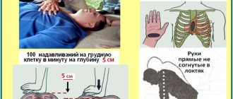
Urgent measures
- It is necessary, first of all, to stop arterial bleeding by pressing the artery to the bone above the site of injury, applying an arterial tourniquet or twisting it above the site of injury. In this case, you should record the time when the tourniquet was applied.
- Assess the state of vitality of the body (determine the presence and nature of the pulse over the peripheral and central arteries, the degree of depression of consciousness, airway patency, the effectiveness of external respiration function).
- Ensure the correct position of the victim's body. In an unconscious state, he should be turned on his side, his head thrown back and the upper half of his body slightly lowered. A separate position is required for patients with fractures of the spine (on a hard surface) and pelvic bones (with legs bent at the joints and spread apart). It is contraindicated to throw the head back in patients with a cervical spine injury!
- Provide immobilization of injured limbs with standard splints or available material.
- Apply a bandage to the wound. In case of venous or capillary bleeding, a compressive bandage has a hemostatic effect. For open pneumothorax, an adhesive bandage seals the chest.
- Anesthetize the patient. Narcotic and non-narcotic analgesics are used (1% promedol solution, 50% analgin solution).
In patients with decompensated shock, narcotic analgesics can suppress the respiratory center. In addition, excluding the stimulating influence of pain reduces the activity of the adrenal glands; in patients with BCC deficiency, this can lead to threatening hypotension. The high probability of these complications requires continuous monitoring of patients. If it is impossible to provide such supervision (for example, in case of mass arrival of victims, their transportation), for the purpose of pain relief, it is advisable to use a medium for non-inhalation anesthesia - ketamine (2-3 ml of 5% solution, administered intramuscularly). It exhibits an analgesic effect, stabilizes blood pressure and does not suppress the respiratory center. To prevent the unwanted hallucinatory effects of ketamine, it is advisable to combine it with the administration of 1-2 ml of a 0.5% sibazon solution.
10-20 ml of 0.5% novocaine solution is injected into the fracture sites. As part of a specialized team, a nurse performs venipuncture and catheterizes peripheral vessels, assists the doctor with catheterization of arteries and main veins, prepares systems for intravenous infusions, measures arterial and central venous pressure, records an electrocardiogram, provides oxygen therapy and artificial ventilation to the victim, etc.
Forecast
The prognosis depends on the degree of shock: in the first degree it is favorable, in the second it is doubtful, in the rest it is not favorable. Despite the improvement of anti-shock therapy methods, mortality in severe injuries has decreased slightly. As stated above, shock is a life-threatening condition, but death does not occur from painful shock. The main cause of death from injuries is blood loss in the absence of medical attention. Pain aggravates shock and is not its main cause, much less the cause of death.
Death can also occur after recovery from shock due to multiple organ failure. The immediate cause of death is heart, kidney and liver failure , increasing respiratory failure and coagulopathic disorders.
If death is prevented during the first week, then the threat of generalization of wound infection ( wound sepsis , pneumonia ) is of decisive importance in the patient’s condition. It is infectious complications that occur at a later date that affect the survival of patients. This threat persists for several weeks, as victims of the trauma experience severe metabolic disorders and impaired immunity .
List of sources
- Bagnenko S. F., Lapshin V. N., Shah B. N. Hemodynamic depression in victims with combined trauma in the acute period of traumatic illness is the basis for subsequent hypoxic changes and reperfusion injuries // Efferent Therapy, 2004. Vol. 1, No. 5 . pp. 23–34.
- Shestopalov A. E., Pasko V. G. Volume replacement therapy for acute blood loss in victims with severe combined trauma // Difficult Patient. 2005. No. 4. pp. 17–23.
- Gural K.A., Klyuchevsky V.V., Dambaev G.Ts. Human traumatic shock // International Journal of Experimental Education. – 2011. – No. 12. – P. 15-16.
- Singaevsky, A.B. Current problems of modern severe trauma // Abstracts of the All-Russian Scientific Conference. - St. Petersburg, 2001. - pp. 106-107.
- Sheiko V.D. Surgery of injuries during polytrauma in peacetime and wartime / Textbook. — Poltava ASMI LLC. — 2015. 558 p.