Macular degeneration of the retina is one of the causes of irreversible loss of central vision in people over 55-60 years of age. In 2007, age-related macular degeneration was responsible for 8.7% of all cases of blindness on the planet. According to the current trend, this number of cases is expected to double by 2021.
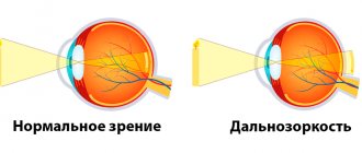
The reason for the decline of visual functions is degeneration of the macula - the most significant part of the retina, responsible for the acuity, sharpness and level of central object vision required for visual work or reading text at close range or driving a vehicle, while peripheral vision in patients such as usually does not suffer at all.
Macular degeneration leads to loss of objective vision, a decrease in overall performance and subsequent disability of the patient, which determines the high socio-medical significance of the disease. At the same time, macular degeneration of the retina can provoke both a slow gradual decrease in vision over several years, and rapid, within just a few months, loss of vision, which depends on the form of age-related macular degeneration and the severity of the disease.
What is the essence of age-related retinal degeneration?
To understand the essence of the pathological process, it is necessary to navigate the structure of the light-sensitive part of the eyeball - the retina. The retina is located at the back of the organ of vision and consists of two main layers. The inner layer is made of special light-sensitive cells - rods and cones. These cells act as receptors - they react to the light signal entering the retina and transmit data about it to the optic nerve. Cones help you see objects in daylight and also form color vision. The rods, in turn, are responsible for twilight vision. The outer layer of retinal cells is made of retinal pigment epithelium, which performs a protective function and is involved in the nutrition of light-sensitive receptors.
| The structure of the retina. Photoreceptors |
The macula or macula is a small part of the retina that is responsible for central vision. The macular region has the highest density of photoreceptors. In the very center there is a special depression - the central fovea or fovea, made only by cones. It is the central fovea that is the main point responsible for human object vision.
| The structure of the eye. Macula |
Age-related macular degeneration of the retina affects this particular area, which is accompanied by a decrease in central object vision, up to irreversible blindness. Age-related macular degeneration is characterized by the deposition of cellular breakdown products between the retina and the choroid (choroid). This process is also associated simultaneously with morphologically determined hyper- and hypopigmentation of the retina. Such initial changes do not yet cause deterioration and reduction in visual acuity. However, further progression of the disease determines clinically significant changes. There are two forms of age-related macular degeneration of the retina, which will be discussed in detail below.
What is macular degeneration?
AMD is the leading cause of central vision loss and social blindness in people over 50 years of age. The macula, the area of the retina located closer to the center, is mainly affected. It is she who is primarily responsible for our ability to distinguish small objects, read, and estimate short distances with high accuracy.
With age, the supply of essential substances to the fundus tissues is often disrupted. Next, a cascade of pathological changes is launched: abnormal vessels are formed, blood and plasma seep through their walls, and damaged areas of the retina become scarred. As a result, central vision disappears.
As a result, serious restrictions appear in a person’s daily life - the inability to drive a car, do usual work, pay for purchases in a store, etc. All this can contribute to the development of depression and mental disorders. It is important to understand that even in the worst case scenario, complete blindness does not occur.
Dry form of macular degeneration
Atrophic or dry age-related macular degeneration accounts for about 85%-90% of cases of this disease and occurs with equal frequency among male and female patients.
This form of the disease is characterized by the deposition of cellular breakdown products, so-called drusen, between pigment epithelial cells and Bruch's membrane. Bruch's membrane is an acellular formation consisting of 5 layers and acts as a barrier between the retina and the choroid. Nutrients and oxygen necessary for normal functioning diffuse through Bruch's membrane to the pigment epithelium and photosensitive receptors of the retina. Metabolic products, on the contrary, are transported from the retina to the choroid.
With age, Bruch's membrane undergoes significant morphological changes, which include thickening, calcification, and degeneration of collagen and elastin fibers. Incomplete elimination and accumulation of metabolic products of lipid nature also occur. Deposits of metabolic products consisting of lipofuscin are called drusen. Drusen are the earliest indicator of macular degeneration of the retina and come in two varieties - soft and hard.
| Macular degeneration. Dry form |
Hard drusen are small, round deposits with well-defined boundaries. They are often a sign of age-related changes in the retina, but do not cause clinically significant visual impairment. As macular degeneration of the eye progresses, small single deposits accumulate into larger formations - soft drusen.
The appearance of soft confluent drusen is associated with an unfavorable prognosis for maintaining high visual vision. There is a disconnection between the retina and the choroid, which disrupts the nutrition of all cellular layers of the retina, causes degradation of photoreceptors and gradual replacement of damaged retinal cells with scar connective tissue.
Geographic atrophy is the final stage of dry macular degeneration, in which large areas of atrophy and death of the retinal pigment epithelium and proliferation of connective tissue are visualized. Such processes lead to significant loss of central vision, which can progress over years. Slow decay of visual functions and a decrease in central vision significantly reduce the patient’s ability to visually work, but not as pronounced as with the wet form of macular degeneration.
Causes and risk factors
The start of the development of pathology is a violation of tissue nutrition, which occurs in the fundus through very small vessels. What could contribute to this?
- Heredity
is the main factor. - Gender
- macular degeneration is diagnosed in women 2 times more often than in men. - Age
- the average age of patients ranges from 55 to 80 years. - Unbalanced diet
- cholesterol plaques are deposited on the walls of blood vessels, narrowing the already small lumen. - Cardiovascular diseases
- for example, in patients with hypertension, the blood vessels constantly receive microtraumatic lesions, which the body “overgrows” with the same plaques, impeding the flow of blood. - Diabetes
. - Smoking
. - Exposure to direct sunlight and ultraviolet radiation
- prolonged and intense exposure to the retina of the eye can cause microscopic burns of the macular area. - Unfavorable environmental conditions
. - Injuries and previous eye diseases
. - Alcohol consumption
- even one-time consumption leads to aggregation (sticking together) of red blood cells, which form “clusters” of several or several dozen cells. Such a lump clogs the lumen of the vessel, which has a much smaller diameter. With regular consumption of alcoholic beverages, vascular disorders become irreversible.
If the process is not stopped at the initial stage, it will progress and in the foreseeable future may lead to loss of central vision.
Wet macular degeneration
Neovascular or wet age-related degeneration of the macula and posterior pole of the eye is an alternative pathway for the development of a pathological process in which the separation of the retinal pigment epithelium and choroid is accompanied by an increase in the concentration of vascular endothelial growth factor. This biologically active substance stimulates angiogenesis, that is, the ingrowth of newly formed vessels under the retina in the projection of the central macular zone.
Neovascularization processes are accompanied by vasodilation, impaired vascular permeability and migration of endothelial cells. Newly formed vessels, penetrating into the subretinal space, destroy the anatomical barrier in the form of Bruch's membrane between the choroid and the retina and form a kind of vascular network, called the “subretinal neovascular membrane”. The wall of the newly formed vessels is functionally defective, which leads to leakage of fluid, plasma and blood cells under the central zone of the retina and is accompanied by subretinal hemorrhages of varying volumes into the macula.
| Age-related macular degeneration of the retina. Wet form |
The constant presence of blood and fluid under the retina ultimately leads to the separation of Bruch's membrane, pigment epithelium and the photosensitive layer of the retina from each other, with subsequent disruption of the structure and function of photoreceptors, their degenerative degeneration, fibroglial transformation of tissue in the macular zone into a single scar conglomerate. Over time, a specific ridge forms in the area of the macula, surrounded by scar tissue and small hemorrhages.
Clinically, the processes described above are manifested by disturbances in central vision and the appearance of dark spots (scotomas) before the eyes. Thus, choroidal subretinal neovascularization, being a reparative reaction of the body aimed at improving the trophism of the central retina, increasing the supply of oxygen and nutrients to the macula, leads to the progression of the disease and the inevitable loss of objective vision.
The wet form of age-related macular degeneration of the retina often develops in a short time - the disease can greatly impair the patient's quality of life in a few months or even weeks.
How is AMD diagnosed?
Changes in vision can be determined at home yourself using a simple test that uses the Amsler grid. This test is intended both to identify diseases of the central retina and to monitor the dynamics of treatment for existing pathology of the central retina. The Amsler test should be placed at a distance of 30 cm from the eye, and the other eye should be covered with your hand, then focus on the bold dot in the center of the test. If you find any changes, mark them on the Amsler test or sketch how you see them, and take them with you to an appointment with an ophthalmologist.
Causes of development of age-related macular degeneration
Until now, scientists have not been able to identify the only reliable cause of age-related macular degeneration of the eyes. However, age-related macular degeneration is believed to have a direct correlation with the age of the patients. Thus, in patients of the middle age group, the disease occurs in only 2% of cases; at the age of 65-75 years, the disease is detected in 20% of patients. And when people reach the 75-year barrier, the risk of developing the disease increases by 35%, that is, age-related macular degeneration of the retina is diagnosed in every third person. That is why, according to most researchers, the main reason for the development of the disease is age.
However, there are a lot of predisposing factors that, when combined with hereditary predisposition, determine the high risks of this pathology. Some of them will be listed below:
- The risk of vision loss associated with macular degeneration in smokers is twice as high as in people without this bad habit.
- Arterial hypertension, various cardiac pathologies, alcohol abuse, obesity, and Alzheimer's disease increase the likelihood of developing degenerative changes in the retina.
- Hereditary predisposition, especially in combination with smoking, should increase suspicion for age-related macular degeneration of the retina.
All these factors cannot directly influence the condition of the retina. However, the biochemical reactions induced by them are the basis of macular degeneration of the eyes.
The retina is thought to be particularly susceptible to oxidative stress due to chronic exposure to visible light and high oxygen concentrations. The discovery of the role of oxidative stress in the development of macular degeneration determined the possibilities of preventive treatment with antioxidants of individuals with an increased risk of age-related macular degeneration of the retina. This issue will be discussed in more detail in the section on therapeutic options.
Symptoms of AMD
As a rule, the patient simply does not notice the onset of the disease due to the absence of any “acute” vision problems. But, given that retinal dystrophy affects central and color vision, the standard sequence of disease development can be described as follows.
- First, the brightness and contrast of color perception are lost and visible lines are distorted, and the patient also lacks the illumination that was sufficient before.
- At the next stage, when reading and writing texts with the usual glasses, the Patient notices the loss of individual letters or entire words.
- Then there is a loss of visual acuity at near and far distances.
- Later, as the disease progresses, a spot appears in front of the eye, at first translucent, then completely opaque, sharply worsening vision, depriving the Patient of the ability to distinguish objects and people’s faces.
Age-related macular degeneration is a disease of a paired organ, the damage extends to both eyes. Most often, retinal dystrophy manifests itself and progresses more actively in one eye. The manifestation of the disease in the fellow eye may not occur immediately, even 5-8 years after the diagnosis of AMD. If the Patient does not undergo regular check-ups with an ophthalmologist, he himself may not immediately notice any vision problems that have arisen, since the better-seeing eye takes on the additional load.
Symptoms of age-related macular degeneration
The initial stages of age-related macular degeneration, especially if only one eye is involved in the pathological process, are most often asymptomatic. There are also no pain sensations that could cause discomfort and prompt a person to visit an ophthalmologist. Age-related macular degeneration of the retina has many symptoms that affect the daily life of patients, the main of which are the following:
- Reduction in varying degrees, up to complete loss, of object vision with the formation of a spot or spots of gray or black colors in the central field of vision. Distortion of the image in the form of metamorphopsia - the objects in question have an elongated shape, a larger or smaller size than they actually are, broken straight lines. These symptoms are the most common and characteristic of pathology of the macular zone.
- Cloudy and defective central vision causes problems with reading, writing, driving, watching TV, and recognizing faces.
| Healthy person's vision | Vision of a patient with retinal degeneration |
- Impaired contrast sensitivity. It becomes difficult for patients to distinguish the textures of environmental objects and their changes. For example, such people may not notice small changes under their feet in the form of a drop in the sidewalk or a step. This increases the risk of falls and injury. Difficulties arise in differentiating colors that are similar in color range.
- Poor tolerance to changes in light levels. Difficulties are caused by walking or driving a car at sunset or dawn, as well as moving from a well-lit room to a darker one.
- Need for more lighting. Patients with age-related macular degeneration need brighter light for reading, cooking, and performing daily tasks.
- Impaired perception of distances. People cannot adequately estimate the distance between objects, skipping steps or tripping over a threshold when walking.
Dry macular degeneration, as a rule, is characterized by a slow decrease in object vision, a gradual increase in symptoms and the development of blurred images when viewing objects both near and from afar. Over time, central vision becomes increasingly blurred, and this area increases in size as the disease progresses.
Wet age-related macular degeneration is characterized by a sharp increase in disease symptoms and causes much more rapid vision loss, sometimes even within a few weeks.
Modern methods of diagnosing the disease
The examination always begins with a conversation, clarifying the details of the disease and complaints of patients who are suspected of age-related macular degeneration of the retina. The symptoms presented by the patient are quite characteristic and typical, which allows one to assume the nature of the pathology, which is subsequently confirmed by standard ophthalmological examinations and instrumental diagnostic methods.
- First of all, a fundus examination or fundoscopy is performed. During visual assessment, characteristic drusen are clearly visible in the form of pale yellow dots. In the wet form of the pathology, abnormal choroidal vessels, as well as foci of local hemorrhage, are well differentiated.
| Fundus of the eye with macular degeneration |
- Amsler grid. The Amsler test is the simplest and most functional test for diagnosing the condition of the central visual field, and is often used for self-monitoring. If a patient has macular degeneration, the visible lines appear broken and wavy, and there are gray or dark spots in the field of vision.
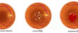
| Normal vision | Dry macular degeneration | Wet retinal degeneration |
- Fluorescein angiography is performed if choroidal neovascularization is suspected. Hypofluorescent changes are usually associated with hemorrhages and pigmentary hyperplasia. The causes of hyperfluorescent changes are more numerous and include soft and hard drusen, a network of newly formed vessels, atrophy of the pigment epithelium, and subretinal fibrosis.
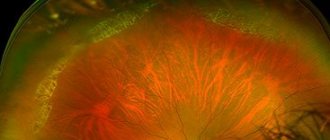
| Macular degeneration with massive subretinal hemorrhage |
- Optical coherence tomography is a highly effective non-invasive diagnostic method that allows you to detect the presence of intra- and subretinal fluid, as well as evaluate the effect of the treatment.
| Optical coherence tomography of the macula of a healthy eye |
| Optical coherence tomography for age-related macular degeneration of the retina |
Age-related degeneration of the macula and posterior pole requires constant monitoring in order to timely identify the progression of the disease or the transition of the disease from a dry form to a more aggressive wet one.
Dynamic instrumental observation 2-3 times a year in patients suffering from age-related macular degeneration of the retina can significantly improve the prognosis and promptly stop the irreversible decline in central object vision.
If the patient has a complicated medical history, but in the absence of clinical signs of the disease, control examinations of the fundus and dynamic monitoring of the results of instrumental examination for early detection of hard or soft drusen 1-2 times a year are usually recommended.
What diagnostic examination for AMD is carried out in the clinic?
In addition to routine diagnostic examination methods for retinal dystrophy, such as determining visual acuity, biomicroscopy, examination of the fundus (ophthalmoscopy), determination of visual fields (perimetry), we use modern computerized methods for diagnostic examination of the retina. Among them, the most informative for AMD is optical coherence tomography. This study allows us to identify the earliest changes that appear in macular degeneration of the retina. Optical coherence tomography (OCT) allows you to identify changes within the tissue structures of the retina and determine the form of macular degeneration.
Particular importance is attached to OCT in cases where there is a discrepancy between visual acuity and the fundus picture obtained with conventional ophthalmoscopic examination. In addition, this study is prescribed to monitor the effectiveness of the treatment. In addition to OCT, in some cases we prescribe fluorescein angiography of the retina (FAG) - this allows us to use intravenous dye (fluorescein) to diagnose changes in the structure of retinal vessels, which is necessary to identify the source of edema when prescribing laser coagulation of the retina. All these studies make it possible to clarify the diagnosis, stage of the disease, and choose the right treatment tactics.
How to treat dry macular degeneration?
Unfortunately, there is no therapeutic intervention that would be able to stop the progression or cure patients with dry macular degeneration of the eyes. Taking into account the theory of oxidative stress, patients with a large number of drusen, pigmentary changes or geographic atrophy are advised to take antioxidants according to various regimens.
The goal of this treatment for age-related macular degeneration of the retina is to neutralize oxygen free radicals that provoke pathological reactions. Dosages and individual dosage regimens are determined by the doctor. The main components of such therapeutic regimens are vitamin C, vitamin E, zinc oxide, lutein, beta-carotene, vitamin A, copper oxide. Patients are also usually advised to stop smoking and eat foods rich in polyunsaturated omega-3 fatty acids.
The mechanism of development of macular degeneration
The nutrition through the existing vessels of the eye becomes insufficient. The body begins to grow additional blood vessels. In fact, new growths are obtained that do not have all the properties of healthy tissues. Plasma passes through their vascular walls, carrying out large molecules of cholesterol and fats. They accumulate under the retina, making it difficult to nourish it. This also includes hemorrhages from “defective” newly formed vessels, which lead to the proliferation of connective tissue and the appearance of microscars.
As a result of these processes, the retina is exposed to numerous (albeit microscopic) lesions and eventually ceases to cope with its functions. However, if you intervene in time, it is possible to eliminate or at least significantly reduce the consequences of such lesions. For example, with timely and competent treatment of wet macular degeneration of the eye, the prognosis is quite favorable.
Macular degeneration - wet form: treatment of pathology
Treatment of the wet form of age-related macular degeneration of the retina is aimed at inhibiting the processes of subretinal neovascularization, preventing and treating complications.
Angiogenesis inhibitors
Inhibition of angiogenesis is currently one of the most effective treatments for wet macular degeneration of the retina. The basis of the therapeutic effect is the intravitreal administration of antiangiogenic drugs, that is, drugs that block vascular growth factor and, accordingly, suppress the process of subretinal neovascularization.
| Treatment of macular degeneration with angiogenesis inhibitors |
The most commonly used in practice are Pegaptanib (Macugen), Bevacizumab (Avastin), Ranibizumab (Lucentis) and Aflibercept (Aylia). The use of drugs in this group for age-related macular degeneration helps stop the growth of pathological blood vessels, thereby reducing the risk of vision loss. Over the past decade, numerous clinical studies have confirmed their high effectiveness in patients diagnosed with wet age-related macular degeneration.
Treatment with angiogenesis inhibitors allowed them not only to stabilize, but also to improve visual function. Significant disadvantages of this type of treatment are the invasive nature of the intervention, a decrease in the effect of treatment if it is abandoned, and the significant cost of treatment, especially taking into account the need for a course of intravitreal injections to achieve a clinically significant result.
Laser coagulation for macular and posterior pole degeneration
Laser treatment methods are indicated for the presence of subretinal neovascular membrane in patients who have been diagnosed with age-related macular degeneration. Treatment consists of laser coagulation of the neovascular membrane located extrafoveolar to the central fovea of the retina.
| Laser treatment for retinal degeneration |
The purpose of laser coagulation is to stop blood flow in newly formed vessels due to the coagulating effect of laser radiation on their wall. The main disadvantage of this treatment for macular degeneration is the presence of an undesirable damaging effect on the photoreceptors of the retina, which significantly limits the indications for the use of laser treatment, both in terms of the localization of the pathological process and the magnitude of object vision.
Photodynamic therapy
When treating the wet form of age-related macular degeneration of the retina, photodynamic therapy is a worthy alternative to laser treatment. Often, photodynamic therapy is an even more effective way to combat the phenomena of wet degeneration of the macula and posterior pole of the eye compared to the above treatment methods.
The clinical result of treatment is due to the laser effect on newly formed vessels and blocking blood flow in them. The light-sensitive drug “Visudin” used in photodynamic therapy accumulates only in areas of neovascularization. Laser irradiation of "Visudin" accumulated in newly formed vessels leads to the formation of a blood clot in them and obliteration of the lumen, as a result of which blood flow in the neovascular vascular network completely stops.
| Photodynamic therapy for macular degeneration |
The undoubted advantage of photodynamic therapy compared to laser treatment is its exclusive effect only on newly formed vessels without damaging the photoreceptor cells of the retina. It is also necessary to note the possibility of combined use of photodynamic therapy in combination with other methods of treating the wet form of age-related macular degeneration of the retina.
Transpupillary thermotherapy
Transpupillary thermotherapy is one of the safe and effective methods of treating age-related macular degeneration of the retina, which has proven itself in the wet form of macular degeneration with the presence of hidden choroidal neovascularization of any type, including subfoveal localization.
Carrying out transpupillary thermotherapy in the macular region of the retina does not lead to coagulation and photochemical damage to photoreceptor cells, since the main objective of the method is to reduce blood flow in the choroid as a result of irradiation with an infrared laser.
Transpupillary thermotherapy, as a rule, is an alternative to photodynamic therapy when there is no positive therapeutic effect of the latter.
Treatment with MACDEL devices
For macular degeneration of the retina, treatment is aimed at stopping the formation of new vessels, which helps slow down further progression of the disease and prevent loss of vision. An effective result is provided by taking antioxidants in combination using the MAKDEL-08 device, made on the basis of a helium-neon laser. Retinal tissue is stimulated by low radiation energy, due to which there is no visible destruction and the antioxidant activity of cells increases.
In addition, under the influence of laser beams, a speckle structure is formed, which the patient perceives as chaotically moving “grain”. Looking at it has a beneficial effect on the eyes, reduces tension, and improves nutrition of the retina.
Macular degeneration of the retina - treatment of macular degeneration
Macular degeneration is a medical term that refers to eye diseases.
It combines a group of pathologies associated with retinal damage and central vision impairment. The lack of comprehensive treatment is very dangerous; it leads to a partial or complete loss of the ability to see the world around us. The main cause of dystrophy is insufficient blood supply to the retina of the visual organ. Most often, pathological changes occur in two eyes, which significantly worsens the patient’s quality of life. The age period for the development of the disease is 45-50 years. This disease is very difficult to treat; restoration of vision at an advanced stage is almost impossible.
Elderly people need to undergo regular examination by an ophthalmologist, who will help to identify retinal dystrophy in time. Comprehensive treatment will stop its detachment and loss of central vision.
Why does AMD occur?
With macular degeneration, the photoreceptors of the macula are affected, resulting in impaired central vision. Macular degeneration can occur due to a large number of reasons, one of the main ones being dystrophic changes in the vessels of the retina. Disruption of normal oxygen transport through altered vessels causes oxygen starvation of retinal cells.
The risk of AMD increases with age: studies show that the highest incidence of macular degeneration is observed in the age group over 60 years. In addition to age, risk factors include excess weight and smoking, gender (women get sick more often than men), as well as detection of AMD in close relatives. The results of numerous scientific studies allow us to say with confidence that macular degeneration is a genetically inherited disease.
Disease prevention
In order to prevent the occurrence of AMD, it is recommended to systematically undergo an ophthalmological examination, especially after 45 years. Experts also advise protecting your eyes from excessive exposure to ultraviolet radiation (using sunglasses). It has been proven that people who spend a lot of time in the sun are 4 times more likely to be diagnosed with macular degeneration. Ophthalmologists strongly advise giving up cigarettes. It has been proven that smoking significantly increases the risk of developing and progressing AMD (about 5 times). In addition, doctors recommend limiting the consumption of fatty foods and monitoring cholesterol levels (high cholesterol levels impair blood circulation in the eyes). You should include foods that are good for your eyesight in your diet: blueberries, lettuce, cabbage, spinach, fish, eggs, grapefruit and others. In autumn and winter, it is advisable to take vitamin complexes. In general, experts advise leading an active lifestyle, spending more time outdoors and, as far as possible, reducing visual stress.
Our website offers a wide selection of contact lenses, multifunctional solutions and moisturizing eye drops. You can purchase world bestsellers at a competitive price. Fast delivery of products and high level of service Ochkov.Net will pleasantly surprise you!
Advantages of treatment at MGK
The modern equipment used in our clinic allows our doctors to conduct examinations quickly, painlessly and with comfort for the patient, while it is possible to identify any pathologies of the visual apparatus at the initial stage, when there are no symptoms of the disease.
The clinic's doctors have extensive practical experience in treating eye diseases, including optic nerve atrophy. MGC uses the widest range of treatment procedures to preserve vision in patients with optic nerve atrophy. A comprehensive treatment program has been developed using drug therapy, hardware techniques, hirudotherapy, and physiotherapeutic treatment. Our specialists are ready to provide highly professional assistance to patients with the most severe forms of optic atrophy.
The specialists of our clinic have developed course treatment programs for patients with macular degeneration, aimed at preserving and restoring visual functions for 10 and 15 days, including the use of medicinal leeches.
To improve the quality of treatment and achieve better results, the clinic operates a hospital (including a day hospital), where patients can receive all necessary treatment in a timely manner and in full, while under the supervision of the attending physician.
If you have questions, you can ask our specialists using the feedback form on the website or by phone in Moscow 8 (800) 777-38 81 and (daily from 9:00 to 21:00).
Author:
Yakovleva Yulia Valerievna 5/5 (1 rating)
Honey. portal: