The human body is a very complex structure. Some areas of the body, for example, the paranasal sinuses, are hidden from human eyes, but this does not make them any less significant. The paranasal sinuses, also known as the paranasal sinuses, play an important role in the human breathing process, and any inflammation of them can lead to a malfunction of other organs.
Computed tomography of the sinuses, or CT of the nasal sinuses, is a high-precision research method that is widely used in ENT practice and allows one to obtain an image of the nose and paranasal sinuses, as well as adjacent areas, using X-rays. A CT scan of the nose helps to see the problem from the inside: the method allows you to diagnose diseases of the nasal cavity, discern pathology in the maxillary sinuses, as well as in other paranasal sinuses.
How tomography of the nasal sinuses is performed, in what cases this research method is prescribed, for whom the procedure is contraindicated and whether there are alternative CT methods - we will talk about all this in our article.
Sinuses and their diseases
The paranasal sinuses are cavities in the skull filled with air. The walls of the sinuses are covered with mucous membrane. The sinuses are connected to the nasal cavity by special canals - anastomoses. Sinuses play an important role in the human body. Thanks to these cavities, the weight of the skull is reduced. With their help, the inhaled air is moistened and heated to the desired temperature before entering the lungs. On the mucous membrane of the cavities there are tiny cilia that trap harmful particles and prevent them from penetrating further into the body. The sinuses are involved in speech production and smell recognition. Therefore, any inflammation or swelling can lead to a malfunction of several important systems and functions of the body.
There are four types of paranasal sinuses:
- maxillary (maxillary), the lesion of which is called sinusitis;
- frontal - when they become inflamed they speak of frontal sinusitis;
- wedge-shaped - when it is inflamed, a diagnosis of “sphenoiditis” is made;
- cells of the ethmoid labyrinth - its inflammation is called ethmoiditis.
Is it possible to take antibiotics for sinusitis?
Antibiotics are prescribed to treat various forms of sinusitis. The most effective groups of drugs are considered to be:
- Penicillins are a combination of amoxicillin and clavulanic acid
- Cephalosporins - powder or saline solution that suppresses inflammation
- Macrolides are antibacterial drugs of natural and semi-synthetic origin that contain a lactone ring in their structure
- Fluoroquinolones are substances that have a wide spectrum of action against gram-positive and gram-negative flora, aerobes and anaerobes, and atypical bacteria
All medications must be prescribed by your attending physician. Improper use of antibiotics can cause serious problems in the functioning of the body.
CT scan of the paranasal sinuses: what is it?
CT scan of the paranasal sinuses is a type of X-ray tomography. During the examination, X-rays pass through different areas and tissues of the body. The X-ray tube takes many pictures in different planes, then the computer analyzes the information received and creates a layer-by-layer 3D image of the desired area.
A computed tomograph is widely used in ENT practice and helps the otorhinolaryngologist to see in detail and clearly the condition of the paranasal sinuses and adjacent areas. CT scan of the nose shows abnormalities in the structure of the nasal cavity; helps to recognize the beginnings of the inflammatory process or determine the extent of its spread. A computed tomogram of the sinuses allows the ENT doctor to draw correct conclusions about the patient’s health status, correctly diagnose the disease and prescribe appropriate treatment.
Causes of sinus inflammation
The following factors can provoke severe inflammation of the sinuses:
- viral (adenoviruses, influenza virus, herpes) and bacterial (staphylococcus, streptococcus) infection. Pathogenic microflora, entering the sinuses, begins to actively multiply in them, the anastomosis swells, the mucus that accumulates in the sinuses cannot come out of them, stagnates, pus appears, and the person begins to experience symptoms of severe inflammation.
- structural features of the nose (for example, a deviated nasal septum);
- nasal polyps;
- adenoids;
- tendency to allergies;
- hypothermia of the body;
- dental diseases (for example, carious teeth, periodontal disease);
- sinusitis that occurs due to untreated teeth is called odontogenic;
- mechanical injuries of the nose;
- weakened immunity.
CT scan of the sinuses: indications and contraindications
Computed tomography is performed in the following cases:
- when x-rays contradict symptoms and diagnosis;
- when the inflammatory process is chronic;
- for sinusitis (for example, for sinusitis, a CT scan of the maxillary sinuses is prescribed);
- in the presence of headaches along with nasal congestion;
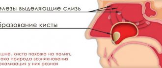
- if you suspect the presence of neoplasms (cysts, polyps, tumors);
- to study existing tumors, compare their growth dynamics and adjust the existing treatment plan;
- for injuries and damage to the face, skull fractures, etc.;
- in the presence of a foreign body in the nasal cavity and sinuses;
- with bleeding from the nasal cavity of unknown etiology;
- with difficulty in nasal breathing.
This type of examination differs from conventional x-rays in that it is more accurate and visual. The CT scan of the sinuses shows and recognizes inflammation and tumor development at the earliest stages. This is especially important when the ethmoidal labyrinth and sphenoid sinus are affected, when there is a high probability of the inflammatory process spreading to the brain.
A CT scan of the paranasal sinuses allows the ENT doctor to thoroughly examine the condition of the sinuses and adjacent areas. But despite the fact that CT shows accurate results, and this type of research seems to be the most effective and optimal, the method has a number of contraindications.
How is sinusitis treated?
When the first symptoms of the disease appear, you should consult a specialist. The doctor will conduct a survey, examination and diagnosis. After which a treatment strategy will be prescribed. It’s better not to delay going to a specialist. Then the disease can provoke serious complications or become chronic. It is also recommended to visit a specialist periodically as a preventative measure.
How is sinusitis treated? After taking anamnesis, the specialist prescribes a treatment strategy. Treatment can take several forms - conventional therapy, physical therapy or surgery. In the first case, the patient is prescribed a number of medications that should suppress inflammation and eliminate pain.
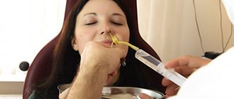
Physiotherapy is prescribed as a concomitant method of treatment. One of these methods is warming up: it is carried out using a blue lamp. Surgical intervention is the most extreme method prescribed in advanced forms of the disease. A puncture or puncture of the sinus is made with a special needle to diagnose and extract purulent masses.
Contraindications:
- pregnancy (instead of tomography it is recommended to do a sinus scan);
- body weight more than 200 kg;
- thyroid diseases;
- kidney diseases;
- severe diabetes mellitus;
- multiple myeloma;
- allergy to the contrast agent administered during CT scanning.
A tomogram of the paranasal sinuses is not recommended for children, since the procedure requires perseverance and the need to lie still for several minutes. It is extremely difficult to force a child to do this.
Preventing sinus inflammation
There are no specific preventive measures that will certainly protect you from sinus inflammation. But with the help of simple rules you can reduce the risk of developing sinusitis and strengthen the body.
- Don't get too cold. Try to dress appropriately for the weather, and take an umbrella with you in rainy weather.
- Pay attention to a runny nose. Even a harmless runny nose can cause disruption of sinus ventilation and inflammation.
- Treat infectious diseases of the nasopharynx correctly and on time to prevent pathogenic flora from reaching the sinuses.
- Visit your dentist for preventive care so as not to miss the formation of caries and cure it in time.
- If you have allergic reactions, avoid interaction with allergens.
- Solve the problem of nasal breathing surgically if you have a severely deviated nasal septum and enlarged inferior turbinates.
- Rinse your nose with saline solutions during epidemics and active flowering of plants. Try to avoid crowded places as much as possible during the height of seasonal illnesses.
- Strengthen your immune system: walk more, eat right, play sports. Say goodbye to bad habits without regret.
- Take vitamin complexes.
- Maintain a comfortable level of humidity and temperature at home, especially during the height of the heating season. A moist nasal mucosa is easier to cope with viruses and bacteria than a dry nasal mucosa.
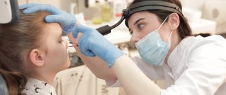
How the research is carried out
No special preparation is required for the procedure. You must remove all metal objects or clothing with metal elements. The patient is placed on the couch and his body is secured with belts, and his head is placed on a special headrest. You should not move during the entire procedure, as movement may affect the accuracy of the result. If the patient cannot lie still (for example, children or people with mental disabilities), the doctor may use anesthesia.
If the CT procedure requires the injection of a contrast agent, such as iodine, into the patient’s blood, you should refrain from eating for several hours before the procedure.
Then the couch on which the patient lies is pushed into the tomograph. After this, the doctor retires to another room where a computer is installed that records the tomograph signals. All this time, with the help of sound equipment, the doctor and the patient are in touch. If the patient feels any discomfort, he reports it, and the doctor hears it. The study itself lasts no more than ten minutes.
Then the specialist deciphers the tomogram and gives the patient an x-ray and a disk recording the results of the study, as well as a medical report.
CT, as a method of radiation diagnostics, involves radiation exposure to the body. Therefore, it is recommended to do CT scans no more than three times a year. The cost of this study varies from 3,000 to 5,000 rubles.
Complications of sinusitis
If you consult an ENT doctor in a timely manner, sinusitis occurs without complications. But if the patient ignores medical help and self-medicates, the disease can take unpleasant and even dangerous turns.
Complications of sinusitis are divided into two categories: orbital and intracranial.
Orbital complications include diseases of the orbit that occur against the background of inflammatory processes in the sinuses. They are explained by the anatomical proximity of the sinuses and orbits. The main signs that the orbit is involved in the inflammatory process are the following symptoms: swelling and redness of the eyelids, protrusion of the eyeball (exophthalmos), difficulty moving the eyeball, pain when pressing on it.
Orbital complications include:
- swelling and inflammation of the tissue of the eyelids and eye orbit;
- periostitis of the orbit;
- orbital abscess;
- abscess of the eyelid;
- phlegmon of the orbit;
- thrombosis of veins of the orbital tissue.
Intracranial complications are diseases in which there is an infection from the paranasal sinuses into the membranes of the brain. These are the most dangerous types of complications as they can be fatal. Signs of intracranial complications of sinusitis are headache, nausea, dizziness, photophobia, high body temperature, stiff neck, when the patient’s chin rises up and it is impossible to lower the head down.
Such complications include:
- rhinogenic meningitis;
- arachnoiditis;
- brain abscess;
- thrombosis of the cavernous sinus.
The prognosis for the development of intracranial complications is not always favorable. Much depends on the speed of receiving competent medical care. Any delay may result in the death of the patient.
Medical Internet conferences
1. Anyutin R.G., Kulikov L.S., Nersesyan M.V. Morphological features of the mucous membrane of the sphenoid sinus in patients with sphenoiditis // Ros. rhinology. - 2005. - No. 4. -P.21-22. 2. Arefieva N.A. Immunology, immunopathology and problems of immunotherapy in rhinology. – Ufa: B.I., 1997. – 114 p. 3. Ashurov A.M., Mirazizov K.D. Anatomical features of the sphenoid sinus on computer and magnetic resonance imaging // Ros. rhinology. - 2002. - No. 2. — P. 23-24. 4. Boenko S.K., Rodin V.I., Lvov L.M. Clinic, diagnosis and treatment of patients with sphenoiditis // Journal. ear, nose and throat diseases. – 1989. – No. 4. – pp. 87-93. 5. Boyenko S.K., Lvov L.M., Danilchenko S.A., Buryak L.A., Tsygankov V.A. The significance of the structural features of the sphenoid sinuses in the development of sphenoiditis // Vestn. otorhinolaryngology. – 1992. – No. 4. – pp. 16-18. 6. Boenko S.K., Lvov L.M., Boenko D.S., Klimov Z.T. Klinchasta sinus: is the black box clean? // Journal. ear, nose and throat ailments. – 2002. – No. 3-s. – pp. 93-94. 7. Buchatsky M.V. Observation of diencephalic syndrome in a patient with chronic sphenoiditis // Journal. ear, nose and throat diseases. – 1977. – No. 6. – pp. 87-88. 8. Bykova V.P. The mucous membrane of the nose and paranasal sinuses as an immune barrier of the upper respiratory tract // Ros. rhinology. – 1993. – No. 1. – pp. 40-46. 9. Bykova V.P. Structural foundations of mucosal immunity // Ros. rhinology. – 1999. -№1. – P. 5-9. 10. Volkov A.G., Eroshenko A.Yu., Popel S.E. Our techniques in the diagnosis and treatment of sphenoiditis // Ros. rhinology. - 2003. - No. 2. - P.45. 11. Goffman R.V., Polezhaev A.V., Cherebillo I.Yu. Endoscopic topographic-anatomical landmarks of the sphenoid sinus with various options for its pneumatization // Ros. rhinology. - 2002. - No. 2. — P. 64-66. 12. Grineva V.A. Physiotherapeutic possibilities for treating patients with sphenoiditis // Journal. ear, nose and throat ailments. — 1997. -№5. — P.69-73. Shevrygin B.V., Shadyev Kh.D. Anatomical and topographical data on the natural opening of the main sinus in children // Journal. ear, nose and throat diseases. – 1973. -No. 2. – P. 46-48. 13. Maykova-Stroganova V.S., Rokhlin D.G. - Quote according to Kiselev A.S. Chronic sphenoiditis. – St. Petersburg: Publishing house. VmedA, 1997. – P. 5. 14. Neiman L.V. The significance of morphological features in the pathology and surgery of the main sinus // Vestn. otorhinolaryngology. – 1948.- No. 3. – pp. 29-38. 15. Shevrygin B.V., Agaev G.V. Endonasal microsurgery and anatomical and topographic data on the natural opening of the sphenoid sinus // Journal. ear, nose and throat diseases. – 1987. – No. 4. – P. 34-37. 16. Panfilova G.V., Shpak N.I., Polovkin E.M. Pneumatization of the main sinus and its additional formations in patients with neuritis and optic nerve atrophy // Oftalmol. magazine – 1972. – No. 5. – pp. 350-355.
17. Pie A.V. Morphofunctional characteristics of the mucous membrane of the human sphenoid sinus in normal conditions and with sphenoiditis (macroscopic, microscopic, histological, electron microscopic and morphometric studies): Abstract. dis. Ph.D. honey. Sci. – Kharkov, 1992. – 23 p. 18. Brandwein M. Histopathology of sinonasal fungal disease // Otolaryngol. Clin. North. – 1993. – Vol. 26. - 35. 19. Braun J. et al. - Quote according to Piskunov S.Z. et al. Isolated lesions of the sphenoid sinus. – Kursk: B.I., 2004. – 151 p. 20. Davidson F. Hyperplastic rhinosinusitis // Ann. Otol. – 1982. – Vol. 82, No. 5. – P. 703-708.
21. Deans J. Welch A. Acute isolated sphenoid disease: Adisease with complication // J. Laryngol., Otol. – 1991. – Vol. 105. – P. 1072-1074. 22. Dessi P., Moulin G., Bartoli JM, Canroni M. Procidens intrasphenoidale de l'artere carotide terne. Etude tomodensitometrique de 300 sinus//Press Medicale. - 1994. - Vol. 23, N13. — P. 616-617.
23. Drettner B. Pathophysiology of paranasal sinuses with clinical implications // Clin. Otolaryngol. 1980. - Vol. 5. – P. 277-284. 24. Landolt AM, Gammert C. Transsphenoidal surgery for pituitary adenomas // Aesculap scientific information.-1986.-March.-P.6-15. 25. Lew D., Southwick F. Montgomery W., A., Backer A. Sphenoid sinusis // New Eng. Med. – 1983. – Vol. 309. – P. 1149-1154. 26. Onodi A. Die topographishe Anatomi Nasenhоhle und ihren Nevenhohlen // Нandbush der speziellen Chirurgie des Оhres. – Wurzburg, 1912 – S. 51-125. 27. Sugita R., Oguri J., Fujimaki G. et al. Microorganisms detected in unilateral sinusitis // Otol. - 1987. - Vol. 80, N3. - P. 397-405. 28. Thomas RD, Oliverio P. The uncovered internal carotid artery in the sphenoid sinus: CT derection // J. Comput. Assist. Tomogr. - 1994.18, N4. - P. 650-651. 29. White J. Paranasal sinus infections // Jn: Ballnger J. Diseases of the Nose, Throat, Ear, Head Neck. – Philadelphia, 1991. – P. 184-202.
How do the paranasal sinuses work?
The nose is one of the most important human organs. This system is complex and quite fragile, which leads to frequent illnesses. But without the maxillary sinuses, normal nose function would be impossible. They perform the following functions:
- provide full breathing;
- warm and humidify the inhaled air;
- clean the inhaled air from dust and allergens;
- allow you to recognize many aromas and smells;
- form an individual timbre and other voice parameters.
The sinuses have other tasks as well. For example, they quickly respond to changes in environmental pressure (act as baroreceptor organs) and reliably protect the eyeballs, tooth roots and skull bones from injury.