Causes of the disease
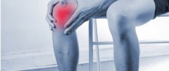
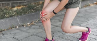
Osteoarthritis especially often develops in the knee joint. The appearance of degenerative-dystrophic changes in it is facilitated by its complex structure, frequent injuries and the heavy load that the knee joint experiences throughout a person’s life. There are several etiological factors that result in the disease. It can be:
- age-related changes;
- injuries (domestic and sports);
- obesity;
- hormonal changes and metabolic disorders;
- congenital pathologies of the musculoskeletal system;
- autoimmune diseases.
Osteoarthritis of the knee joint that occurs in old age is considered primary. Secondary is a lesion that develops against the background of (or after) another disease.
Development mechanism
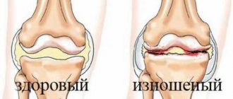
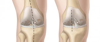
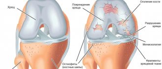
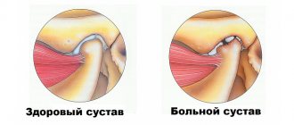
Normally, the articular surfaces of the tibia and femur are covered with densely elastic hyaline cartilage. Its main function is to reduce the friction force during flexion and extension of the knee joint, reducing shock loads when walking. Under the influence of external or internal provoking factors, blood circulation in small intraosseous vessels through which nutrients enter the cartilage is disrupted. It becomes dry, rough, and loses its ability to retain the moisture necessary for restoration. Now the cartilaginous surfaces do not shift smoothly, but “cling” to each other. Constant microtraumas lead to thinning of the cartilage.
Course of the disease
The disease develops gradually and unnoticed by the patient. During the course of the disease, there are 3 degrees of severity:
- 1st degree - pain appears only during prolonged and intense physical exertion. They are short-lived and go away quickly after rest. Usually the patient does not attach any importance to them, does not consult a doctor, and the disease progresses to the next stage;
- 2nd degree - even with minor loads, noticeable pain occurs in the knee joint. When moving, clicks and crunching are heard, swelling and limping appear;
- 3rd degree - the pain becomes constant, because of it the patient loses sleep and cannot step on the sore leg. Joint swelling may be accompanied by joint deformation.
A feature of the disease is that osteoarthritis of the knee joint is very difficult to diagnose before the onset of stage 2. Even an x-ray may not show changes in the joint. Therefore, a comprehensive examination is necessary for timely diagnosis.
Symptoms of pathology
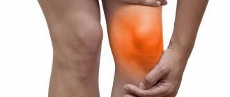
At the initial stage, the disease may not manifest itself clinically at all. As it progresses, the first discomfort appears when going up or down stairs, after running, intense sports training or long walks.
When visiting a doctor, patients complain of some stiffness of movement immediately after waking up, a feeling of “tightening” on the outside of the knee. These sensations disappear quickly and are rarely accompanied by pain.
No external changes in the joint are observed. Only occasionally does the knee look a little swollen. Swelling of the skin may indicate synovitis, an inflammation of the synovium. Others with its specific symptoms are a feeling of heaviness and limited mobility.
Treatment of osteoarthritis
Treatment of the disease may include both conservative therapy and surgical intervention. Osteoarthritis of the knee joint of grade 1 and 2 responds best to conservative therapy. Such treatment consists of the use of medications, physiotherapeutic and kinesitherapy methods.
Medications used include analgesics (painkillers), non-steroidal anti-inflammatory drugs, chondroprotectors, hyaluronic acid preparations and sometimes hormones. At degrees 1 and 2 of osteoarthritis, it is possible to take medications in tablets. If they are ineffective, they switch to injections. These same drugs are used topically - in the form of ointments and gels. In physiotherapy, electrophoresis with drugs, ultrasound, magnetic therapy, paraffin, and ozokerite have proven themselves well. It is mandatory to perform physical therapy exercises and exercise on special simulators, conducted under the guidance of a doctor.
Therapeutic exercise for osteoarthritis
Therapeutic exercise is your best friend in the treatment of osteoarthritis in the early stages. Sports are excluded during illness.
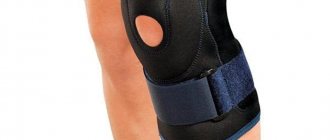
In the exercise therapy room, the patient is introduced to special means of knee protection - fixing bandages. They contribute to uniform load on the joint. Be sure to wear bandages on both knees, even if only one hurts!
Effective exercises for knee osteoarthritis that you can do at home.
| Initial position | Actions (perform smoothly, without straining the joints) | Quantity |
| Stand vertically with your back to the wall, arms down. | Touch the wall with your palms, shoulder blades and buttocks. Slowly rise onto your toes. Descend. | 5-7 seconds, 15-20 times. |
| Lying down, feet rest on the floor, legs bent. | Raise your right leg to knee height, straighten, hold, and lower smoothly. Repeat with your left leg. | 7 seconds, 10 times on each knee. |
Diagnostic methods
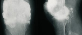
The basis for making a diagnosis are patient complaints, results of external examination and radiography. This instrumental study is the most informative, but with grade 1 osteoarthritis, characteristic signs may be absent. At the final stage of the disease, radiological stage 1 reveals single osteophytes, narrowing of the joint space and compaction of the subchondral zone.
If radiographic images are uninformative, MRI, CT, and ultrasound may be prescribed. The resulting images clearly visualize cartilage tissue, ligamentous-tendon apparatus, blood vessels, and nerve trunks.
Differential diagnosis is carried out to exclude damage to the knee joint by arthritis, synovitis, bursitis, tendonitis, and tendovaginitis.
Causes of deforming arthrosis of the knee
Without normal lubrication, the joint “dries out,” cracks and loses height, exposing the heads of the bones. In this case, the end plate of the articular surface of the bone remains defenseless; overstimulation of the numerous nerve endings that are located in it causes pain and discomfort.
The following factors or a combination of them can cause deforming arthrosis of the knee:
- the presence of joint diseases (and knee diseases in particular) in relatives;
- genetic disorders associated with the formation of abnormal, unstable cartilage cells or their accelerated death;
- congenital and acquired malformations of the musculoskeletal system (flat feet, joint hypermobility, dysplasia, scoliosis, kyphosis and others);
- excessive professional, household or sports stress;
- microtraumas and injuries of the knee joint and meniscus, operations on it, leg fractures;
- blood supply disorders (varicose veins, atherosclerosis, thrombosis and other vascular diseases), their consequences (osteochondritis dissecans), as well as other causes of prolonged spasms in the legs;
- inflammatory diseases of joints and periarticular tissues (synovitis, bursitis, tendonitis, arthritis), incl. autoimmune nature (rheumatoid, psoriatic arthritis);
- metabolic disorders (gout, diabetes);
- age-related processes of aging of joints and leaching of calcium from bones;
- hormonal disruptions and changes in hormonal levels (for example, associated with a lack of estrogen in women);
- hypovitaminosis;
- excess weight (observed in ⅔ patients);
- physical inactivity.
But the main reason that deforming arthrosis of the knee is so common lies in its structure. The knee joint has only one axis (plane) of movement. Therefore, the volume of permissible movements is very limited. One awkward turn can injure the periarticular tissues and trigger arthrosis changes - after all, the sore knee will be subject to daily stress.
The reasons for the development of deforming arthrosis of the knee can be a large number of factors.
Prevention and prognosis of defarthrosis
It is easier to prevent any disease than to treat it later. Even if a person has a predisposition to disease in both the right and left knee joints, it is possible to prevent the development of pathology before serious problems arise.
It is necessary to adhere to a healthy lifestyle. Office work, unhealthy diet, excess weight - all this contributes to the development of arthrosis, as well as excessive stress. Exercise, although absolutely necessary as a preventive measure, should be reasonable. An hour-long walk at a brisk pace is better than squats and weight lifting, especially when it comes to a woman’s health.
Persons suffering from systemic and endocrine diseases should carefully monitor the condition of their legs. Any discomfort is a reason to consult a doctor as soon as possible. The disease does not progress so quickly, so with timely treatment, complete remission and a return to a normal lifestyle can be achieved.
In general, the prognosis depends on the stage and degree of development of the pathology. The earlier the disease is diagnosed and measures taken, the better arthrosis is treatable. At the first stage, the cartilage can be completely restored, but at the third stage, joint replacement will most likely be required. A timely visit to the doctor is the key to a healthy knee joint.
Pathogenesis
The pathogenesis of osteoarthritis (OA) is based on pathological changes in the molecular structure of hyaline cartilage, in which processes of remodeling (synthesis/degradation) of the basis of cartilaginous tissue, the extracellular matrix . A key role in this process is played by highly differentiated cells of cartilage tissue - chondrocytes , which begin to produce low-molecular proteins of the interstitial cartilage tissue (matrix), which reduces the shock-absorbing properties of the knee joint cartilage.
of proteoglycans in the surrounding cartilage matrix and react negatively to their changes. It is known that the state of cartilage tissue is determined by the balance of anabolic/catabolic processes. At the same time, the speed/intensity of catabolic processes increases under the influence of cytokines (interleukin-1, tumor necrosis factor-α), metalloproteinases (stromelysin, collagenase), cyclooxygenase-2 , which are produced by chondrocytes, cells of the subchondral bone and synovial membrane. Also in the process of restoration of cartilage tissue, a significant role is played by the reparative activity of chondrocytes, which is based on insulin-like/transforming growth factors and morphogenetically altered cartilage/bone proteins.
As a result of the increase in degenerative processes, the cartilage loosens/softens, cracks appear, and the articular surfaces of the bone, due to the destruction of cartilage tissue, begin to experience an unevenly distributed increased mechanical load. This contributes to the appearance of a zone of dynamic overload in the subchondral bone, which causes redistribution disturbances of microcirculation, the development of subchondral osteosclerosis , changes in the curvature of joint surfaces, and the formation of osteophytes (marginal osteochondral growths).
A significant role in the pathogenesis is played by synovitis , characterized by moderately expressed exudative/proliferative reactions in the form of hyperplasia and mononuclear infiltration of the synovial membrane, most manifested in places of attachment to cartilage with subsequent transition to lipomatosis / sclerosis . Exudative-proliferative reactions in the synovial membrane and subchondral bone occur against the background of a disorder of regional microcirculation and hemodynamics with the gradual development of tissue hypoxia . osteophytosis appear in the subchondral bone , and the structure of the changes becomes irreversible. The pathogenesis of OA is schematically presented below.
What complications can there be?
Osteoarthritis progresses slowly but persistently. Its course is complicated by secondary reactive synovitis, spontaneous hemorrhages into the joint cavity (hemarthrosis), osteonecrosis of the femoral condyle, and external subluxations of the patella. At the 4th radiographic stage, the joint space fuses, which leads to complete or partial immobilization.
Endoprosthetics - a modern alternative to surgery
At the second and even third stage of arthrosis, progressive doctors recommend their patients intra-articular injection of a synovial fluid substitute, for example the drug Noltrex. The procedure is carried out in a dressing room, under local anesthesia, for 20-30 minutes. The full course involves several administrations at weekly intervals.
After a short period of time, the person forgets about the pain, mobility is restored, and the result lasts for 1-2 years. Endoprosthetics of synovial fluid is a relatively new treatment for arthrosis, however, due to its long-term effect and the absence of side effects, it is increasingly preferred instead of drugs based on hyaluronic acid.
Treatment with Noltrex is focused on long-term effects rather than temporary relief of symptoms.
The second degree of arthrosis is a certain intermediate stage. The period when it is not too late to start treatment, recover and return to a healthy lifestyle without pain and restrictions. The main thing is not to miss the first signs and take action in time, since otherwise the consequences for the joints can be unpredictable.
Drug therapy
Since the pain that occurs is not severe, analgesics in the form of tablets or injection solutions are rarely used. To eliminate mild discomfort, patients are prescribed ointments, gels, creams, and balms for local application to the knee joint. Therapeutic regimens may include drugs from the following groups:
- means to improve blood circulation - Pentoxifylline, Xanthinol nicotinate;
- preparations with a complex of B vitamins - Milgamma, Combilipen, Neuromultivit;
- balanced complexes of vitamins and microelements - Supradin, Complivit, Vitrum, Centrum, Multitabs.
Systemic chondroprotectors Teraflex, Structum, Artra, Dona, Chondroxide are mandatory for long-term course use (from 3 months). The active ingredients of these products stimulate partial restoration of damaged cartilage.
| External agents for the treatment of grade 1 osteoarthritis of the knee | Therapeutic effect |
| Non-steroidal anti-inflammatory drugs (Voltaren, Fastum, Artrosilene, Nise, Ketorol, Diclofenac, Indomethacin) | Stop inflammatory processes, reduce the severity of pain, stimulate the resorption of edema |
| Ointments with a warming effect (Viprosal, Finalgon, Apizartron, Nayatox) | Eliminate morning swelling and stiffness of movement, eliminate pain |
Surgery
For grade 1 osteoarthritis, surgery is not required. Arthroplasty, aimed at restoring the size, shape and conformity of the surfaces of the knee joint to each other, is carried out with multiple bone growths (osteophytes). Such changes are not typical for this stage of the disease.
Exercise therapy
Dosed physical activity is the most effective method of treating osteoarthritis at the initial stage of development. The main objectives of exercise therapy are to strengthen the muscles of the knee and improve the blood supply to its structures with nutrients. A set of exercises is compiled by a physical therapy doctor immediately after making a diagnosis. The patient’s age, physical fitness, and general health must be taken into account.
Physical education for grade 1 knee osteoarthritis.
The following exercises are most often recommended for patients:
- flexion and extension of the legs at the knee joints while lying on the back;
- imitation of cycling in a lying or sitting position with emphasis on arms extended back;
- in a standing position, swing your legs back and forth, from side to side;
- shallow squats;
- walking around the room with your knees raised high.
The appearance of even a slight painful sensation is a signal to stop exercising. You can start training only after an hour break. During exercises, movements should be smooth, slightly slow, without jerking.
Massage
At the beginning of treatment, about 5 sessions of classical massage are performed. When performing them, all traditional massage movements are used - kneading, rubbing, clapping, vibration. To improve the patient’s well-being, vacuum massage, including hardware massage, is also used. Before installing plastic or glass jars, air is removed from them to ensure better contact with the skin. This stimulates blood flow to the knee and the production of bioactive substances with an analgesic effect. The therapeutic effect of the procedure is enhanced when the massage therapist begins to smoothly move the cups and perform kneading and rubbing with them.
Manual therapy
The help of a chiropractor is used for osteoarthritis accompanied by increased muscle tension. The doctor acts on the spasmodic skeletal muscles with his fingers. The muscles relax, which improves blood circulation and lymph flow.
In addition to common massage movements, the chiropractor uses superficial and deep palpation techniques.
Nutrition
Since the development of osteoarthritis is associated with a deterioration in the blood supply to tissues, the goal of the diet is to restore optimal blood circulation. To do this, you should exclude from your diet foods high in fat, which is deposited as cholesterol blocks on the walls of blood vessels. You need to give up semi-finished products and fast food, and use vegetable oils when cooking, not margarine or cooking oil. The daily menu must include fresh vegetables and fruits, fermented milk products, and cereal porridges.
Traditional methods of treatment
After the main treatment, during the rehabilitation period, patients are allowed to use folk remedies. These are ointments, rubs, infusions, decoctions that accelerate the cleansing of the joint from tissue breakdown products and mineral salts, and have a mild warming effect. What folk remedies are advisable to use for grade 1 osteoarthritis:
- Herb tea. Pour a teaspoon of corn silk, St. John's wort, sage, elecampane into a ceramic teapot, and pour 2 cups of boiling water. After an hour, strain, drink 100 ml three times a day;
- compress. Mash a fresh leaf of horseradish well, grease with honey, and apply to the sore knee for an hour.
Herbal collection.
In spring you can start preparing a healthy tincture. You need to collect, put in a jar and pour vodka over young leaves or flowers of calendula, dandelion, plantain, clover, shepherd's purse, chamomile, coltsfoot. By the end of autumn, the tincture for rubbing on the knee is ready.
