This site was made by experts: toxicologists, narcologists, hepatologists. Strictly scientific. Tested experimentally.
Author of this article, expert: gastroenterologist Daniela Sergeevna Purgina
This article was edited by gastroenterologist Daniela Sergeevna Purgina.
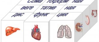
Sphincter of Oddi dysfunction (SDO) is a disease in which adequate contractile activity of the sphincter, which is located at the junction of the common bile and pancreatic ducts into the duodenum, is disrupted.
Why do we need the sphincter of Oddi
The bulk of bile is produced in the liver, and then passes through the ducts to the gallbladder. During food intake, the gallbladder contracts and the accumulated bile enters the duodenum through the ducts, where it is mixed with food and then goes to the small intestine, where the main processes of digestion occur. The timeliness of bile entering the duodenum is controlled by the sphincter of Oddi.
Being constantly in a state of increased tone, the sphincter prevents bile and pancreatic juice from entering the small intestine (although a small amount of discharge is observed even between meals). As soon as partially processed food begins to flow from the stomach into the small intestine, the sphincter of Oddi relaxes, directing small portions of the contents of the ducts into the lumen of the duodenum.
This smooth muscle can perform its function from a few seconds to a minute. The duration of work is affected by the intensity of digestion. In addition, the closed passage prevents the entry of small intestinal contents into the pancreatic and bile ducts.
What causes dysfunction?
With Oddi dysfunction, the flow of bile and pancreatic secretions is disrupted, resulting in a disorder of the digestive mechanism. Why could this happen? For different reasons. Pathology can be of different nature:
- Organic. Disruption of the functioning of an organ of an organic nature occurs due to inflammation, as well as pathologies such as hyperplasia or fibrosis of the duodenal mucosa, strictures of the biliary tract, and undiagnosed stones of the common bile duct.
- Functional.
Functional reasons may include:
- gastritis;
- diseases of the liver and biliary tract;
- pancreatitis;
- peptic ulcer;
- intestinal diseases;
- diabetes;
- autoimmune diseases;
- treatment of diseases with hormonal drugs;
- pathologies of the adrenal glands;
- thyroid diseases;
- postoperative period after removal of part of the intestine, stomach.
Very often, this complication occurs after cholecystectomy: surgery to remove the gallbladder. The reason is that after such a surgical intervention, a significant increase in pressure occurs in the entire biliary tract, even with a weak contraction of the sphincter of Oddi. Either a spasm of this smooth muscle can occur, or vice versa, constant relaxation, in which bile enters the lumen of the duodenum all the time (and not just when needed).
Yu.V. Vasiliev
TsNIIG, Moscow
It is known that chronic pancreatitis is a group of diseases (variants), the appearance of which is based on certain factors, usually considered as etiological, morphological and/or clinical criteria determining the development of the disease. The latter are very similar in the early stages, but as chronic pancreatitis progresses, depending on the appearance of exocrine and/or endocrine pancreatic insufficiency and/or various complications, some differences appear [1-3]. Chronic pancreatitis, as is known, can develop due to various reasons, including due to obstruction of the pancreatic duct due to cicatricial papillary stenosis or due to impaired motility of the sphincter of Oddi. In particular, according to some researchers [12], sphincter of Oddi dysfunction was detected in 20 out of 68 patients with chronic pancreatitis. A similar relationship is possible between abnormal pancreatobiliary fusion and recurrent pancreatitis associated with sphincter of Oddi dysfunction in children and adolescents [6]. In principle, the term “dysfunction” is usually considered as a violation or disorder of the functions of an organ, mainly of a qualitative nature. The sphincter of Oddi is a fibromuscular formation surrounding the terminal sections of the common bile duct, the main pancreatic duct (Wirsung's duct) and the common channel in the area where they pass through the wall of the duodenum. Main functional features of the sphincter of Oddi:
• regulation of the flow of bile and pancreatic secretions into the duodenum; • preventing the flow of contents from the duodenum into the common bile and Wirsung ducts; • ensuring the filling of the gallbladder with bile.
Currently, “Dysfunctional disorders of the biliary tract” are increasingly being considered from various points of view (Rome, 1999). In this case, dysfunction of the biliary tract is conventionally divided into two types: dysfunction of the gallbladder and dysfunction of the sphincter of Oddi. Dysfunction of the sphincter of Oddi most often refers to a benign clinical condition of non-calculous etiology, manifested by a violation of the passage of bile and pancreatic secretions at the level of the junction of the common bile duct and the Wirsung duct. Violation of the secretion of pancreatic juice when passing through the sphincter of Oddi into the duodenum is known to lead to an increase in pressure in the pancreatic ducts - one of the main reasons for the development of chronic pancreatitis and the appearance of clinical symptoms, primarily pain. Dysfunction of the pancreatic sphincter of the papilla of Vater, which is a variant of dysfunction of the sphincter of Oddi, may be the cause of the development of pancreatitis or the cause of so-called pancreatitis-like pain. With dysfunction of the sphincter of Oddi in 77% of patients with presumed “idiopathic” pancreatitis, further examination reveals an increase in basal pressure in the sphincter of the Wirsung duct. Chronic pancreatitis combined with dysfunction of the sphincter of Oddi, according to some data [11, 12], occurs 4 times more often (relative risk is 4.6) compared to chronic pancreatitis without dysfunction of the sphincter of Oddi, which can develop in both men and women. among women. However, women aged 35-60 years who have previously undergone cholecystectomy for calculous cholecystitis suffer more often than men. Among the causes of “extrapancreatic” pain in chronic pancreatitis, stenosis or spasm of the sphincter of Oddi due to edema and impaired motility, as well as compression of the ducts by a cyst or pseudocyst of the pancreas (as they increase in volume) are often identified. The cause of the development of chronic pancreatitis can be either primary or secondary sclerosing cholangitis. The latter is associated with inflammatory changes and sclerosis of the terminal portion of the common bile duct, leading to its narrowing, manifested by edema, prolonged spasm, and later obstruction of the Wirsung duct due to papillary stenosis, leading to the development and progression of chronic pancreatitis. Among the factors that contribute to increased intracavitary pressure in the duodenum, duodenal dyskinesia and duodenal hypertension are often identified. Duodenal hypertension syndrome is usually understood as a clinical symptom complex manifested by abdominal pain and symptoms of dyspepsia resulting from increased intraductal pressure and impaired transit of chyme through the duodenum. Most often, in the initial period of the disease, patients with chronic pancreatitis are bothered by pain in the upper abdomen, and to a lesser extent, dyspeptic disorders [1, 3]. In principle, the pain syndrome in chronic pancreatitis is characterized by variability in the appearance of pain in frequency and duration during different periods of the course of chronic pancreatitis, including depending on the functional state of the pancreas, which often makes it difficult to determine the exact localization of a possible pathological focus leading to the appearance of pain. That is why the treatment of patients with chronic pancreatitis, combined with dysfunction of the sphincter of Oddi, often becomes nonspecific, aimed primarily at eliminating the symptoms of the disease that are most disturbing to specific patients. As exocrine pancreatic insufficiency develops, there is a tendency for pain intensity to decrease, but when complications occur, more or less periodic occurrence or intensification of pain is possible [1-3]. In 10-15% of cases, a “painless” course of chronic pancreatitis is possible, first manifested by the appearance of functional pancreatic insufficiency.
Clinical manifestations of sphincter of Oddi dysfunction Clinical manifestations of this disease are quite nonspecific, the main of which during the period of deterioration of the patients’ condition are nausea, vomiting, pain and/or flatulence. Manifestations of sphincter of Oddi dysfunction, such as colic, are not a typical symptom of biliary dyskinesia. More characteristic, according to the observations of some researchers [5], in this case is rather a feeling of heaviness in the upper abdomen. In this case, the basal pressure of the sphincter of Oddi does not exceed 40 mm Hg. Art. However, when chronic pancreatitis is combined with dysfunction of the sphincter of Oddi, clinical manifestations can be very variable. In particular, in some cases the pain may be similar to the pain that occurs during attacks of chronic pancreatitis; in other cases, biliary-type pain is possible, which is considered characteristic of pain that occurs with diseases of the biliary tract; sometimes the pain is similar to an attack of biliary colic, but unclear, difficult to explain pain in the upper abdomen is also possible. One of the diagnostic signs is the occurrence of pain of unknown origin, in which there may be an increase in the levels of serum liver enzymes, observed in some cases with the so-called idiopathic pancreatitis. In some patients with dysfunction of the sphincter of Oddi, along with the appearance of pain, upon palpation of the abdomen, pain occurs in certain parts of the abdomen; Some patients may experience chills and fever. During the period between attacks of pain, the patient's condition may be quite satisfactory. The difference in the type and severity of clinical manifestations in sphincter of Oddi dysfunction obviously forced some researchers to somehow systematize clinical data. At the International Meeting of Gastroenterologists (Rome, 1999), it was decided to identify diagnostic criteria for sphincter of Oddi dysfunction, which conditionally included a complex of functional disorders lasting more than three months, the main clinical symptoms of which are:
• recurrent attacks of severe or moderate pain lasting 20 minutes or more, localized in the epigastric region and/or in the right hypochondrium (biliary type); • pain localized in the left hypochondrium, decreasing in intensity when the body is tilted forward (pancreatic type); • girdle pain (combined type). In this case, pain often occurs at a certain time (the onset of occurrence is after eating; the appearance of pain at night), nausea and/or vomiting may occur.
According to the same meeting (Rome, 1999), it was proposed to distinguish:
• primary biliary dysfunctions caused by functional disorders of the biliary system, manifested by disturbances in the outflow of bile and/or pancreatic secretions into the duodenum; • secondary dyskinesia of the biliary tract, combined with organic changes in the sphincter of Oddi, gall bladder or with various diseases of the abdominal organs, which would allow systematizing various data obtained by different researchers.
Examination of patients with suspected dysfunction of the sphincter of Oddi Clinical, laboratory and instrumental examination of patients with sphincter dysfunction, as well as examination of patients with chronic pancreatitis, includes a study of symptoms, medical history and life, as well as a physical examination. During a laboratory examination, the detection of changes in the levels of liver enzymes (with an increase of two times or more), an increase in the levels of AST and/or alkaline phosphatase in two or more studies, as well as a change in the concentration of bicarbonates in the pancreatic secretion indicates dysfunction of the sphincter of Oddi in chronic pancreatitis. Identification of columnar epithelial cells not stained with bile (not imbibed) indicates the presence of duodenitis accompanying spasms of the sphincter of Oddi; identification of imbibed cells is an indication of a spasm of the sphincter of Oddi caused by inflammatory changes in the common bile duct. If necessary, it is advisable to carry out endoscopic manometry of the sphincter of Oddi, performed during endoscopic cholangiopancreatography (EPCP), pancreatic ductography, traditional ultrasound and/or endoscopic ultrasound, scintigraphy of the liver and biliary tract and/or EPC, according to the results of which, in identified cases of dysfunction of the sphincter of Oddi, a slowdown is noted arrival of contrast agent (more than 5 minutes). Non-invasive methods such as ultrasound assessment of the diameter of the pancreatic duct under secretin stimulation are less informative and have relatively low sensitivity. In 2/3 of patients with dysfunction of the sphincter of Oddi, an increase in the basal tone of the parasympathetic sphincter is detected. Unfortunately, carrying out some instrumental methods for examining the nipple of Vater in routine practice, for various reasons, is not always possible and even sometimes turns out to be unjustified. Thus, according to some observations [5], the assessment of the relationship between effectiveness and risk does not confirm the advisability of conducting manometry for diagnostic purposes and endoscopic papillotomy for therapeutic purposes. Certain technical difficulties in carrying out the above instrumental studies, the likelihood of side effects (with endoscopic papillotomy - 1-2% of cases) or complications (with endoscopic manometry - more than 10% of cases), as well as deaths (with endoscopic papillotomy - 0.8% ) is one of the reasons preventing the introduction into widespread practice of invasive instrumental methods for examining the papilla of Vater and the pancreas and, accordingly, the timely recognition of sphincter of Oddi dysfunction.
Treatment of patients The main approach to the treatment of chronic pancreatitis, including those combined with dysfunction of the sphincter of Oddi, is to eliminate pain and symptoms of dyspepsia, eliminate inflammatory changes in the pancreas and sphincter of Oddi, as well as concomitant lesions of other organs, prevent complications, improve quality of life sick. In the process of treatment, it is necessary, first of all, to eliminate (reduce) the intensity and frequency of pain, ensure “unloading” of the pancreas and restore digestive function. Moreover, depending on the condition of the patients, both conservative and surgical methods, including endoscopic ones, can be used in treatment. During treatment, it is advisable to recommend that patients follow a low-fat diet and avoid drinking alcoholic beverages. When chronic pancreatitis is combined with dysfunction of the sphincter of Oddi, depending on the condition, in the complex treatment of patients it is also advisable to use antisecretory and antispasmodic therapy. As an antisecretory drug that enhances the effect of enzyme preparations in eliminating (reducing the severity and frequency of occurrence) pain and signs of exocrine pancreatic insufficiency that occurs in patients with chronic pancreatitis, it is advisable to use omeprazole in capsules of 20 mg once or twice a day for 30-40 minutes before eating. The main pharmacodynamic effect of omeprazole is inhibition of the enzyme H+, K+-ATPase in the parietal cells of the gastric mucosa, which allows not only to reduce acid formation in the stomach, but also to reduce acidification of the contents of the duodenum, and also to protect the enzymes used in the treatment of patients from destruction in the stomach drugs. This advantage of omeprazole makes it possible to effectively use it in the treatment of patients with chronic pancreatitis, including those combined with dysfunction of the sphincter of Oddi, peptic ulcer and gastroesophageal reflux disease. Taking into account the known fact - treatment of patients with chronic pancreatitis with proton pump inhibitors, one of which is omeprazole, and enzyme preparations often requires a long time (often more than 8 weeks) - the possible financial cost of the drug therapy is one of the first places. There is no doubt that the use of omeprazole in the treatment of patients in such situations, given its low financial cost, makes it accessible to all segments of the population. The division of the causes of pain and dyspeptic disorders into “pancreatic” and “extra-pancreatic” is quite arbitrary. There is an undoubted connection between the development of pain and dyspeptic disorders, which are more or less interconnected, but lead to the appearance of pain and symptoms of dyspepsia. To increase the effectiveness of treatment of patients with chronic pancreatitis combined with dysfunction of the sphincter of Oddi, it is advisable to use plenalgin. Plenalgin is a combined drug belonging to the group of analgesic and antispasmodic agents, containing a non-narcotic analgesic - metamizole sodium (analgin), a myotropic antispasmodic agent - pitofenone hydrochloride and a choline-blocking agent - fenpiverine bromide. Metamizole sodium has an analgesic, antipyretic and anti-inflammatory effect; pitefenone hydrochloride (like papaverine) has a direct myotropic effect on the smooth muscles of internal organs, causing it to relax; Fenpiverine, due to its choline-blocking effect, has an additional relaxing effect on smooth muscles. The above qualities of plenalgin in the treatment of patients with chronic pancreatitis, including those combined with dyskinesia of the sphincter of Oddi, allow this drug to be used to eliminate (reduce the intensity) of pain by relaxing the spasm of the sphincter of Oddi, and also, if necessary, to eliminate fever.
Endoscopic operations for sphincter of Oddi dysfunction The possibilities of using endoscopic operations for sphincter of Oddi dysfunction are quite controversial, largely due to the fact that only the immediate results of treatment of patients are often taken into account. The information below often indicates the insufficient effectiveness of endoscopic operations for dysfunction of the sphincter of Oddi, despite some optimistic statements. In particular, after endoscopic biliary sphincterotomy there is a high risk of developing pancreatitis, however, installation of a stent in the pancreatic duct, according to the observations of some researchers [11], significantly reduces the risk of developing pancreatitis in patients with pancreatic sphincter hypertension who have undergone endoscopic sphincterotomy. According to other researchers [8], improvement in the condition during long-term observation of patients with sphincter of Oddi dysfunction, who had previously undergone endoscopic sphincterotomy, is observed more often than in patients with suspected sphincter of Oddi dysfunction, but with normal endoscopic manometry data, who have not undergone endoscopic sphincterotomy. However, in almost all cases, despite endoscopic sphincterotomy, patients continue to experience pain that is comparable to most chronic pain disorders and which suggests a multifactorial cause for the pain. Pancreatic sphincterotomy is accompanied by the same complications as biliary sphincterotomy, with the exception of a higher (2-4 times) incidence of pancreatitis. Prophylactic installation of a stent in the pancreatic duct reduces the risk of developing such pancreatitis, according to one observation [7], by approximately 50%. According to other observations [9], endoscopic pancreatic sphincterotomy reliably leads to confirmed clinical improvement in half of the patients (follow-up period - 16 months). Endoscopic sphincterotomy turns out to be ineffective [13] in approximately half of patients with sphincter of Oddi dysfunction type III, i.e. in patients without clinical signs of biliary obstruction (normal liver function tests, normal diameter of the bile ducts, unchanged drainage time during ECP), in addition However, performing this endoscopic operation is associated with a certain risk. To increase the effectiveness of treatment, it was proposed [13] to pre-treat patients with sphincter of Oddi dysfunction with single injections of botulinum toxin in a dose of 100 mouse units in the area of the papilla of Vater with assessment of results after 6 weeks (endoscopic manometry made it possible to confirm the presence of type III sphincter of Oddi dysfunction). According to these researchers, the effectiveness of a preliminary injection of botulinum toxin into the area of the papilla of Vater can serve as a prognostic factor to predict the long-term clinical effect of subsequent endoscopic sphincterotomy in patients with sphincter of Oddi dysfunction. When placing stents in patients with chronic pancreatitis, manifested by constant moderate pain and severe strictures of the pancreatic duct, an initial positive clinical effect is achieved in eliminating symptoms in the majority of patients (49 out of 51 patients), complications - in 9 cases (18%). However, dysfunction of the sphincter of Oddi after stent placement still remains a common late complication (in 27 of 49 patients, 55%). Pancreatic drainage, according to some data [10], led to clinical improvement in 40 of 49 cases (82%), while in 22 of 40 patients the positive effect persisted after stent removal (average follow-up duration was 28.5 months). Obviously, the results of endoscopic operations largely depend on the selection of patients, the experience of endoscopists and, no less important, on the multi-cause nature of pain in patients with chronic pancreatitis combined with dysfunction of the sphincter of Oddi.
Conclusion When choosing one or another treatment option for patients with chronic pancreatitis with suspected dysfunction of the sphincter of Oddi, it is necessary, first of all, to identify or exclude its presence. Considering the multifactorial nature of the possible causes of pain and dyspeptic disorders, which are considered characteristic of chronic pancreatitis with exocrine pancreatic insufficiency, the main of which are steatorrhea, creatorrhea and amilorrhea, treatment of patients with chronic pancreatitis combined with dysfunction of the sphincter of Oddi should be comprehensive, targeted, especially in period of severe exacerbation, for possible causes of clinical symptoms. The combined use in the treatment of patients as an antisecretory drug omeprazole and as an antispasmodic and analgesic drug plenalgin will increase the efficiency and reduce the cost of treating patients.
Literature 1. Vasiliev Yu.V. Chronic pancreatitis: diagnosis, treatment of patients // Attending physician. 2004. No. 2. P. 10-13. 2. Vasiliev Yu.V. Differentiated approach to antisecretory therapy of chronic pancreatitis combined with peptic ulcer and gastroesophageal reflux disease // RMJ. 2005. T. 7. No. 2. P. 57-60. 3. Vasiliev Yu.V. Pain syndrome in chronic pancreatitis: drug treatment of patients // Farmateka. 2005. No. 14. P. 44-48. 4. Vasiliev Yu.V. Autoimmune pancreatitis // Experimental and clinical gastroenterology. 2005. No. 2. P. 83-86. 5. Leishner U. Practical guide to diseases of the biliary tract. M.: "GEOTAR-MED", 2001. 260 p. 6. Guelrud M., Morers C., Rodriguez M. et al. Sphincter of Oddi dysfunction in children with recurrent pancreatitis and anomalous pancreaticobillary union: an etiologic concept // Gastrointest. Endosc. 1999. Vol. 50. P. 194-199. 7. Lehman GA, Sherman S. Hypertonicity of the pancreatic sphincter // Can. J. Gastroenterol. 1998. Vol. 12. P. 333-337. 8. Limder JD, Klapow JC, Linder SD et al. Incomplete response to endoscopic sphincterotomy in patients with sphincter of Oddi dyspynction: Evidence for chronic pain disorder // Am. J. Gastroenterol. 2003. Vol. 98. P. 1739-1743. 9. Okolo PI Pasricha PJ, Kallo AN Wheat are the long-term results of endo-scopic pancreatic sphincterotomy? //Gastrointest.Endosc. 2000. Vol. 52. P. 15-19. 10. Smits V., Badiga AM, Rauws E, AJ et al. Long-term results of the use of pancreatic stents in chronic pancreatitis // Gastri-intest.Endosc. 1995. Vol. 42. P. 451-467. 11. Tamasky PR, Palesch YY, Cunningham JT et al. Pancreatic stentig pre-vents pancreatitis after biliary sphincterotomy in patients with sphincter of Oddi dysfunction // Gastroenterology. 1998. Vol. 115. P. 1519-1524. 12. Tarnasky PR, Hoffman B, Afbakken L et al. Dysfunction of the sphincter of Oddi in connection with chronic pancreatitis // Am. J. Gastroenterol. 1997. Vol. 92. P. 1125-1129. 13. Wehrmann T., Seifert H., Seipp M. et al. Endoscopic injections of botulinum toxin in the treatment of biliary dysfunction of the sphincter of Oddi // Endo-scopy. 1998. Vol. 30. P. 702-707.
Symptoms of the disease
Symptoms of this disorder can manifest in different ways. Most often, patients are concerned about:
- pain in the stomach (epigastric region) and right hypochondrium. The pain may last for 30 minutes or more in a row, alternating with pain-free periods;
- over the course of 12 months, the patient has experienced at least one such pain attack (or more);
- persistent pain makes it difficult to do any work and requires seeing a doctor;
- however, there are no structural changes in organs that could explain the development of pain. Ultrasound of the liver, pancreas, gall bladder shows normal.
Patients note that the first attacks of pain appear 2–3 hours after eating food.
Forms of the disease
Doctors divide patients with DSO into 2 large groups:
- With dysfunction of the bile segment. Such patients make up the majority.
- With disorders of the pancreatic segment of the mucous membrane.
For a long time, doctors have identified the following types of sphincter of Oddi dysfunction:
- Biliary I - characterized by moderate or severe pain that spreads to the right shoulder, back, and sometimes neck. The duration of the attack is at least 20 minutes. With ERCP (endoscopic retrograde cholangiopancreatography, joint examination with a probe and radiography), an increase in contrast release time is diagnosed: more than 45 minutes. The common bile duct becomes wider by 12 mm or more, and the level of transaminases and (or) alkaline phosphatase increases by 2 times.
- Biliary II - sphincter dysfunction according to biliary type II is characterized by a classic attack of biliary pain, as well as the manifestation of one or two signs of type I. The presence of both structural and functional disorders is noted. According to a manometric study, this variant of DSO is diagnosed in 50–63% of cases.
- Biliary III - characteristic attacks of biliary pain without other signs. Only functions are impaired, without any more severe deviations. This is the rarest variety.
- Pancreatic - pain typical of chronic pancreatitis (in the stomach area, radiating to the back). When bending forward, the pain decreases. There is a high level of amylase and lipase enzymes in the blood plasma. This type, according to manometric studies, is detected in 39–90% of patients.
In 2021, the IV revision of the Rome criteria for functional disorders of the biliary tract took place. As a result, of the three biliary subtypes of DSO, only one was left: the second, as the most typical and most treatable. However, some doctors continue to use the expanded classification for convenience and greater accuracy.
Pancreatic DSO
Clinically it can manifest itself as recurrent chronic pancreatitis. For this form of DSO, 3 types have also been identified [11].
- The first type: pain of a pancreatic nature, amylase or lipase is 1.5-2 times higher than normal, dilatation of the pancreatic duct is more than 6 mm in the head and 5 mm in the body of the pancreas.
- The second type: pain of a pancreatic nature and 1 of the following signs - amylase or lipase is 1.5-2 times higher than normal, dilatation of the pancreatic duct is more than 6 mm in the head and 5 mm in the body of the pancreas.
- Third type: only a typical attack of pancreatic pain.
What examinations need to be completed
Dysfunction of the sphincter of Oddi is diagnosed by excluding other causes that may cause such symptoms: cholelithiasis, choledocholithiasis, pancreatitis.
Laboratory research methods include:
- Biochemical blood test: ALT, AST, GGTP, alkaline phosphatase, bilirubin, total and pancreatic amylase.
- Fecal elastase to exclude exocrine pancreatic insufficiency.
Instrumental research methods:
- Ultrasound examination It is possible to perform ultrasound with a sample, but this is technically difficult to perform, since such accurate visualization is often difficult. Using this method, you can determine the size of the common bile duct and pancreatic duct. Measurements are recorded every 15 minutes, the total time period is 1 hour. A clear sign of sphincter of Oddi dysfunction will be an expansion of more than 2 mm.
- Cholescintigraphy, which is used to determine the period of movement of the isotope with bile from the liver to the small intestine. With DSO, a delay in this transit is diagnosed.
- ERCP (retrograde cholangiopancreatography). The patient is injected with contrast using a probe, after which an x-ray is taken. If an expansion of the common bile duct is detected by more than 12 mm, and the release of the contrast takes more than 45 minutes, then the diagnosis is confirmed.
- Endoscopic manometry of the sphincter of Oddi is the most accurate method for directly measuring sphincter tone. If the pressure in the common bile duct is 10 mm Hg. higher than the pressure in the duodenum, then the pathology is not diagnosed, this is the norm. In the case of spastic contraction of the sphincter, the pressure rises to approximately 110 mm Hg. This method is technically complex, complications are possible after implementation, and there are also a number of contraindications, so it is used infrequently.
- The “gold standard” is MR cholangiography, which allows for a detailed assessment of the patency of the bile ducts.
Treatment
The approach to the treatment of such patients must be differentiated because the patient group is very heterogeneous. The main clinical manifestation of the syndrome is pain, and it is by its reduction that the success of treatment can be judged. Because In a number of patients with DSO, the occurrence of pain syndrome is associated with the intake of fatty foods and alcohol; it is rational to prescribe a diet similar to that for cholelithiasis. Drug treatment consists of prescribing nitrates and calcium channel blockers. For example, nifidepine proved its effectiveness in a placebo-controlled study [6]. Nitrates reduce the tone of the sphincter of Oddi in humans and animals [2], however, I have not seen any controlled studies of the effectiveness of this group of drugs in DSO. Recently, we have been trying to make wider use of antispasmodics that selectively act on the sphincter of Oddi - dicetel and duspatalin in standard dosages. They solved the unwanted cardiovascular effects inherent in the first two drugs, but there is not enough information about the degree of their effectiveness in DSO. From personal experience, the drugs lead to relief of the symptoms of “biliary” and “pancreatic” pain in a number of patients with the dyskinetic form of DSO. The positive effect of these drugs may be associated with their effect on intestinal smooth muscles, given that the combination of DSO with other dysmotor disorders of a functional nature is very likely [13]. In addition, a positive clinical effect is sometimes achieved by prescribing lyolytic therapy with ursodeoxycholic acid drugs at a dosage of 10 mg/kg body weight per day for 3-6 months. One of the possible explanations for the positive result of using this drug is the dissolution of microliths that are not detected by ultrasound.
Endoscopic sphincterotomy (EST). This is a very effective intervention that greatly facilitates the lives of patients, provided that patients are selected correctly. It is absolutely indicated for patients of group 1 and patients of groups 2-3, subject to increased basal pressure of the sphincter of Oddi. This conclusion was made based on a study of the effectiveness of PST in patients of different groups. Thus, in patients of group 1, a positive effect from sphincterotomy is observed in the vast majority of cases and, interestingly, was observed even in patients of this group with normal basal pressure in the sphincter [9]. In patients of group 2 and with increased basal pressure in the sphincter of Oddi, PST led to a positive clinical result in approximately 90% of cases; in the absence of this criterion, the intervention was ineffective [3]. Geenen JT et al. randomized patients with elevated and normal sphincter pressure to PST and “sham” PST. The percentage of patients with improvement after real and sham PST in the normotensive group did not differ significantly (42% and 33%, respectively), whereas in patients with pressure above 40 mm H2O, sphincterotomy resulted in a much higher percentage of clinical improvement than sham sphincterotomy (91% and 25% respectively) [3]. Patients in group 3 have the lowest effectiveness of PST, even with increased pressure in the sphincter [13]. Because In our work, we have decided on the possibility of measuring pressure, then in the 2nd groups we perform PST when strictures are detected on RCP or when the emptying of the common bile duct is slowed down, as well as in patients of this group with ineffective drug therapy and severe pain. We try not to perform PST in patients of the 3rd group (considering that in this group the percentage of cases of increased basal pressure in the sphincter of Oddi is small and, therefore, only a small part of patients will benefit from performing PST; in addition, they have a higher risk of developing pancreatitis and less effective PST). As for the pancreatic form of DSO, we perform PST only if biliary-pancreatic reflux is detected.
We have not used surgical sphincterotomy, sphincteroplasty and sphincter excision, described as a treatment for DSO, in our practice. In any case, only manometric data that accurately verifies DSO and the ineffectiveness of all the methods described above can justify such highly invasive interventions.
Injection of botulinum toxin into the sphincter can be effective in a number of patients [14]. However, the question of the degree and duration of the positive effect remains open.