Malignant tumors of the gallbladder account for about 4% of cancers of the gastrointestinal tract, which are formed from epithelial cells. The mechanism of development of the disease is not fully understood. In 1% of patients, gallbladder cancer develops as a complication after surgery. In 90% of cases, it can be a consequence of cholelithiasis left without proper attention. There is also a correlation with the gender of patients - women are 3 times more likely to develop cancer of the gallbladder and bile ducts than men. Up to 80% of tumors in these organs are adenocarcinomas, that is, neoplasms from glandular epithelial cells. They metastasize to neighboring structures - the liver, hepatic, duodenal and pancreatic lymph nodes.
Clinical picture
Like many oncological diseases, cancer of the gallbladder and ducts develops rapidly and aggressively, often when the diagnosis is made, the tumor has already metastasized to neighboring organs. As a rule, it is diagnosed during the preparation of the patient for surgery for gallstone disease.
The very first clinical symptom is periodic pain in the right hypochondrium, digestive disorders, nausea and vomiting. As the tumor grows, ascites and carcinomatosis of the abdominal organs can also develop.
Risk factors
There is no more or less coherent unified theory of the development of gallbladder cancer. It is assumed that both the carcinogenic effect of some components of bile and proliferation - increased cell division of cells in the gallbladder mucosa against the background of chronic inflammation, supported by stones accumulating in it. In chronic cholecystitis, polyps of the mucous membrane are often detected, and the beginning of any polyp and cancer is given by increased division of epithelial cells of the mucous membrane compared to normal.
One study even showed a pattern in the risk of gallbladder cancer depending on the diameter of the stones in it. For stones from 2 to 3 cm in diameter, the risk increased almost two and a half times, for stones more than 3 cm in diameter - ten times. It is difficult to judge how true this is, because there may be not one or two stones in the gall bladder, but many more and all of different sizes. However, irritation and even injury to the mucous membrane from stones is possible, although most people suffering from calculous cholecystitis do not suffer from gallbladder cancer.
There is an assumption that the development of cancer is promoted by stagnation of bile and inflammation of the ducts in the liver, and diseases of the pancreas. The role of nutrition cannot be ruled out, in particular excess fats and carbohydrates with insufficient fiber, as well as obesity and, of course, smoking. A higher incidence of illness was noted among workers in the metallurgical industry and some hazardous industries where β-naphthylamine and benzidine are used. Why women get sick more often is also not explained, but a connection with hormones is assumed.
Classification of the disease
The most widely used international classification of oncological diseases is TNM, derived from the Latin terms tumor, nodus and metastasis. The first letter describes the size of the tumor and the degree of its growth into healthy tissue, the second - damage to the lymph nodes, the third - the presence of metastases. Within each characteristic there is also a separate gradation of the attribute, for example:
- TIS or carcinoma in situ - the tumor does not extend beyond the lesion;
- T1 - the muscle layers and mucous membrane of the gallbladder are affected;
- T2 - the tumor grows into the perimuscular connective tissue;
- T3 - the neoplasm metastasizes to the visceral peritoneum and regional organs, for example, to the liver;
- T4 - the tumor grows into the liver by more than 2 cm, or spreads to at least two nearby organs: omentum, stomach, duodenum, pancreas, colon, extrahepatic bile ducts;
- N1 - metastasis involves the porta hepatis and lymph nodes of the cystic and common bile ducts;
- N2 - metastases spread to the lymph nodes of the head of the pancreas, portal vein, superior mesenteric and celiac arteries, duodenum.
Publications in the media
Benign tumors of the gallbladder are rare (papillomas, adenomyomas, fibromas, lipomas, fibroids, myxomas and carcinoids).
Gallbladder cancer accounts for 4% of the total number of malignant epithelial neoplasms of the gastrointestinal tract. This tumor is detected in 1% of patients undergoing operations on the gallbladder and biliary tract. In women, gallbladder cancer is diagnosed 3 times more often
• Etiology unknown. 90% of cancer patients suffer from gallstones. About 80% of all gallbladder carcinomas are adenocarcinomas. Characterized by local invasive growth in liver tissue. Tumors metastasize to the pancreas, duodenal and hepatic lymph nodes
• Clinical picture •• Gallbladder cancer is very aggressive, often metastasizing to distant organs even before the clinical manifestation of the disease •• Pain in the right upper quadrant of the abdomen, often nausea and vomiting •• Carcinomatosis of the abdominal organs, ascites often develops •• Diagnosis is rare installed before surgery, usually performed for cholelithiasis.
• TNM classification (see also Tumor, stages) •• Tis - carcinoma in situ •• T1 - tumor invades the mucous membrane or muscle layer of the bladder wall •• T2 - tumor spreads to the perimuscular connective tissue, but does not invade the visceral peritoneum or into liver •• T3 - the tumor grows into the visceral peritoneum or directly grows into one neighboring organ (grows into the liver no more than 2 cm) •• T4 - the presence of one of the following signs: the tumor grows into the liver more than 2 cm; the tumor grows into more than two neighboring organs (stomach, duodenum, colon, pancreas, omentum, extrahepatic bile ducts) •• N1 - metastases in the lymph nodes near the cystic and common bile ducts and/or hilum of the liver •• N2 - metastases in the lymph nodes nodes located near the head of the pancreas, duodenum, portal vein, celiac and superior mesenteric arteries.
• Grouping by stage • Stage 0: TisN0M0 • Stage I: T1N0M0 • Stage II: T2N0M0 • Stage III •• T3N0M0 •• T1–3N1M0 • Stage IV •• T4N0–1M0 •• T1–4N2M0 •• T1–4N0–2M1 .
• Treatment. There is a chance of cure in cases where a gallbladder tumor is discovered by chance, for example, during a cholecystectomy undertaken for other reasons. If the tumor extends beyond the gallbladder, it is necessary to perform cholecystectomy with marginal liver resection and regional lymphadenectomy.
• The prognosis for gallbladder cancer is poor due to the advanced stage of the tumor. The most common occurrence is liver invasion, which occurs in approximately 70% of patients undergoing surgery. Overall 5-year survival rate is less than 10%. If there is obstruction of the common bile duct (by a tumor or lymph nodes), it is possible to perform symptomatic intervention: stenting with an endoprosthesis, hepaticojejunostomy, transhepatic cholangiostomy.
Malignant tumors of the common bile duct • Etiology . An increased risk is noted in patients with sclerosing cholangitis, choledocholithiasis, ulcerative colitis, or in long-term contact with benzene or toluene derivatives. Parasitic invasion of the biliary system, especially by trematodes of the genus Clonorchis, determines the high incidence of cholangiocarcinomas in Asia. Quite often, tumors develop in a common bile duct cyst (congenital cystic dilatation).
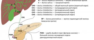
• Pathological anatomy . Cholangiocarcinoma (most tumors are scirrhous or papillary type) - severe fibrosis often complicates diagnosis •• On macroscopic examination, cholangiocarcinoma is a tumor-like formation involving part of the bile ducts. It is often difficult to distinguish cholangiocarcinoma from sclerosing cholangitis •• Localization of the tumor - distal parts of the common bile duct (1/3 of cases), common hepatic or cystic duct (1/3 of cases), right or left hepatic duct.
Damage to the confluence of the right and left bile ducts is defined as Klatskin tumor •• This type of cancer metastasizes to regional lymph nodes (16% of cases), liver (10%). Direct tumor growth into the liver is possible (14%).
• Clinical picture •• Due to the small diameter of the ducts, signs of duct obstruction appear even with small sizes of the primary tumor. Severe jaundice, itchy skin, lack of appetite, weight loss and constant aching pain in the right upper quadrant of the abdomen develop.
• Laboratory data . The content of serum alkaline phosphatase, direct and total bilirubin was significantly increased. The content of serum transaminases was less significantly changed.
• Diagnosis can be made using percutaneous transhepatic cholangiography or endoscopic retrograde cholangiopancreatography. Both tests allow biopsy of suspicious areas of tissue for histological examination.
• TNM classification (see also Tumor, stages) •• Tis - carcinoma in situ •• T1 - tumor invades the subepithelial connective tissue or muscular connective tissue layer •• T2 - tumor spreads to the perimuscular connective tissue •• T3 - tumor spreads to adjacent structures: liver, pancreas, duodenum, gallbladder, colon, stomach •• N1 - metastases in the lymph nodes near the cystic and common bile ducts and/or porta hepatis (i.e. in the hepatoduodenal ligament) •• N2 - metastases in the lymph nodes located near the head of the pancreas, duodenum, celiac and superior mesenteric arteries, posterior peripancreatoduodenal lymph nodes.
• Grouping by stage • Stage 0: TisN0M0 • Stage I: T1N0M0 • Stage II: T2N0M0 • Stage III: T1–2N1–2M0 • Stage IV •• T3N0–2M0 •• T1–3N0–2M1.
• Treatment is surgical, but tumor resectability does not exceed 10% •• For tumors of the distal parts of the common bile duct, pancreaticoduodenectomy (Whipple procedure) is performed with further restoration of the patency of the biliary tract and gastrointestinal tract. If the tumor is localized in more proximal parts, the tumor is excised followed by reconstruction of the common bile duct. The average life expectancy of patients after surgery is 23 months. Radiation therapy in the postoperative period can increase life expectancy •• If the tumor cannot be removed, it is tunneled. A permanent drainage is carried out through the resulting channel, opening on one side into the intrahepatic ducts proximal to the tumor, and on the other hand into the common bile duct distal to the tumor ••• It is possible to introduce external drainage during percutaneous transhepatic cholangiography ••• For palliative purposes, U- drainage is indicated shaped tube according to Praderi. Both ends of the drainage are brought to the skin: one is similar to the drainage of the common bile duct, the other - through the tumor tissue, liver parenchyma and abdominal wall ••• The advantage of U-shaped drainages is that they can be replaced in case of obstruction with tissue detritus. A new drainage tube is sutured to the end of the previously installed one and replacement is carried out by tightening the drainage ••• Installation of drainage increases life expectancy by 6–19 months.
• Prognosis •• Distant metastasis of common bile duct cancer usually occurs late and, as a rule, is not the direct cause of death •• Causes of death ••• Biliary cirrhosis due to inadequate outflow of bile ••• Intrahepatic infection with the formation of abscesses ••• General exhaustion, decreased reactivity of the body ••• Sepsis.
ICD-10 • C23 Malignant neoplasm of the gallbladder • C24 Malignant neoplasm of other and unspecified parts of the biliary tract • D13.4 Benign neoplasm of the liver
Treatment tactics and survival prognosis
Treatment for gallbladder cancer is extremely rare because the disease is almost impossible to diagnose at an early stage. As a rule, a tumor is discovered during diagnostic and therapeutic procedures indicated for other reasons, for example, during cholecystectomy. If metastasis extends beyond the gallbladder, cholecystectomy is performed. To prevent the development of cancer due to residual diseased epithelial cells, a lobe of the liver and regional lymph nodes are removed.
The chances of a full recovery, however, are low. In advanced cases, the survival prognosis is extremely low, since very often the tumor progresses, growing into the liver. They are observed in 70% of operated patients. If the bile duct is blocked by a tumor or inflamed lymph nodes, endoprosthesis replacement (stenting), transhepatic cholangiostomy or hepaticojejunostomy is performed. However, only 10% of patients manage to prolong life by a maximum of 5 years.
Common bile duct cancer
The etiology of the disease reveals a close connection between the development of bile duct tumors and other pathologies: sclerosing cholangitis, ulcerative colitis, choledocholithiasis. Also at risk are patients in contact with toluene and benzene derivatives. In Asian countries, the development of cholangiocarcin is often provoked by parasitic invasions of the biliary system organs, for example, Clonorchis trematodes. In most cases, the tumor develops from a congenital cystic dilatation of the bile ducts. Diagnosis of papillary and scirrhous tumors, or cholangiocarcinoma, is often difficult due to fibrosis. The study of its macroscopic structure makes it possible to detect the involvement of the bile ducts in the cancer process. In approximately one third of cases, the tumor develops in the distal portions of the common duct, cystic duct, or hepatic duct. If the tumor is localized at the confluence of the left and right ducts, the disease is called Klatskin tumor. In 10% of cases, such cancer metastasizes to the liver, in 16% to regional lymph nodes. Also, in 14% of cases, the tumor can grow into the liver.
Causes of the disease
There are certain factors that increase the risk of developing gastric cancer:
- age 65 years and older;
- hereditary factor;
- excessive alcohol consumption, smoking, exposure to harmful chemicals;
- diseases that cause inflammation and scarring (colitis, primary sclerosing cholangitis).
- liver fluke infection.
Almost 2/3 of cases of gastric cancer develop with a long previous course of chronic cholecystitis or cholelithiasis. Carcinogenesis is promoted by injury to the mucous layer by moving stones.
Symptoms and diagnosis of the disease
The bile ducts have a small diameter, so even with a minimal tumor size, signs of blockage begin to appear. They are expressed in aching pain in the right hypochondrium, jaundice, lack of appetite, sudden loss of body weight, and itchy skin. Laboratory diagnostics can detect an increase in direct and total bilirubin, serum alkaline phosphatase, and a slight change in the concentration of serum transaminases. Instrumental studies are also informative - endoscopic retrograde cholangiopancreatography and percutaneous transhepatic cholangiography. Both methods also involve the collection of affected tissue for histology.
Establishing diagnosis
It is recommended to carefully collect complaints and medical history from patients to determine factors that influence the choice of treatment tactics. A physical examination is required to assess nutritional status to determine functionality.
Diagnosis of gastric cancer includes:
- Laboratory research. They are carried out at primary signs of the disease and regularly during treatment to predict the course and determine risk factors. Laboratory diagnostics include a CBC (complete blood count) and a biochemical test to check liver function.
- Instrumental diagnostics (ultrasound and CT). The structure of the gallbladder, bile ducts, as well as the size and location of the tumor are determined.
Additional studies - endoscopy (choledochoscopy, endoscopy). Direct contrast methods and angiographic examination are recommended. For morphological verification of the diagnosis, a tumor biopsy is indicated.
TNM classification of the disease
In accordance with the international classification of stages of development of oncological diseases, the following types of cholangiocarcinomas are distinguished:
- TIS or carcinoma in situ - the tumor does not extend beyond the affected organ;
- T1 - malignant tumor spreads in the muscular-connective layer of the organ or in the subepithelial connective tissue;
- T2 - carcinoma grows into the perimuscular connective tissue;
- T3 - carcinoma metastasizes to adjacent structures;
- N1 - the tumor metastasizes to the lymph nodes in the hepatoduodenal ligament;
- N2 - metastasis spreads to the lymph nodes near the celiac and mesenteric arteries, head of the pancreas, duodenum and peripancreatoduodenal lymph nodes.
Treatment tactics and survival prognosis for common bile duct cancer
Only surgical removal of the tumor can increase the chance of recovery, and its resectability is no more than 10%. For resection of distal cancer, the so-called Whipple operation is performed - pancreaticoduodenectomy, after which the patency of the biliary tract and gastrointestinal tract is restored. If the tumor is localized in the proximal parts, restoration of the general bile flow is required after removal of the tumor. When tumor removal is not possible, tunneling is performed using drainage. On one side it opens into the common bile duct, on the other - into the intrahepatic ducts. External drainage is sometimes used in percutaneous transhepatic cholangiography. After surgery, life expectancy increases by an average of 23 months. Chemotherapy helps to slightly extend this period.
According to Praderi, drainage is performed with a U-shaped tube. The manipulation is carried out for palliative purposes, with its ends being exposed to the skin. In cases of blockage, drainage can be replaced with tissue debris. The new tube is sutured at one end to the old one, and replacement is done by pulling it up. Drainage makes it possible to prolong the patient’s life by 6-19 months.
The prognosis for cancer of the common bile duct is disappointing. Metastasis occurs quite late. The causes of death in this disease can usually be general exhaustion, sepsis, liver infection, abscesses and biliary cirrhosis.


