A pinched nerve is most often not dangerous: rest and treatment will help relieve the compression and restore nerve function within a few days or weeks. But in rare cases, the situation is so serious that surgical intervention is necessary.
A pinched nerve is a condition in which too much pressure is placed on a nerve by nearby tissue. These can be muscles, cartilage, tendons, bones. As a result, the functioning of the nerve is disrupted. First of all, this is manifested by pain in the corresponding part of the body, as well as a decrease in muscle strength and tone, a feeling of numbness, tingling, and decreased sensitivity.
The most typical example of pinched nerves is compression of the spinal cord roots by a herniated disc. If this happens in the lumbar region, then sciatica usually occurs - pain that spreads along the back of the leg, along the sciatic nerve. Another fairly common condition is carpal tunnel syndrome, where the tissue in the wrist area swells and puts pressure on a nerve.
Nerve compression is a very common condition and one of the most common reasons for visiting doctors. According to American scientists, the pathology occurs in 85 people out of one hundred thousand adults. It can happen at any age, but most often occurs in people over 50 years of age. The most common cause is arthrosis and degenerative changes in the spinal column.
Why does pinched nerve occur?
The causes of compression of nerves in the body vary widely. Main risk factors:
- soft tissue injuries, bone damage;
- inflammation and swelling in surrounding tissues;
- herniated intervertebral discs;
- osteochondrosis, arthrosis of intervertebral joints and other degenerative changes in the spinal column;
- repeated frequent repetitive movements - for example, in people who type on a keyboard;
- intense physical activity, sports - especially if a person does it professionally;
- obesity – since extra pounds place increased stress on the joints and spine;
- rheumatoid arthritis is a disease that leads to inflammation in the joints and compression of nearby nerves;
- pregnancy - during it, body weight increases, the load on the musculoskeletal system increases and at the same time the connective tissue softens so that the woman’s pelvis can pass the fetus;
- diabetes mellitus - does not directly lead to nerve compression, but increases the risk of developing this condition;
- thyroid disease increases the risk of developing carpal tunnel syndrome;
- bone spurs (osteophytes) – growths on the bones that occur with degenerative joint diseases;
- prolonged bed rest;
- cicatricial changes in tissues after injuries, burns.
Another risk factor is gender. Women are more likely to experience carpal tunnel syndrome. This is probably due to the fact that their channel is normally narrower than that of men. Of course, heredity also plays a role. Some people are more likely to develop degenerative processes in the spine and joints - this is influenced by certain gene variants that affect the characteristics of connective tissue.
What symptoms should you look out for?
Pinched nerves can occur throughout the body, and the manifestation will depend on where the compression occurs. The most common conditions:
- Compression of the nerves of the neck is what is most often called cervical radiculopathy.
- Compression of nerves in the lumbar region is lumbar radiculopathy.
- Compression of the sciatic nerve – sciatica.
- Compression of the ulnar nerve.
- Carpal tunnel syndrome.
Let's look at the eight most common manifestations.
Pain or burning sensation spreading down the leg
The largest nerve in the human body, the sciatic, emerges from the lower part of the spine. It passes through the lumbosacral region, the buttock and then along the back of the entire leg. Its branches provide muscle movement and skin sensitivity. Compression of the sciatic nerve can be caused by such reasons as intervertebral hernias and spinal stenosis in the lumbar region, trauma, spondylolisthesis (a condition in which the upper vertebra seems to slip from the lower one), piriformis syndrome (when this pelvic muscle spasms and compresses the nerve) , tumors of the spinal column. In medical parlance, compression of the sciatic nerve is called sciatica or radiculopathy. It manifests itself in the form of pain along the back of the leg, along the nerve - dull, aching or sharp, like lumbago.
Pain radiating from the neck down the arm
Just as the sciatic nerve descends from the lumbar spine to the lower limb, the nerves of the arm are formed by roots that are located in the cervical spine. When they are squeezed, the pain spreads down the arm. Pain sensations can be localized in the area of the upper arms, upper arms, forearms, hands, and upper back.
In general, if we talk about compression of nerves caused by pathologies of the spine, the lumbar region most often suffers: it is located at the bottom, and it bears heavy loads. In addition, there is high mobility here. In second place is the cervical spine, due to its high mobility and the fact that it has to withstand the weight of the skull. In the thoracic region, disturbances occur much less frequently, because there is minimal mobility and the vertebrae are fixed by the ribs.
Weakness in the legs
While walking, the nerves of the lower extremities work at lightning speed. They must quickly transmit signals that cause specific muscle groups to contract and relax. Severe compression of the nerve leads to poor signal transmission. As a result, weakness occurs in the leg muscles, their tone decreases, and gait is disrupted. This is already quite a formidable symptom. Patients often describe it with the phrase: “Doctor, one of my legs literally drags while walking.”
Decreased hand grip strength
The nerves of the hands provide muscle contractions, precise coordination of movements, and fine motor skills. When pinched, these functions are disrupted, and the person notices that everything is literally falling out of his hands. Particularly difficult is when writing and performing other tasks that require small movements.
Feeling numb
In parallel with motor ones, sensory nerve fibers also suffer. They stop sending impulses to the brain, and a feeling of numbness occurs - like after an injection of an anesthetic, or “freezing” the mouth during dental treatment. In the most severe cases, sensitivity in a certain area is completely lost - this indicates that the nerve has suffered greatly.
Tingling feeling
Unpleasant sensations in the form of tingling, burning, and crawling sensations are called paresthesia in medical terms. They occur when the nerve is irritated and incompletely compressed. This feeling can be compared to the sensations in the hand when it was “rested” in a dream or after being hit with the elbow in the place where the nerve passes. Paresthesia in the hand is often the first sign of carpal tunnel syndrome.
Dysfunction of the rectum and bladder
The nerves that exit the lumbar spine control not only the muscles, but also the internal organs, in particular the bladder and rectum. Loss of control over urination and bowel movements indicates that the compression has caused severe nerve damage and the patient needs immediate medical attention. This condition may manifest as urinary and fecal incontinence or other disorders.
Pain that changes in intensity as the person changes position
Here sciatica can again be cited as a clear example. The patient usually feels better when he sits, leaning forward, or lies on his back. And in the position on the side, the pain syndrome, on the contrary, intensifies. This causes problems with sleep. When a person assumes a certain position to relieve pain, this is called antalgic position.
With malignant tumors and metastases in the spine, the pain is usually long-lasting, painful, and intensifies during rest and night sleep.
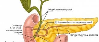
Baliar dysfunction with pathology of the gallbladder and sphincter of Oddi
Author: Yu.V.
Vasiliev , Doctor of Medical Sciences, Professor, Central Research Institute of Gastroenterology, Moscow
Recently, the term “biliary dysfunction” is increasingly mentioned in the domestic medical literature. This is due to the widespread prevalence of functional disorders of the biliary tract, which, according to various estimates, ranges from 12 to 58%. Moreover, among women they occur 2–3 times more often than among men.
Functional diseases of the biliary tract are a complex of clinical symptoms that develop as a result of motor-tonic dysfunction of the gallbladder, bile ducts and sphincters. Rome III criteria (2006) distinguish the following functional disorders of the biliary tract: E1 - functional disorders of the gastrointestinal tract, E2 - functional biliary disorder of the sphincter of Oddi, E3 - functional pancreatic disorder of the sphincter of Oddi.
In accordance with ICD-10, dysfunction of the biliary tract can be classified into two categories K82.8 (dyskinesia of the gallbladder and cystic duct) and K83.4 (spasm of the sphincter of Oddi).
Functional disorders of the gallbladder and sphincter apparatus of the biliary tract are characterized by spontaneity and a variety of clinical manifestations, duration of the course, and complexity of diagnosis, which ultimately determines the high demand of patients for medical help.
One of the features of the biliary system is the closely interconnected anatomical location of the gallbladder and duodenum. Violation of neurohumoral regulation and disruption of the concentration function of the gallbladder are the main factors contributing to the formation of biliary insufficiency. The parasympathetic and sympathetic parts of the autonomic nervous system, as well as the endocrine system, take part in the regulation of the motor activity of the biliary system, providing a synchronized sequence of contraction and relaxation of the gallbladder and the sphincter apparatus of the biliary system.
It has been shown that moderate irritation of the vagus nerve causes coordinated activity of the gallbladder and sphincters, and severe irritation causes spastic contraction with delayed bile evacuation. Stimulation of the sympathetic nerve helps relax the gallbladder. Of the gastrointestinal hormones, cholecystokinin - pancreazimin (CCK-PZ) has the maximum effect, which, along with contraction of the gallbladder, promotes relaxation of the sphincter of Oddi. The incentive for the production of CCK-PZ is fatty foods, and the nervous regulatory influences are the pressure gradient and its change.
Considering the fact that the process of bile formation is continuous (the daily flow of bile is on average 500–1,200 ml), and the flow of bile into the duodenum occurs only during meals, the importance of the coordinated actions of the gallbladder, which performs the reservoir function, and the sphincter the biliary tract apparatus, which ensures the timely and required amount of bile entering the intestines.
Violation of neurohumoral regulation contributes to the development of chronic stagnation of bile in the gallbladder and disruption of its evacuation into the duodenum. And a violation of the concentration of bile in the gallbladder against the background of a violation of the mechanisms of the sphincter apparatus increases the likelihood of the development of inflammatory and dystrophic pathological changes in the mucous membrane of the gallbladder, which contributes to impaired absorption of lipids, evacuation of pancreatic secretions from the pancreas to the duodenum. Functional disorders of the gallbladder
Gallbladder dysfunction is a disorder of gallbladder motility, manifested by biliary pain due to initial metabolic disorders (saturation of bile with cholesterol) or primary disturbances of gallbladder motility in the absence, at least initially, of abnormalities in the composition of bile.
In this case, all of the following criteria must be present: - biliary pain; — preserved gallbladder; — absence of gallstones, biliary sludge, microlithiasis, and other organic diseases; — gallbladder ejection fraction less than 40%; - no changes in the biochemical blood test: normal levels of transaminases (ALAT, ASAT), conjugated bilirubin and pancreatic enzymes (amylase/lipase).
With hyperkinetic disorders, colicky pain of varying intensity occurs, without irradiation or with irradiation to the right, to the back, sometimes to the left half of the abdomen (with the involvement of the pancreatic ductal system). With hypokinesia, dull pain in the right hypochondrium, a feeling of pressure, and fullness are noted, which intensify with changes in body position and with an increase in intra-abdominal pressure, which changes the pressure gradient for bile flow. Common manifestations for various forms of dysfunction are bitterness in the mouth, bloating, and unstable stools.
Thus, the central symptom of gallbladder dysfunction is the “biliary” type of pain, and the only objective characteristic that ultrasound can record is delayed emptying of the gallbladder or its increased size. A very important objective symptom of impaired gallbladder motility is the ultrasonic phenomenon of “sludge” (sediment), which can be presented in two variants: a) diffuse, b) parietal. Diagnostic criteria for gallbladder dysfunction are episodes of severe or persistent pain localized in the epigastrium or in the right upper quadrant of the abdomen, characterized by: - episodes lasting 30 minutes or more; — development at least once in the previous 12 months; - permanent character.
Functional disorders of the sphincter of Oddi. Dysfunction of the sphincter of Oddi The main functional features of the sphincter of Oddi: - regulation of the flow of bile and pancreatic secretions into the duodenum; - preventing the flow of contents from the duodenum into the common bile and Wirsung ducts.
Clinical manifestations of sphincter of Oddi dysfunction—nausea, vomiting, pain, and/or flatulence—are rather nonspecific. Manifestations of sphincter of Oddi dysfunction, such as colic, are not a typical symptom of biliary dyskinesia. More typical, according to the observations of some researchers, for this disease is a feeling of heaviness in the upper half of the abdomen. In this case, the basal pressure of the sphincter of Oddi does not exceed 40 mm Hg. However, when chronic pancreatitis (CP) is combined with sphincter of Oddi dysfunction, clinical manifestations can be very variable. Sometimes it looks like an attack of biliary colic, but unclear, difficult to explain pain in the upper abdomen is also possible. One of the diagnostic signs is the occurrence of pain of unknown origin, in which there may be an increase in the levels of serum liver enzymes, observed in some cases with the so-called idiopathic pancreatitis.
In some patients with dysfunction of the sphincter of Oddi, along with the appearance of pain, upon palpation of the abdomen, pain occurs in certain parts of the abdomen; Some patients may experience chills and fever. During the period between attacks of pain, the condition of patients can be quite satisfactory. The difference in the type and severity of clinical manifestations in sphincter of Oddi dysfunction obviously forced some researchers to somehow systematize clinical data. At the International Meeting of Gastroenterologists (Rome, 1999), it was decided to identify diagnostic criteria for sphincter of Oddi dysfunction, which conditionally included a complex of functional disorders lasting more than 3 months, the main clinical symptoms of which are the following: 1) recurrent attacks of severe or moderate pain lasting 20 and more than minutes, localized in the epigastric region and/or in the right hypochondrium (biliary type); 2) pain localized in the left hypochondrium, decreasing in intensity when the body is tilted forward (pancreatic type); 3) girdle pain (combined type).
In this case, pain often occurs at a certain time (the onset of occurrence is after eating; the appearance of pain at night), nausea and/or vomiting may occur.
Examination of patients with suspected sphincter of Oddi dysfunction
Clinical, laboratory and instrumental examination of patients with sphincter dysfunction includes the study of symptoms, medical history and life, as well as a physical examination of patients. During a laboratory examination of patients, the detection of changes in the levels of liver enzymes (with an increase of 2 times or more), an increase in the levels of AST and/or alkaline phosphatase in two or more studies, as well as a change in the concentration of bicarbonates in the pancreatic secretion indicates dysfunction of the sphincter of Oddi in CP. Identification of columnar epithelial cells not stained with bile (non-imbibed) indicates the presence of duodenitis accompanying spasms of the sphincter of Oddi; identification of imbibed cells is an indication of a spasm of the sphincter of Oddi caused by inflammatory changes in the common bile duct.
If necessary, it is advisable to conduct endoscopic manometry of the sphincter of Oddi, performed during endoscopic cholangiopancreatography (EPCP), pancreatic ductography, traditional ultrasound and/or endoscopic ultrasound, scintigraphy of the liver and biliary tract and/or EPC, based on the results of which in identified cases of sphincter dysfunction Oddi there is a slowdown in the flow of contrast agent (more than 5 minutes). Non-invasive methods such as ultrasound assessment of the diameter of the pancreatic duct under secretin stimulation are less informative and have relatively low sensitivity. In 2/3 of patients with dysfunction of the sphincter of Oddi, an increase in the basal tone of the parasympathetic sphincter is detected.
Unfortunately, carrying out some instrumental methods for examining the nipple of Vater in routine practice, for various reasons, is not always possible and even sometimes turns out to be unjustified. Yes, according to some observations. assessment of the “efficacy-risk” ratio does not confirm the advisability of manometry for diagnostic purposes and endoscopic papillotomy for therapeutic purposes. Certain technical difficulties in carrying out the above instrumental studies, the likelihood of side effects (with endoscopic papillotomy - 1–2% of cases) or complications (with endoscopic manometry - more than 10% of cases), as well as deaths (with endoscopic papillotomy - 0.8% ) is one of the reasons hindering the introduction into widespread practice of invasive instrumental methods for examining the papilla of Vater and the pancreas and, accordingly, the timely recognition of sphincter of Oddi dysfunction.
Treatment of patients
Diet therapy occupies a significant place in the treatment of patients with functional diseases of the biliary system, because Maintaining proper nutrition, taking into account the nature of motor disorders, contributes to faster rehabilitation of patients and improves the quality of life. A diet with frequent meals of small amounts of food (5-6 meals a day) is recommended, which leads to regular emptying of the gallbladder and normalizes pressure in the ductal system of the biliary tract and duodenum. Patients are allowed to eat late meals shortly before bedtime. When selecting a dietary diet, the influence of individual nutrients on the normalization of the motor function of the gallbladder and biliary tract is taken into account. Thus, with the hyperkinetic type of dysfunction, the consumption of foods that stimulate contraction of the gallbladder should be sharply limited - animal fats, vegetable oils, rich meat, fish and mushroom broths. With hypotension of the gallbladder, patients usually tolerate weak meat and fish broths, cream, sour cream, vegetable oils, and soft-boiled eggs well. Vegetable oil is prescribed in a teaspoon 2-3 times a day 30 minutes before meals for 2-3 weeks.
Principles of therapy
1. Increased contractile function of the gallbladder (if it is insufficient). 2. Decreased contractile function of the gallbladder (with its hyperfunction). 3. Restoring the tone of the sphincter system. 4. Restoring pressure in the duodenum, which normalizes the pressure gradient in the biliary tract. 5. Restoration of adequate outflow of bile and pancreatic juices into the duodenum.
For dysfunction caused by increased tone of the sphincters of the biliary system, antispasmodics are used. Both non-selective and selective M1-anticholinergic blockers are used as antispasmodics. It should be noted that when taking this group of drugs, a number of side effects may occur: dry mouth, urinary retention, blurred vision, tachycardia, constipation, drowsiness. The combination of the relatively low therapeutic efficacy of drugs in this group with a wide range of side effects limits their use, especially in the treatment of sphincter of Oddi dysfunction. Myotropic antispasmodics include drotaverine, bencyclane, mebeverine, otilonium citrate, trimebutine or hymecromone, which have a selective antispasmodic effect on the sphincter of Oddi.
When treating hypofunction of the gallbladder, drugs are used that enhance its motility. For this purpose, choleretics can be used, which include drugs containing bile and bile acids; synthetic drugs (oxamide, hydroxymethylnicotinamide, cyqualone), some herbal drugs (flamin, corn silk, etc.), as well as cholekinetics, such as magnesium sulfate, olive oil and other oils, sorbitol, xylitol, holosas, etc.
In the treatment of biliary dysfunction, a significant place is given to ursodeoxycholic acid (UDCA), which affects certain links in the pathogenesis of developing cholestasis. The main mechanisms of action of UDCA: 1) anticholestatic: UDCA suppresses the secretion of toxic bile acids into bile, their absorption in the ileum and thereby promotes their excretion from the body; 2) cytoprotective: due to the presence of hydrophilicity, UDCA improves the fluidity of the phospholipid bilayer of the hepatocyte membrane, restores the structure of cells and protects them from damage; 3) hypocholesterolemic: decreased synthesis of cholesterol in the liver, decreased secretion into bile and absorption in the intestine; 4) litholytic: reducing the lithogenicity of bile due to the formation of liquid crystals with cholesterol molecules, preventing the formation and dissolution of gallstones; 5) immunomodulatory: under the influence of UDCA, the synthesis of immunocompetent IgM (and to a lesser extent IgG and IgA) is reduced, the expression of histocompatibility antigens on hepatocytes and cholangiocytes is reduced, which in turn prevents the activation of cytotoxic T lymphocytes, and also reduces the production of autoantibodies and promotes reduction of immunopathological reactions; 6) anti-apoptotic: by reducing the concentration of ionized Ca in cells, the release of cytochrome C from mitochondria is blocked, which in turn prevents activation of caspases and, accordingly, apoptosis of cholangiocytes.
In clinical practice, a new representative of UDCA, the drug Ursodez, has become available to outpatient doctors. The drug is well tolerated by patients and is used in both adults and children (with a body weight of 34 kg). This drug is effective in the complex treatment of biliary dysfunction. The use of UDCA (Ursodez) in an average daily dose of 10–15 mg per 1 kg of body weight reduces dyscholia. The dose and duration of treatment with Ursodez are determined by the degree of biliary insufficiency and the dynamics of its changes during the therapy. Thus, for biliary insufficiency of stage I, UDCA is prescribed at a dose of 7–10 mg/kg for at least 1–2 months; in grade II – the dose is increased to 10–15 mg/kg for at least 3 months; for grade III biliary insufficiency, Ursodez is prescribed at a dose of 15 mg/kg or higher.
It is advisable to adjust the dose after leveling the lithogenic properties of bile, gradually reducing it over 3–4 months, and then completely stop taking the drug. It is necessary to carry out a biochemical study of bile at intervals of 1–2 times a year to determine the level of cholesterol and bile acids in it. When taking UDCA, side effects are rare and do not exceed 2–5%.
Conclusion
The increased attention of gastroenterologists to functional disorders of the biliary system and the entire gastrointestinal tract is not accidental. This is due to the increasing prevalence of melon changes among the population, as well as the emergence of the possibility of successful treatment, prevention or delaying the development of organic pathology that is more severe in prognosis.
The use of modern methods for diagnosing biliary tract dysfunction, taking into account the clinical features of the course of the disease, now makes it possible to timely and accurately diagnose this pathology in most patients.
Medicines with different and sometimes combined mechanisms of action, depending on the pathogenetic characteristics of a particular disorder, make it possible to select more adequate therapy that can significantly improve the well-being and quality of life of patients with functional disorders of the biliary tract. Literature
1. Vasiliev Yu.V. Chronic pancreatitis: diagnosis, treatment of patients // Attending physician. - 2004. - No. 2. - P. 10–13. 2. Vasiliev Yu.V. Differentiated approach to antisecretory therapy of chronic pancreatitis combined with peptic ulcer and gastroesophageal reflux disease. // RMJ. - 2005. - T.7. - No. 2. - P. 57–60. 3. Vasiliev Yu.V. Pain syndrome in chronic pancreatitis: drug treatment of patients // Farmateka. - 2005. - No. 4. – pp. 44–48. 4. Vasiliev Yu.V. Autoimmune pancreatitis // Experiment. and clinical gastroenterology. – 2005. – No. 2. — pp. 83–86. 5. Leishner U. Practical guide to diseases of the biliary tract. M., “GEOTAR-MED” - 2001. - 260 p. 6. Maksimov V.A., Chernyshev A.L. et al. Biliary insufficiency. M., 2008. – 231 p. 7. Minushkin O.N. Biliary dysfunction: definition, classification, diagnostics, treatment. - Attending doctor. – 2004, September No. 7. — P. 50–53. 8. Ilchenko A.A., Orlova Yu.N. Use of hepabene in patients with chronic cholecystitis. Materials of the 3rd Russian Scientific Forum “St. Petersburg - Gastro-2001”. Gastrobulletin No. 2–3. 2001. P. 39. 9. Vikhrova T.V. Biliary sludge and its clinical significance. Diss. Ph.D. honey. Sci. M.: 2003. 115 p. 10. Nemtsov L.M. Dysmotility of the gallbladder in biliary pathology (clinical and pathophysiological characteristics and correction). Vitebsk: VSMU, 2004. 183 p. 11. Maev I.V., Samsonov A.A., Salova L.M. and others. Diagnosis and treatment of biliary tract diseases. Tutorial. M.: GOU VUNMC Ministry of Health of the Russian Federation, 2003. 96 p. 12. Ilchenko A.A. Cholelithiasis. M.: “Anacharsis”, 2004. 200 p.
When do you need to see a doctor urgently?
The following symptoms should be the reason to immediately consult a doctor:
- very severe pain, especially if it occurs acutely after an injury;
- pain that does not go away within several days, despite the fact that the person kept rest and took painkillers;
- pain in the spine, arm, leg, which is accompanied by an increase in body temperature;
- deterioration of sensitivity, paresthesia, decrease in muscle tone, strength.
To understand the causes of nerve compression, the doctor may prescribe different diagnostic methods. The most commonly used are electroneuromyography (study of the passage of electrical impulses in nerves and muscles), ultrasound (preferably using a high-resolution device), computed tomography and magnetic resonance imaging.
Treatment technologies
After processing all the data, three options are possible: the doctor will prescribe a special diet for you; will prescribe complex drug treatment to eliminate the causative agent of the disease; or will also prescribe additional treatment, taking into account the existence of other pathologies (diseases of the cardiovascular system, respiratory, urinary, etc.)
Acute diseases of the liver and gallbladder are completely curable in most cases, but chronic diseases are almost impossible to completely cure. During chronic treatment in our clinic, doctors achieve rapid relief of exacerbation, long-term remission, preservation of maximum liver function (that is, compensation for liver failure), and the gall bladder.
Timely diagnosis and prompt initiation of treatment will reduce medical intervention, as well as the consequences of the disease, to a minimum. Contacting a gastroenterologist at our clinic guarantees you qualified diagnostic and therapeutic services. You will be treated according to an individual program.
How are pinched nerves treated?
The first measure in such cases is rest for the part of the body in which the pain is bothering. Your doctor may prescribe a splint to limit movement in the limb. For example, for carpal tunnel syndrome, such a splint is recommended to be worn around the clock.
Drug treatment is with nonsteroidal anti-inflammatory drugs (NSAIDs), such as ibuprofen or naproxen. These drugs help relieve pain in the acute period. If the pain is very severe, the doctor may prescribe injections of glucocorticosteroids - preparations of adrenal hormones. They have a more pronounced analgesic and anti-inflammatory effect.
After a period of immobilization, physical therapy begins. There are special exercises that help strengthen the muscles in the affected area or relieve their spasm. Due to this, compression is reduced. An exercise therapist may recommend changes in daily activities, such as avoiding certain repetitive movements. Physiotherapy and therapeutic massage are used.
Typically, if conservative treatment does not help within a few weeks or months, surgery is indicated. The type of surgery depends on where the nerve compression occurs and what is causing it. For example, this may include removing bone spurs, a herniated disc, or cutting a ligament to free a nerve for carpal tunnel syndrome.