the whole list
Peptic ulcer disease is a chronic disease that occurs with alternating periods of exacerbation and remission, the main symptom of which is the formation of a defect (ulcer) in the wall of the stomach and/or duodenum.
An ulcer is a defect in the mucous membrane that has a clear boundary. A superficial ulcer is called erosion. Peptic ulcer disease is detected in 5–10% of the adult population.
Depending on the location, ulcers are divided into gastric and duodenal (duodenum - duodenum).
The ratio of gastric and duodenal ulcers is approximately 1: 4. Young people have a lower incidence of gastric ulcers compared to duodenal ulcers (1: 13). Conversely, in middle-aged and elderly patients the frequency of gastric ulcers increases.
Along with peptic ulcer disease as an independent disease, it is now customary to distinguish secondary symptomatic ulcers of the stomach and duodenum that occur under the influence of stress, circulatory disorders, taking antipyretics, painkillers (acetylsalicylic acid, indomethacin, diclofenac, ibuprofen) and others.
Depending on the cause, there are:
- stress ulcers (for example, with myocardial infarction, widespread burns, after neurosurgery, etc.);
- drug-induced ulcers (taking non-steroidal anti-inflammatory drugs, hormones and other medications);
- endocrine ulcers (with Zollinger-Ellison syndrome, hyperparathyroidism);
- ulcers in certain diseases of internal organs (Crohn's disease, atherosclerosis of the branches of the abdominal aorta, etc.).
Stomach ulcer, its types and stages
Gastric ulcer (hereinafter referred to as PUD) is a disease in which a mucosal defect occurs and the submucosal layer may be affected. This is a chronic disease that occurs in waves, with periods of exacerbation and remission.
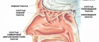
An ulcer can be localized in different parts of the stomach:
- subcardial and cardiac - in the upper part;
- antrum - in the lower section;
- in the body - in the middle part;
- in the pyloric canal - at the transition to the duodenum.
The size of the ulcer is:
- small - up to 5 mm in diameter;
- medium - in diameter 6-19 mm;
- large - 20-30 mm in diameter;
- gigantic - more than 30 mm in diameter.
There are also ulcers associated with the bacterium Helicobacter pylori (hereinafter referred to as HP) and those not associated with it.
The disease occurs in several stages:
- exacerbations - the occurrence of a defect in the mucous membrane;
- scarring - the formation of connective tissue at the site of the ulcer;
- remission - healing of the defect.
The stage of cicatricial and ulcerative deformation of the organ is distinguished separately.
Diagnostics
- General clinical analysis of blood and urine
- Analysis of stool for coprogram
- Fecal occult blood test
- Biochemical blood test (liver tests, cholesterol, alkaline phosphatase)
- ECG
- X-ray of the chest organs in 2 projections and X-ray of the abdominal organs (to exclude perforation of ulcers)
- X-ray of the esophagus, stomach with barium mixture
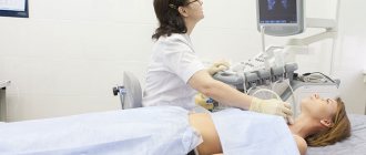
- Ultrasound of the hepatobiliary system
- 24-hour pH monitoring in the lower esophagus and stomach
- EGDS
- Non-invasive tests for the determination of Helicobacter pylori (respiratory)
Main causes
The cause of a defect in the gastric mucosa is considered to be an imbalance between the aggressive secretions of the stomach and the protective qualities of the mucous membrane. To digest food, the stomach secretes pepsin and hydrochloric acid. To prevent them from destroying the stomach, mucus forms on its surface, protecting the organ. If more acid is produced and less mucus is produced, an ulcer may form. The background for such a pathology may be a decrease in gastric motility, a violation of the regenerative - restorative properties of the mucous membrane against the background of certain diseases.
The main etiological cause of ulcer formation is damage to the mucous membrane of the HP. This bacterium successfully survives in an acidic environment and produces an enzyme that causes an inflammatory reaction with subsequent formation of a defect. Helicobacter pylori is detected in 82% of patients suffering from gastric ulcers.
Another reason may be uncontrolled use of anti-inflammatory non-steroidal drugs, such as aspirin.
Provoking factors may be:
- smoking and alcohol abuse;
- constant consumption of spicy food;
- stressful situations;
- hereditary predisposition;
- dry food;
- eating disorder.
Gastric ulcers often develop against the background of certain diseases, such as atrophic gastritis and hormonal pathologies.
Prevention and recommendations
It is necessary to exclude those factors that lead to the development of peptic ulcers and monitor your health in the presence of a hereditary predisposition, even if nothing bothers you.
Risk factors for developing peptic ulcers:
- Helicobacter pylori infection
- hereditary predisposition
- blood type I (0)
- bad habits (tobacco smoking, drinking alcohol and its surrogates)
- increased stomach acidity
- violation of working and rest conditions, frequent stress
- taking ulcerogenic medications (hormones, ibuprofen, Nise, Nurofen, diclofenac and others)
- non-compliance with food intake
- diseases of the gastrointestinal tract (cholecystitis, pancreatitis, gastritis, gastroduodenitis)
- chronic obstructive pulmonary disease, systemic diseases, chronic renal failure.
Symptoms of the disease
Peptic ulcer usually manifests itself more often in spring and autumn, during the period of exacerbation of the disease. The first sign of the disease is pain that occurs immediately after eating. This is due to the fact that pepsin and hydrochloric acid released during meals aggressively affect the defect unprotected by mucus. Moreover, the manifestations of pain symptoms increase within an hour after eating. The pain is localized in the epigastric region, in the region of the heart, and can radiate - radiate under the scapula, as in a heart attack. At the height of the pain syndrome, nausea often occurs, which can result in vomiting, which brings relief.
Other dyspeptic manifestations and signs of illness: heartburn, loss of appetite, constipation. A coated tongue may indicate stomach problems. Against this background, the patient quickly loses weight.
Effective dosages of primary therapy drugs
The choice of treatment method is the prerogative of a gastroenterologist. Manufacturers recommend the following effective dosages:
- Tetracycline – 250–500 mg every 6 hours for adult patients. Children over 8 years of age are prescribed 25–50 mg every 6 hours.
- Amoxicillin – 250–500 mg every 8 hours. In severe cases, it is recommended to increase the dose to 1 g per dose.
- Ampicillin – 250–500 mg 4 times a day. If necessary, the amount of the drug is increased to 4 g divided into 4 doses.
- Clarithromycin - dosage is selected individually. The drug is not prescribed to children under 12 years of age. Adults – from 250 mg to 1 g every 12 hours.
- Cifrofloxacin – 250–750 mg morning and evening. The duration of the course is selected individually and can be up to 1 month.
- Metronidazole (Trichopolum) – for ulcers, prescribe 500 mg (2 tablets) 2 times a day. The duration of treatment is at least 7 days as part of complex therapy.
- Omez or omeprazole are drugs with the same active ingredient. There are several recommendations for taking this remedy. Prescribe either 1 tablet in the morning and evening, or 2 tablets once in combination with other drugs for 2 weeks. The drug should be taken 30 minutes before meals.
- De-nol - adult patients and children over 12 years of age are prescribed either 1 tablet every 6 hours, or 2 pieces. – 2 times a day. The drug is taken half an hour before meals. The course lasts from 1 to 2 months. In addition to standard problems with the gastrointestinal tract, bismuth preparations stain the tongue and palate dark.
Side effects of the main therapy drugs are standard. In 1st place are various digestive disorders and dyspeptic symptoms. Other side effects may vary depending on the drug. If unpleasant phenomena develop, you should inform your gastroenterologist.
Important! Antibiotics and metronidazole preparations are incompatible with alcoholic beverages. During treatment, you should not even take tinctures containing alcohol. Failure to comply with this rule can result in a severe hangover even with extremely small doses of ethyl alcohol.
Treatment of the disease
Gastric ulcers are well treated with conservative methods, which include pharmacotherapy and physiotherapeutic procedures. In emergency and some other cases, surgical intervention is indicated.
Drug treatment
The treatment of gastric ulcers uses complex pharmacotherapy, including medications aimed at the root cause of the pathology, relieving irritation of the mucous membrane and creating a protective barrier for gastric juice and relieving pain. The main group of drugs includes:
- Proton pump inhibitors are long-acting drugs that reduce gastric secretory activity.
- Antacids - envelop the mucous membrane, protecting it from the aggressive effects of gastric juice, and reduce its acidity.
- Antibacterial drugs are aimed at eradication (destruction) of Helicobacter pylori. The best effect is achieved when antibiotics are combined. For peptic ulcers, macrolides, antiprotozoal drugs, penicillin and tetracycline antibiotics are prescribed.
- Bismuth preparations relieve inflammation and form an insoluble colloid that creates a protective film when combined with protein.
- Vitamin B5 (Pantothenate) - normalizes the production of hydrochloric acid, stimulates the regeneration of the mucous membrane.
- Methylmethionine sulfonium chloride, also called vitamin U, acts as a cytoprotector that helps reduce gastric secretion and heal gastric ulcers.
Treatment can be supplemented with other drugs aimed at eliminating the causes of ulcerative pathology.
Physiotherapy
Physiotherapy helps ulcer healing and can be used in complex therapy. The mechanisms of self-regulation and adaptation are beneficially affected by:
- laser puncture - exposure of bioactive points with a laser beam;
- EHF therapy - the influence of electromagnetic waves in the millimeter range;
- magnetotherapy - exposure to low-frequency magnetic fields.
Ultrasound therapy is used to improve gastric motility in case of gastric atony. This procedure also improves blood supply to the organ, reduces the number of bacteria and relieves inflammation.
Surgery
Emergency surgery is performed in case of massive bleeding and perforated gastric ulcer. Surgical intervention is also indicated for complicated forms of ulcers in the case of a non-healing ulcer, as well as for malignancy, when the pathology becomes malignant. In some cases, gentle methods are used: laparoscopy or endoscopy.
specialist
Our doctors will answer any questions you may have
Tumasova Anna Valerievna Gastroenterologist
Ask a Question
Diagnosis and differential diagnosis of OHSS and PU
What is the difference between gastroduodenal ulcers in PU and OHSS? To answer this question, we used literature data and our own observations.
At the Main Military Clinical Hospital named after. N.N. Burdenko, where at the time of the study there were 46 specialized bed departments, we observed almost all types of symptomatic ulcers. 762 medical histories of patients with OHSS were analyzed. The distribution of patients by type of gastroduodenal ulcers is presented in Table. 1. In all cases, gastroduodenal ulcers were confirmed endoscopically, during surgery or autopsy. The comparison group included 524 patients suffering from ulcer, similar to the main group in gender and age.
Clinical and morphological features of OHSS
Analysis of the clinical picture allows one to suspect the development of OHSS in patients with an increased risk of their formation. For example, the appearance of heartburn, nausea, discomfort and pain in the epigastrium in people taking acetylsalicylic acid or other NSAIDs for a long time may indicate the development of OHSS.
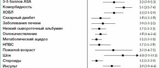
In order to answer the question of what are the morphological and clinical differences between OHSS and gastroduodenal ulcers in ulcers, we compared gastroduodenal ulcers according to various parameters (Fig. 1, 2).
For OHSS, in contrast to ulcers in PU, acute damage to the mucous membrane, localization in the stomach, multiplicity (p < 0.001) and large size (p < 0.05) are more typical. In addition, OHSS was characterized by blurred clinical manifestations and more frequent bleeding with varying degrees of blood loss, which were often the only manifestation of the disease. These signs were significantly more common in OHSS than in PU (p < 0.001). Symptomatic ulcers, like ulcers in PU, were relatively rarely complicated by perforation.
The difficulty of distinguishing between OHSS and PU is further aggravated by the fact that extreme situations in which symptomatic ulcers form can also occur in patients suffering from PU. Taking into account this circumstance, the following variants of OHSS have been identified: 1) acute symptomatic ulcers (erosions); 2) chronic symptomatic ulcers; 3) ulcers provoked by ulcerogenic influences in patients with ulcers.
With the latter option, ulcer remains the main diagnosis, but the extreme situation imposes peculiarities on the course of the provoked relapse, which requires a special approach to treatment. This option includes cases when the patient had indications of ulcers in the anamnesis or when, in response to ulcerogenic effects, single ulcers of typical localization arose (lesser curvature of the stomach, duodenal bulb). Variants of gastroduodenal ulcerations in OHDS are presented in Table. 2.
. Variants of gastroduodenal ulcerations in patients with OHSS.
Stress ulcerations are predominantly acute, but they also easily occur in patients with a history of ulceration, i.e., a provoked relapse of ulceration was observed. Ulcers combined with severe diseases of internal organs were chronic in almost 40%, and in 19% of cases a provoked relapse of ulcer was also noted. Medicines with side ulcerogenic properties more often provoke a relapse of ulcerative disease or implement a genetic predisposition to the disease, but can also cause the formation of acute multiple defects in the mucous membrane of the stomach and duodenum. Most authors only refer to the last option as SGDS.
Features of diagnostics of OHSS
Diagnosis of OHSS due to the blurred clinical symptoms is difficult. As follows from a comparison of intravital diagnosis and sectional data, OHSS are recognized during life only in 43.3% of cases.
Gastrointestinal bleeding (GIB) is a typical complication of OHSS. Ulcerative bleeding is observed in approximately a third of patients when an ulcer develops against the background of a severe somatic illness and in half of the cases of stress ulcers. Often, gastrointestinal tract is the only manifestation of OHSS. However, even severe gastrointestinal tract infections, especially in patients in critical condition, may not be diagnosed and are diagnosed only at autopsy.
Identification of background diseases and pathological processes leading to the development of OHSS is a prerequisite for establishing the correct diagnosis.
X-ray examination is not a fairly reliable method, especially in recognizing erosions and acute superficial ulcers, but it allows one to exclude large ulcers and malignant ulcerations. The endoscopic method provides complete information about the condition of the mucous membrane of the gastroduodenal zone and allows a final diagnosis to be made.
In the analyzed observations, gastroduodenoscopy was performed in 468 patients with OHSS. The results of endoscopic examinations in 121 cases were checked at surgery (18) and autopsy (103). Of these, only 20 (16.5%) endoscopist reports were incomplete. In 16 cases, ulceration with ongoing bleeding was not diagnosed, and in 4 cases, a large gastric ulcer was mistaken for malignant.
Indications for endoscopic examination are: 1) the presence of clinical symptoms indicating the possibility of gastroduodenal ulcerations; 2) a high risk of OHS formation, even in the absence of gastroenterological complaints; 3) signs of bleeding from the upper gastrointestinal tract. Exploratory endoscopic examination can be a justified method for identifying the causes of pain in the epigastrium, gastric dyspepsia in somatically severe patients, in patients who have undergone surgery, trauma, or who have been taking medications for a long time that have ulcerogenic properties. Therefore, preventive endoscopic examinations should be performed more often in patients with a high risk of OHSS, regardless of the presence of gastroenterological complaints. Urgent gastroduodenoscopy is performed for urgent indications when the patient develops symptoms of bleeding from the upper gastrointestinal tract. For patients in critical condition (in the acute phase of myocardial infarction, stroke, in the early postoperative period), endoscopy is prescribed for life-threatening bleeding.
When performing endoscopy for emergency indications, it is advisable to adhere to the following rules. Firstly, the severity of the patient’s condition should not be an absolute contraindication to endoscopic examination; the examination should only be carried out in the most gentle manner possible. This is achieved by good psychological preparation of the patient, thorough anesthesia, the use of anesthesia when necessary, and reducing the duration of the examination.
Secondly, gastroduodenoscopy should be carried out under conditions in which the patient can be provided with appropriate emergency care. Patients in the acute phase of myocardial infarction or stroke are examined in intensive care and intensive care units; For gastrointestinal tract problems that occur after major operations, injuries, burns, endoscopic examination is usually carried out in a dressing room or operating room; non-transportable somatic patients are examined on site in the treatment room or directly in the ward.
During urgent endoscopy, it is necessary to identify the source of the gastrointestinal tract and determine whether it continues. If the gastrointestinal tract continues, an attempt is made to local hemostasis through the endoscope. However, due to the technical difficulties associated with conducting the study without prior preparation and often in conditions of ongoing bleeding, in 8–18% of cases the source of the gastrointestinal tract cannot be detected.
The endoscopic picture of SGDN differs from that of PU.
Identification of background diseases and pathological processes leading to the development of OHSS is a prerequisite for establishing a final diagnosis.
A responsible and difficult task is to carry out a differential diagnosis between large, often “senile” ulcers and malignant ulcerations of the stomach. The final diagnosis is usually established using cytological and histological examination of material from targeted biopsies from the edge and bottom of the ulcer. Often, repeated endoscopic examinations with multiple targeted biopsies are necessary to determine the true nature of the ulcerative lesion of the gastric mucosa.
Diet and bad habits for stomach ulcers
Diet for gastrointestinal ulcers is an essential part of treatment. During the acute period, nutrition should be as gentle as possible, gentle on the mucous membrane and not stimulating the secretory activity of the stomach. In the first two days, preference is given to slimy soups with rice water without vegetables and meat, jelly or warm tea with sugar and crackers. In the next 6 days, table No. 1 is used in therapeutic nutrition.
Authorized Products
General nutrition rules:
- include easily digestible foods in the diet;
- food should be warm
- Do not eat cold or hot foods;
- you need to eat slowly, in small portions
- products need to be crushed and ground;
- Baking, boiling, stewing is allowed;
- fried foods and spices are excluded;
- the break in eating should not be more than 3 hours;
- food must be chewed thoroughly.
The following foods and dishes are allowed for stomach ulcers:
- Slimy soups: with the addition of rice, semolina, oatmeal, you can add cream or butter;
- Slimy porridges: with water or milk from the same cereal, you can add cream;
- Fish and meat: lean beef, veal, turkey, rabbit, chicken. To prepare the meat, you need to boil it, pass it through a meat grinder, add butter, bake it into a soufflé puree, you can cook steamed cutlets, meatballs, meatballs;
- Eggs: soft-boiled, or steam omelet;
- Berries and fruits: baked unsweetened fruits, dried fruit compotes;
- Dairy products: milk, steamed pureed cottage cheese soufflé, cream.
- Drinks: weak tea with added cream or milk, rosehip decoction, milk or fruit jelly.
You can add refined vegetable oil or unsalted butter to dishes.
Prohibited Products
During an exacerbation, foods that irritate the gastric mucosa and increase the production of gastric juice are excluded: fatty meat, baked goods, preserves, canned and smoked foods, confectionery with cream, dishes with vinegar and spices, garlic.
Do not include vegetables with coarse fiber in the diet, as well as radishes, radishes, daikon, cabbage, tomatoes, turnips, and mushrooms.
It is necessary to avoid foods that cause fermentation processes: legumes, sweet fruits and juices, carbonated drinks.
During the treatment period, you are strictly prohibited from smoking and drinking alcohol.
Prevention of peptic ulcers
Stomach ulcers can be avoided if you follow some rules. First of all, give up bad habits, since ethyl alcohol and tobacco reduce the protective properties of the mucous membrane and irritate it.
Diet is no less important in prevention: you need to eat at a certain time, the interval between meals should not exceed 4 hours. You cannot eat dry food, as dry rough food injures the walls of the stomach.
For those who work at night, it is recommended to change jobs to work only during the daytime. It is necessary to avoid stress, treat inflammation of the stomach, hormonal diseases, and visit a gastroenterologist at least once every six months.
Herbal medicine
During the treatment of erosion of the gastric mucosa, decoctions and infusions of medicinal herbs are widely used. The following herbal medicine drugs are indicated:
- St. John's wort
- Chamomile first aid kit
- Peppermint
- Oregano
- Dill
- Shepherd's Purse
- Burdock root
The use of these drugs must be agreed with your doctor. All these herbs have been tested and are widely used in official medicine. Decoctions are made using classical technology. Dry raw materials – 1 teaspoon – are poured with boiling water and left to cool completely.
Important! All herbs have contraindications - for example, pregnancy or a history of cancer. Therefore, do not self-medicate and coordinate the use of herbs with a gastroenterologist.