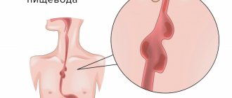
Barrett's esophagus (BE) is a disease in which an area of stratified squamous epithelium of the esophagus is replaced by metaplastic columnar epithelium, resembling the lining of the stomach or small intestine. This condition in medicine is called dysplasia, and its main danger is the high risk of malignancy, i.e. turning into cancer.
- Symptoms
- Forms of Barrett's esophagus
- Causes
- Diagnostics
- Treatment of Barrett's esophagus
- Complications
- Prevention
- Forecast
Symptoms
The symptoms of Barrett's esophagus are similar to gastroesophageal reflux disease, since it is the reflux of gastric contents that is the main cause of the development of this pathology. The patient may be concerned about:
- Heartburn. It can occur after eating, active physical activity, or bending the body. It should be noted that metaplastic epithelium is less sensitive to the action of gastric juice, so in such patients the symptoms of heartburn are mild. But upon questioning, it turns out that previously these symptoms in the patient were more pronounced.
- Belching.
- A burning sensation or pain behind the sternum.
- Dry cough.
- Feeling of heart failure.
- With severe metaplasia, dysphagia may develop - a violation of swallowing and the passage of a food bolus through the esophagus.
- Anemia. It develops rarely when BE is accompanied by erosive changes with hidden chronic bleeding.
General information
Barrett's syndrome was named after the British surgeon who first described ulcers and cancerous degeneration of the lower esophagus. This pathology develops as a complication of gastroesophageal reflux disease (GERD) and is distinguished by the presence in the epithelial layer of the food tube connecting the mouth to the stomach, not flat multilayered, but unusual for the norm - cylindrical small intestinal epithelium.
The disease is considered chronic. Tissue degeneration is asymptomatic, the exact mechanism is not fully understood, but it occurs in the sequence - metaplasia - dysplasia - cancer. Current recommendations boil down to regular endoscopic examinations to identify lesions in the early stages and prevent neoplastic formations.
Forms of Barrett's esophagus
The following morphological variants of Barrett's esophagus are distinguished:
- Cardiac type. It is characterized by a surface in the form of pits (foveolar surface), the cellular composition contains mucin-producing cells.
- Fundal type. With this type of pathology, in addition to mucin-producing cells, there is the presence of cells characteristic of the gastric epithelium - chief and parietal cells.
- Cylindrical cell type. This variant of the pathology histologically resembles the intestinal mucosa, since the epithelium forms folds and, in addition to mucin-producing cells, contains goblet cells.
It is the latter variant of BE that is most susceptible to dysplastic changes and transformation into cancer. Therefore, many authors suggest that only metaplasia containing goblet cells be classified as Barrett's esophagus.
In addition, the disease is classified depending on the extent of the altered area. Here, a distinction is made between short Barrett's esophagus, when the length of the altered section does not exceed 3 cm, and long esophagus, when the length exceeds 3 cm.
Causes
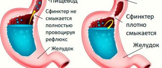
The main cause of esophageal metaplasia is gastroesophageal reflux - the reflux of gastric contents back into the esophagus. At the same time, aggressive gastric juice irritates the mucous membrane and provokes the replacement of stratified squamous epithelium with columnar epithelium that is more resistant to such effects, i.e. metaplasia occurs - the replacement of one type of tissue with another. Further irritating action leads to the fact that the metaplastic epithelium forms a clone of cells with a violation of the system of programmed death (apoptosis). This condition is called dysplasia and subsequently leads to cancer.
Additional risk factors are:
- Esophageal-diaphragmatic hernia. They lead to constant disposition of the stomach and, as a consequence, to constant reflux.
- Obesity. Firstly, obesity leads to increased intra-abdominal pressure, which provokes reflux. Secondly, in this condition there is an increase in the volume of peri-esophageal fiber, which releases pro-inflammatory cytokines that negatively affect the esophageal mucosa.
- Smoking.
- Metabolic syndrome.
Operation
If metaplasia, as well as mild and moderate dysplasia is detected, we are talking about a benign process, in which case organ-preserving surgery is possible. If severe dysplasia or squamous cell carcinoma, a malignant disease, is diagnosed, Lewis surgery is performed. If areas of high-grade neoplasia with a likelihood of invasion are found in the area of metaplasia, then RFA can be performed. In case of deep damage, radical removal of a section of the esophageal mucosa using an endoscopic method is indicated.
Our clinic performs laparoscopy using the unique technique of Professor Puchkov. During the operation, a hiatal hernia is repaired, a physiological valve is created between the esophagus and the stomach, and pathological reflux is eliminated. Recovery occurs in a short time; already six months after the operation there is no need to take medications, there is no need to adhere to any strict diets, and for the rest of your life. The number of relapses has been reduced to 2%. Thanks to the use of modern materials during surgery, after healing there are no scars on the skin of the abdomen, only 3-4 incisions 5-10 mm long, which become invisible over time. By the way, in the clinic it is possible to simultaneously perform operations on the abdominal and pelvic organs during one anesthesia. In one operation, the clinic’s specialists can relieve patients from several pathologies at once.
Diagnostics
To make a diagnosis, an endoscopic examination with a biopsy and subsequent morphological examination of a fragment of suspicious tissue is necessary.
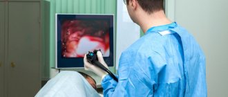
Endoscopy involves examining the wall of the esophagus using a tube equipped with a video camera, a light source and manipulation instruments. The camera transmits an enlarged image to the monitor, which allows the doctor to examine the mucous membrane in more detail. Metaplastic epithelium looks like foci of hyperemia against the background of pearly white normal epithelium of the esophagus. They are also called tongues of flame. Upon closer examination, it is clear that the metaplastic zones are atrophic; blood vessels running in the longitudinal direction are visualized in them.
If the visual picture is in doubt, the doctor can use chromoendoscopy, in which suspicious areas are stained with special dyes, for example, Lugol's solution, methylene blue, 1% acetic acid, etc.
In addition, during endoscopy the following characteristics are described:
- Length of transformed mucosa.
- Relation of the transformation zone to the gastroesophageal junction.
- Level of the proximal border and its position relative to the incisors.
- Presence of strictures.
The main point in diagnosing BE is a morphological study, in which a fragment of the obtained tissue is examined under a microscope. In this case, not only the presence of metaplasia and goblet cells is noted, but also a search is conducted for foci of dysplasia and possible adenocarcinoma of the esophagus. As a rule, a biopsy is used to obtain material, in which a piece of tissue is split off using manipulation instruments.
Questions
How is the procedure performed?
Upper gastrointestinal endoscopy is a safe procedure and involves inserting a thin, flexible endoscope through the mouth. The doctor, carefully moving the endoscope along the esophagus, examines its inner surface for the presence of pathologically changed areas. If they are detected, a biopsy is performed - taking tissue samples from several points; after the procedure is completed, all the resulting material is sent for histology. The duration of the procedure is no more than 15 minutes.
What does a patient experience when diagnosed with Barrett's esophagus?
During the examination, the patient must be in a calm state; it is important that there is no gag reflex, which causes peristaltic contractions. Therefore, the examination is carried out under intravenous sedation, the patient does not experience any unpleasant sensations during the procedure, and the doctor has the opportunity to perform a high-quality examination and make an accurate diagnosis. To perform video fiberoscopy, our clinic uses anesthetics made in the USA.
What is the main advantage of treatment at the Swiss University Hospital?
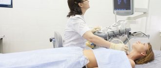
In our clinic, we first perform laparoscopic fundoplication - an operation on the esophagus, after which, 3-4 months later, RFA is performed. However, in some hospitals, surgical treatment is performed in the reverse order. But, as ultrasound studies show, against the background of RFA, swelling of the esophageal wall persists, as a result of which fundoplication at an earlier stage is fraught with stenosis in the area between the esophagus and stomach. Recovery also takes longer, because the reflux of stomach contents still remains uncorrected. Therefore, in our clinic, the first thing we do is get rid of pathological reflux, and only then RFA. Often, after the operation, normalization of the mucous membrane is noted as a result of the body’s own recovery processes, and the need for RFA either disappears, or only 1-2 sessions may be required.
Why is Barrett's esophagus dangerous?
If there is no treatment or if the disease is detected at late stages, there is a high probability of the process becoming malignant. Then the patient will have to undergo long-term treatment, and the prognosis of the disease is not always optimistic.
Treatment of Barrett's esophagus
Drug therapy
As part of drug therapy, long courses of drugs that suppress the secretion of hydrochloric acid are prescribed. First, basic therapy is carried out for 8-12 months, and then they move on to maintenance treatment. The main drugs are proton pump inhibitors, which are prescribed in dosages exceeding recommendations for the treatment of GERD. In addition, antacids that neutralize stomach acid, prokinetics, etc. can be used.
The goal of drug therapy is:
- Control over the secretion of gastric juice, in particular its reduction.
- Reducing the damaging effect of acid on the walls of the esophagus.
- Relieving symptoms and improving the quality of life of such patients.
- Creating conditions for restoration of normal epithelium.
- Reducing the risk of dysplasia and malignant transformation.
Surgery
Antireflux operations are used as part of surgical treatment. They are carried out according to the following indications:
- Presence of a hiatal hernia.
- Insufficiency of the function of the lower esophageal sphincter, proven manometrically and radiologically.
- Ineffectiveness of drug therapy.
The following types of operations are used:
- Fundoplication is the strengthening of the lower esophageal sphincter by creating a cuff around it from the walls of the fundus of the stomach.
- Crurorrhaphy is an operation aimed at suturing the hernial orifice in the diaphragm and strengthening the esophageal-diaphragmatic ligament.
It should be emphasized that surgical treatment does not lead to the abolition of drug therapy. It still has to be continued.
Endoscopic treatment
None of the above methods leads to complete regression of the metaplastic epithelium. Therefore, another stage of treatment is used - endoscopic ablation of the altered mucosa. For this purpose, various technologies are used:
- Argon plasma coagulation.
- Cryoablation.
- Electrocoagulation.
- Photodynamic therapy.
- Laser therapy.
- Radiofrequency ablation.
These methods make it possible to remove not only the metaplastic epithelium, but also its precursor cells - stem cells that have acquired differentiation features characteristic of the intestinal epithelium.
One of the most effective and safe endoscopic treatment technologies is radiofrequency ablation. Its action is based on the thermal effect on the lesions and their destruction. In this case, the doctor can control both the temperature of tissue heating and the depth of exposure. This allows for complete ablation with minimal risks of adverse reactions.
Complications
The main complication of Barrett's esophagus is malignant transformation into adenocarcinoma secondary to dysplasia.
On average, severe dysplasia is diagnosed in 20-25% of patients within 20-23 years after diagnosis. The likelihood of its development correlates with the length of the affected area and the degree of dysplasia:
- With low-grade dysplasia, the risks of malignancy are 0.8-1.9% per year.
- For severe dysplasia - 6-12.2% per year.
In addition, the number of foci of dysplasia is important. With multiple lesions, the likelihood of malignancy is three times higher than with single lesions.
Rehabilitation
Already from the first day, the operated patient gets out of bed, and the next day is allowed to take liquid food. Discharge from the clinic is possible after 1-3 days; a person begins work, as a rule, after 2-3 weeks. Following a strict diet is necessary for two months, then you can switch to a softer one, which should be followed for at least six months.
After RFA, the patient is under medical supervision for several months, during which time he is prescribed gastroscopy. If after surgery for a hernia (HH) the examination shows a decrease in the affected area of the mucous membrane, then the patient continues to be observed and is recommended to undergo FGS every six months. You can get a conclusion about the presence or absence of Barrett's esophagus after a year. If metaplasia is still present, RFA is indicated. In the future, the patient is under the supervision of a doctor, undergoing regular examinations.
Prevention
The main point of preventing Barrett's esophagus is timely diagnosis and treatment of reflux. For early detection of BE, it is recommended to perform esophagoscopy with clarifying diagnostics and targeted biopsy in patients with GERD for more than 5 years. Particular attention should be paid to men over 50 years of age, patients with obesity, GERD for more than 10 years and heartburn for more than 5 years.
General preventive measures, although trivial, also have a positive effect:
- Weight reduction to normal.
- To give up smoking.
- A balanced diet with sufficient amounts of fresh vegetables, fruits, vitamins C and E.
- Refusal of alcohol abuse, especially strong alcoholic drinks.
Healthy lifestyle
The effectiveness of treatment largely depends on compliance with nutritional recommendations and maintaining a healthy lifestyle.
For patients with GERD and Barrett's syndrome, the following recommendations have been developed:
- Weight control. It is necessary to reduce weight in case of obesity and maintain it within the age norm.
- It is necessary to avoid wearing tight belts and tight clothing. There is evidence that excess pressure on the abdominal wall contributes to the worsening of GERD.
- You should avoid eating foods that cause heartburn. Traditionally, it includes spicy, fatty and fried foods. Each patient also has an individual list of foods and drinks that lead to discomfort.
- It is necessary to completely stop smoking and drinking alcohol.
- It is recommended to maintain a drinking regime.
- You should not go to bed immediately after eating. It is better to have dinner 3-4 hours before bedtime.
- Patients who suffer from heartburn at night are advised to raise the head of the bed. It is important that the entire upper half of the body is slightly elevated - pillows will not help.
You can undergo a full examination and begin timely treatment for Barrett's syndrome at the CELT multidisciplinary clinic.